. 2024 Jan 8;18(1):wrae175.
doi: 10.1093/ismejo/wrae175.

Cyanorhodopsin-II represents a yellow-absorbing proton-pumping rhodopsin clade within cyanobacteria

Affiliations

Cyanorhodopsin-II represents a yellow-absorbing proton-pumping rhodopsin clade within cyanobacteria

Masumi Hasegawa-Takano et al. ISME J. .

Abstract

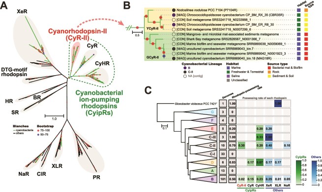
Microbial rhodopsins are prevalent in many cyanobacterial groups as a light-energy-harvesting system in addition to the photosynthetic system. It has been suggested that this dual system allows efficient capture of sunlight energy using complementary ranges of absorption wavelengths. However, the diversity of cyanobacterial rhodopsins, particularly in accumulated metagenomic data, remains underexplored. Here, we used a metagenomic mining approach, which led to the identification of a novel rhodopsin clade unique to cyanobacteria, cyanorhodopsin-II (CyR-II). CyR-IIs function as light-driven outward H+ pumps. CyR-IIs, together with previously identified cyanorhodopsins (CyRs) and cyanobacterial halorhodopsins (CyHRs), constitute cyanobacterial ion-pumping rhodopsins (CyipRs), a phylogenetically distinct family of rhodopsins. The CyR-II clade is further divided into two subclades, YCyR-II and GCyR-II, based on their specific absorption wavelength. YCyR-II absorbed yellow light (λmax = 570 nm), whereas GCyR-II absorbed green light (λmax = 550 nm). X-ray crystallography and mutational analysis revealed that the difference in absorption wavelengths is attributable to slight changes in the side chain structure near the retinal chromophore. The evolutionary trajectory of cyanobacterial rhodopsins suggests that the function and light-absorbing range of these rhodopsins have been adapted to a wide range of habitats with variable light and environmental conditions. Collectively, these findings shed light on the importance of rhodopsins in the evolution and environmental adaptation of cyanobacteria.

Keywords: cyanobacteria; ecology; evolution; microbial rhodopsin.


Conflict of interest statement

The authors declare no competing interests.

Figures

Figure 1
Figure 1
Phylogeny of cyanobacterial rhodopsins and distribution among cyanobacterial lineages: (A) A maximum likelihood tree of amino acid sequences of 102 cyanobacterial and 69 non-cyanobacterial rhodopsins; rhodopsin clades are indicated as follows: NaR, ClR (Cl pumping rhodopsin), XLR, PR, SR (sensory rhodopsin I and sensory rhodopsin II), BR, HR (halorhodopsin), DTG-motif rhodopsin, XeR, CyHR, CyR, and an uncharacterized cyanobacteria-specific clade (CyR-II); CyipRs are phylogenetically distinct cyanobacteria-specific rhodopsins consisting of CyR-II, CyR, and CyHR; the green branches indicate cyanobacterial rhodopsins, and the black branches indicate others; bootstrap probabilities (≥50%) are indicated by colored circles; the scale bar represents the number of substitutions per site; (B) An enlarged view of the novel cyanobacteria-specific clade CyR-II with habitat and source type information; the clade is divided into two subclades: YCyR-II (yellow-absorbing CyR-II) and GCyR-II (green-absorbing CyR-II); the circles in front of the sequence names were colored based on cyanobacterial lineages; names starting with [CON] and [MAG] indicate that the sequence is from the metagenomic contig and MAG, respectively; (C) distribution of rhodopsins in cyanobacterial lineages; the phylogenomic tree was constructed by maximum likelihood estimation based on conserved amino acid sites of 120 ubiquitous single-copy genes of bacteria and collapsed by lineages; “genomes” indicates the number of genomes per lineage, and “rhodopsin-harboring genomes” indicates the number of genomes containing rhodopsin; the numbers within the dashed squares indicate the fraction of genomes that contain each clade of rhodopsins in comparison with the total number of genomes in a lineage.
Figure 2
Figure 2
Characterization of P7104R. (A) Alignment of critical amino acids of light-driven outward H+ pumping rhodopsins and cyanobacterial rhodopsins. Rhodopsin names and clades (also see Fig. 1A) are indicated as follows: P7104R (Nodosilinea nodulosa PCC 7104 rhodopsin, CyR-II), CBR35R (Chroococcidiopsidaceae cyanobacterium CP_BM_RX_35 rhodopsin, CyR-II), MAG18R (uncultured cyanobacterium SRR6869040_bin.18 rhodopsin, CyR-II), N2098R (Calothrix sp. NIES-2098 rhodopsin, CyR), N4075R (Tolypothrix sp. NIES-4075 rhodopsin, CyR), MrHR (Mastigocladopsis repens halorhodopsin, CyHR), SyHR (Synechocystis halorhodopsin, CyHR), ASR (Anabaena sensory rhodopsin, XeR), BR (bacteriorhodopsin, BR), PR (proteorhodopsin, PR), and GR (Gloeobacter rhodopsin, XLR). The “No.” indicates the number of amino acids counted from the first amino acid (“start codon,” also see Fig. S3). The known function of displayed amino acids are as follows: primary proton acceptor (Asp85BR), proton donor (Asp96BR), proton release group (Glu194BR and Glu204BR), counterion (Asp212BR), and Schiff base (Lys216BR). Two carboxylates, Asp (D) and Glu (E), are shown in blue and green, respectively, and Schiff base Lys (K) is shown in red. Specific amino acid residues (motif sequences) crucial for ion transport activity were shown by pink background (No. 85, 89, and 96 in P7104R). The Lys residue (Lys216BR), which forms a Schiff base linkage with the retinal, was highlighted by yellow background. (B) Light-induced changes of the pH of suspensions of E. coli expressing P7104R, CBR35R, and MAG18R. The changes in pH in the absence (solid line) and presence (broken line) of the proton-selective ionophore, 30 μM CCCP, are shown. The measurements were performed under the dark conditions (gray shading) with illumination at 520 ± 10 nm for 3 min (white shading, from 180 sec to 300 sec).
Figure 3
Figure 3
Absorption spectra of CyR-IIs and structure of P7104R. (A) UV–Vis spectra of three CyR-IIs, P7104R (purple line), CBR35R (green line), and MAG18R (red line). The location of amino acids around the retinal chromophore in P7104R (B) and N2098R (C, CyR of Calothrix sp. NIES-2098, PDB code 6LM0). UV–Vis spectra of P7104R mutants (D), rearranged in putative critical amino acids contributing to color-tuning.

References

    1. Miyashita H, Ikemoto H, Kurano Net al. . Chlorophyll d as a major pigment. Nature 1996;383:402–2. 10.1038/383402a0 - DOI
    1. Sanfilippo JE, Garczarek L, Partensky Fet al. . Chromatic acclimation in cyanobacteria: a diverse and widespread process for optimizing photosynthesis. Ann Rev Microbiol 2019;73:407–33. 10.1146/annurev-micro-020518-115738 - DOI - PubMed
    1. Urbach E, Robertson DL, Chisholm SW. Multiple evolutionary origins of prochlorophytes within the cyanobacterial radiation. Nature 1992;355:267–70. 10.1038/355267a0 - DOI - PubMed
    1. Larkum AW, Scaramuzzi C, Cox GCet al. . Light-harvesting chlorophyll c-like pigment in Prochloron. Proc Natl Acad Sci USA 1994;91:679–83. 10.1073/pnas.91.2.679 - DOI - PMC - PubMed
    1. Partensky F, Hess WR, Vaulot D. Prochlorococcus, a marine photosynthetic prokaryote of global significance. Microbiol Mol Biol Rev 1999;63:106–27. 10.1128/MMBR.63.1.106-127.1999 - DOI - PMC - PubMed

LinkOut - more resources