. 2025 Mar;132(3):407-417.
doi: 10.1007/s00702-024-02856-1. Epub 2024 Nov 1.

High correlation of quantitative susceptibility mapping and echo intensity measurements of nigral iron overload in Parkinson's disease

Affiliations

High correlation of quantitative susceptibility mapping and echo intensity measurements of nigral iron overload in Parkinson's disease

Adrian Konstantin Luyken et al. J Neural Transm (Vienna). 2025 Mar.

Abstract

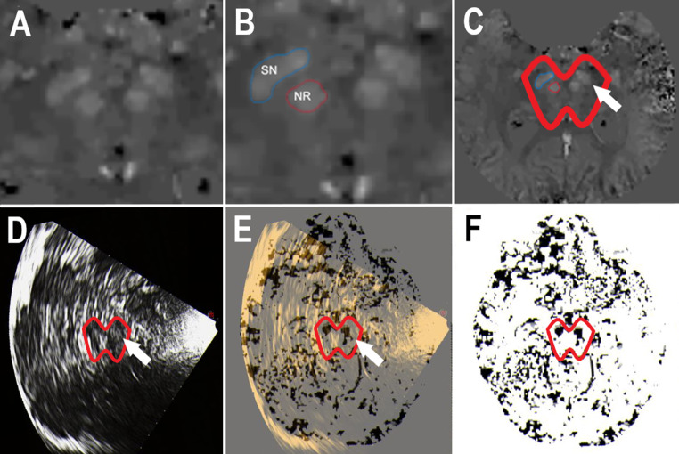
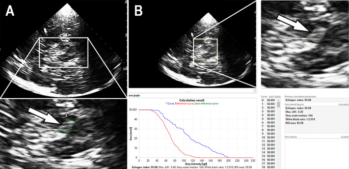
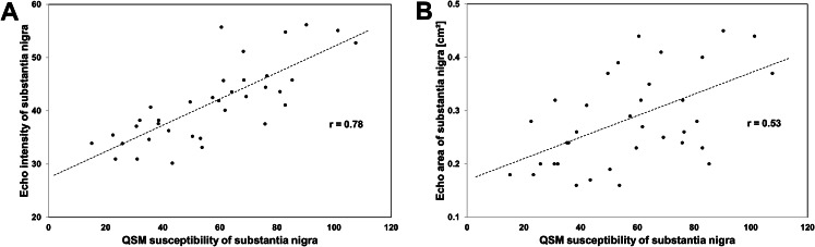
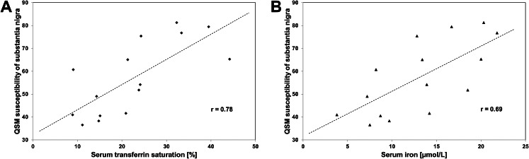
Quantitative susceptibility mapping (QSM) and transcranial sonography (TCS) offer proximal evaluations of iron load in the substantia nigra. Our prospective study aimed to investigate the relationship between QSM and TCS measurements of nigral iron content in patients with Parkinson's disease (PD). In secondary analyses, we wanted to explore the correlation of substantia nigra imaging data with clinical and laboratory findings. Eighteen magnetic resonance imaging and TCS examinations were performed in 15 PD patients at various disease stages. Susceptibility measures of substantia nigra were calculated from referenced QSM maps. Echogenicity of substantia nigra on TCS was measured planimetrically (echogenic area) and by digitized analysis (echo-intensity). Iron-related blood serum parameters were measured. Clinical assessments included the Unified PD Rating Scale and non-motor symptom scales. Substantia nigra susceptibility correlated with echogenic area (Pearson correlation, r = 0.53, p = 0.001) and echo-intensity (r = 0.78, p < 0.001). Individual asymmetry indices correlated between susceptibility and echogenic area measurements (r = 0.50, p = 0.042) and, more clearly, between susceptibility and echo-intensity measurements (r = 0.85, p < 0.001). Substantia nigra susceptibility (individual mean of bilateral measurements) correlated with serum transferrin saturation (Spearman test, r = 0.78, p < 0.001) and, by trend, with serum iron (r = 0.69, p = 0.004). Nigral echogenicity was not clearly related to serum values associated with iron metabolism. Susceptibility and echogenicity measurements were unrelated to PD duration, motor subtype, and severity of motor and non-motor symptoms. The present results support the assumption that iron accumulation is involved in the increase of nigral echogenicity in PD. Nigral echo-intensity probably reflects ferritin-bound iron, e.g. stored in microglia.

Keywords: Magnetic resonance imaging; Parkinson disease; Substantia nigra; Transcranial sonography.


Conflict of interest statement

Declarations. Competing interests: A.K.L., C.L., R.V., H.R.K., S.L., A.M.W., M.W. and M.A.W. do not report competing interests. M.L. has received speaker honoraria for presentations from STADA Pharma. G.K. has received research grants from the French Society of Neuroradiology and the French Society of Radiology. A.S. has received funding from the Deutsche Forschungsgemeinschaft (German Research Association) and the Helmholtz-Association outside the present study. He has received honoraria for presentations/advisory boards/consultations from Esteve, Desitin, Lobsor Pharmaceuticals, STADA, Bial, RG Gesellschaft, Zambon, NovoNordisk and AbbVie outside the present study. He has received royalties from Kohlhammer Verlag and Elsevier Press. He serves as an editorial board member of Stem Cells International. D.D. has received funding from the French Ministry of Health: PHRC grants, French Ministry of Research: ANR, European Preclinical Research: Coen, European Clinical Research: Horizon 2020, the charities: France Parkinson, ARSLA Foundation, Cure Parkinson Trust, the Foundations: University of Lille, Credit Agricole, Bettencourt, de France. He had consultancies: Scientific Advisory Board for Abbvie, Alterity, Orkyn, Air Liquide, Apopharma, Chiesi, Lundbeck, Everpharma and Boston Scientific, PTC Therapeutics, Inflectis, Cajal Neurosciences, AB sciences, Alzprotect, Orion and has equities: InBrain Pharma, InVenis Biotherapies. U.W. has received funding from the German Federal Ministry of Education and Research (BMBF) outside the present study. He has received speaker honoraria and travel grants from Bristol-Myers Squibb, Boehringer Ingelheim Pharma, Daiichi-Sankyo, Ipsen Pharma, Merz Pharmaceuticals, Pfizer Pharma, and an unrestricted research grant from Merz Pharmaceuticals outside the present study. He serves as Joint Editor-in-Chief of the European Journal of Ultrasound (Thieme, Stuttgart, Germany).

Figures

Fig. 1
Fig. 1
Quantitative susceptibility mapping (QSM) of substantia nigra (SN) and topological correspondence with echo signals. (A) MRI susceptibility map of midbrain used for identification of bilateral SN and bilateral red nucleus. (B) MRI susceptibility map of midbrain with outlined ROIs of right-sided SN (blue edging) and nucleus ruber (NR; red edging). (C) MRI susceptibility map of brain corresponding to the scans shown in (A) and (B). The midbrain was surrounded (red line) for better recognition (arrow: left-sided SN). (D) Transcranial brain sonogram of axial transection through caudal midbrain in a patient with Parkinson’s disease. Enlarged echosignal of bilateral SN is visualized. The midbrain was surrounded (red line) for better recognition (arrow: left-sided SN). (E) Exact real-time fusion imaging with transcranial sonography and 3D referenced QSM shows the topological correspondence of SN demarcation by its echogenicity and its susceptibility. The midbrain was surrounded (red line) for better recognition (arrow: left-sided SN). (F) Referenced QSM image, with inversion of the image brightness values for better visualization. Note that the QSM plane shown in (E) and (F) has been reconstructed by the ultrasound system from the imported QSM DICOM 3D dataset according to the ultrasound plane shown in (D) and (E). The midbrain was surrounded (red line) for better recognition
Fig. 2
Fig. 2
Visualization and measurement of substantia nigra echosignals in Parkinson’s patients. (A) Transcranial brain sonogram of axial transection at caudal midbrain level. The lower panel shows the zoomed midbrain in which the substantia nigra echo signals (arrow) on the side ipsilateral to the insonation were manually surrounded for planimetric measurement of echogenic area. (B) Screen shot of the MATLAB-based software tool “Cereb B-Mode Assist” used for digitized quantification of substantia nigra echo-intensity. The right panel shows the zoomed midbrain in which an elliptical ROI of 50 mm2 was placed to cover the echo signals of the substantia nigra (arrow) on the side ipsilateral to the insonation. The automatic analysis results in the curve of the echo-intensity distribution in the ROI compared to a reference curve determined in healthy controls (lower left panel) and the calculated index value of the total substantia nigra echo-intensity (lower right panel)
Fig. 3
Fig. 3
Diagrams showing the relationship between susceptibility and echogenicity measurements of the substantia nigra in Parkinson’s patients. (A) Relationship between susceptibility and digitally analyzed echo-intensity of the substantia nigra (Pearson test, p < 0.001). (B) Relationship between the susceptibility and the planimetrically measured echogenic area of the substantia nigra (p = 0.001)
Fig. 4
Fig. 4
Diagrams showing the relationship between the measurement of circulating iron levels and the susceptibility of the substantia nigra in Parkinson’s patients. (A) Relationship between serum transferrin saturation and substantia nigra susceptibility (individual mean of bilateral measures; Spearman test, p < 0.001). (B) Relationship between serum iron and substantia nigra susceptibility (individual mean of bilateral measures; p = 0.004, not significant after Bonferroni correction with significance level set at p ≤ 0.003)

Similar articles

References

    1. Acosta-Cabronero J, Cardenas-Blanco A, Betts MJ, Butryn M, Valdes-Herrera JP, Galazky I, Nestor PJ (2017) The whole-brain pattern of magnetic susceptibility perturbations in Parkinson’s disease. Brain 140:118–131. 10.1093/brain/aww278 - PubMed
    1. Ahmadi SA, Bötzel K, Levin J et al (2020) Analyzing the co-localization of substantia Nigra hyper-echogenicities and iron accumulation in Parkinson’s disease: a multi-modal atlas study with transcranial ultrasound and MRI. Neuroimage Clin 26:102185. 10.1016/j.nicl.2020.102185 - PMC - PubMed
    1. Azuma M, Hirai T, Yamada K et al (2016) Lateral asymmetry and spatial difference of Iron Deposition in the Substantia Nigra of patients with Parkinson Disease measured with quantitative susceptibility mapping. AJNR Am J Neuroradiol 37:782–788. 10.3174/ajnr.A4645 - PMC - PubMed
    1. Beck AT, Ward CH, Mendelson M, Mock J, Erbaugh J (1961) An inventory for measuring depression. Arch Gen Psychiatry 4:561–571. 10.1001/archpsyc.1961.01710120031004 - PubMed
    1. Becker G, Seufert J, Bogdahn U, Reichmann H, Reiners K (1995) Degeneration of substantia nigra in chronic Parkinson’s disease visualized by transcranial color-coded real-time sonography. Neurology 45:182–184. 10.1212/wnl.45.1.182 - PubMed

LinkOut - more resources