. 2024 Nov;10(44):eado6765.
doi: 10.1126/sciadv.ado6765. Epub 2024 Nov 1.

A dual role for PSIP1/LEDGF in T cell acute lymphoblastic leukemia

Affiliations

A dual role for PSIP1/LEDGF in T cell acute lymphoblastic leukemia

Lisa Demoen et al. Sci Adv. 2024 Nov.

Abstract

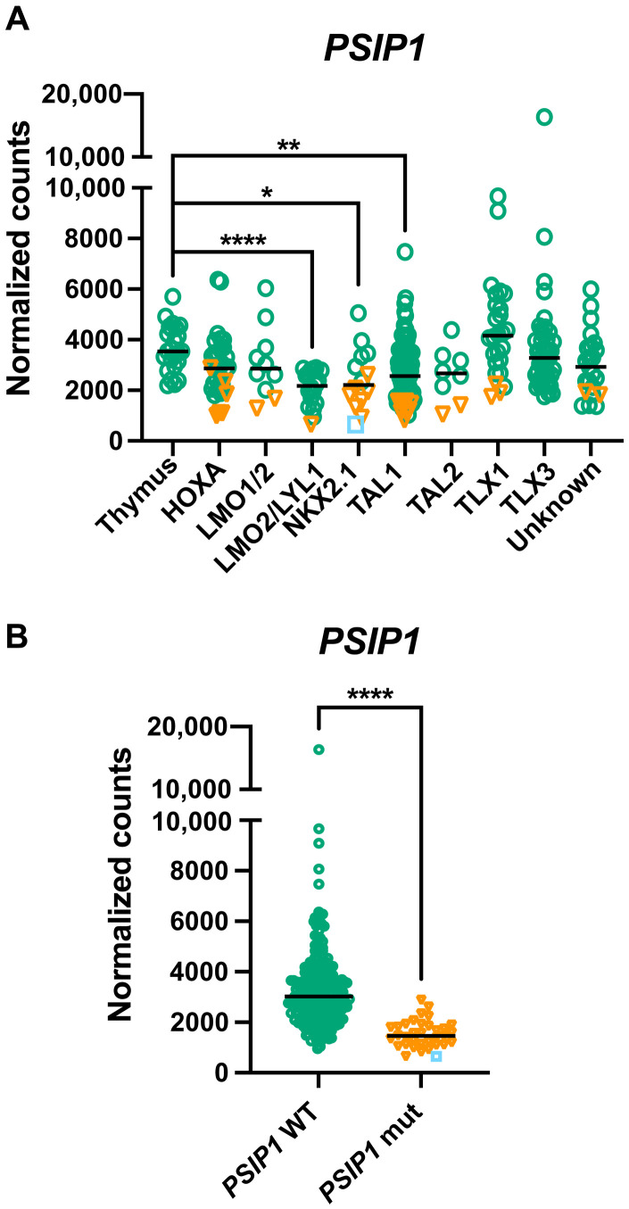
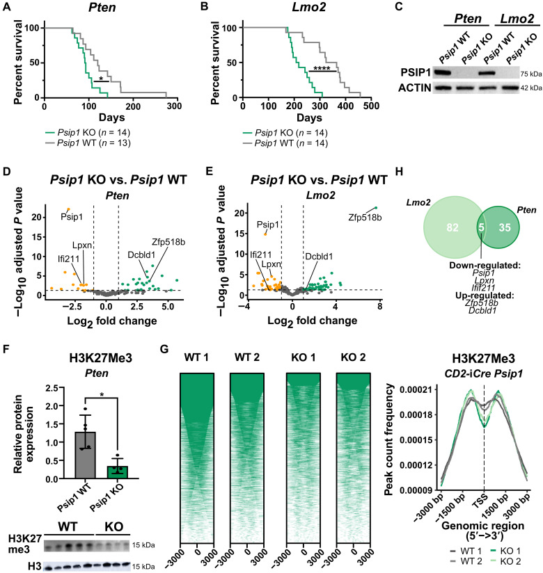
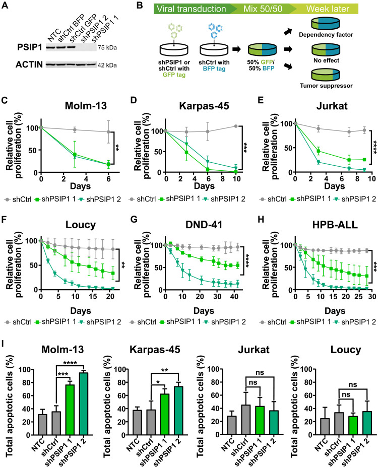
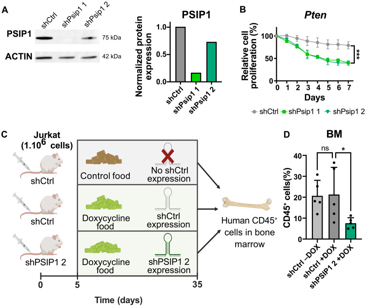
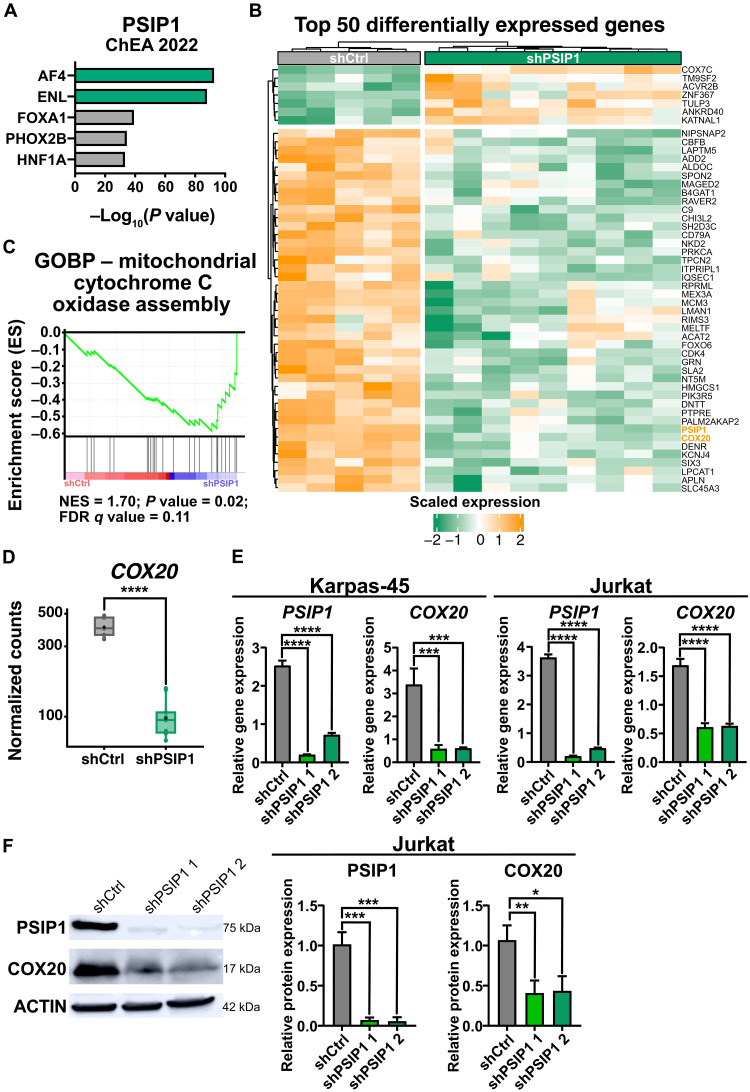
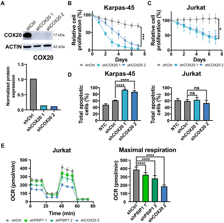
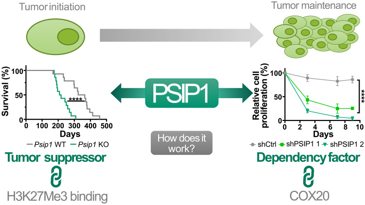
T cell acute lymphoblastic leukemia (T-ALL) is an aggressive hematological malignancy. Current intensified therapeutic protocols coincide with severe side effects, and no salvage therapy is available for primary therapy-resistant or relapsed patients. This highlights the need to identify new therapeutic targets in T-ALL. PSIP1, dispensable for normal hematopoiesis, is a dependency factor in KMT2A-rearranged myeloid leukemia. Nonetheless, loss-of-function mutations suggest a tumor suppressor role for PSIP1 in T-ALL. Here, we demonstrate that the loss of Psip1 accelerates T-ALL initiation in mice which we correlated with reduced H3K27me3 binding. Contrastingly, loss of PSIP1 impaired cell proliferation in several T-ALL cell lines. In cell lines, PSIP1 down-regulation leads to a reduction of COX20, an assembly factor of the cytochrome c oxidase in the mitochondria, and to a reduction in mitochondrial respiration. This indicates that PSIP1 can exert a dual role in the context of T-ALL, either as a tumor suppressor gene during tumor initiation or as a dependency factor in tumor maintenance.


Figures

Fig. 1.
Fig. 1.. PSIP1 is lower expressed in defined T-ALL molecular subgroups and frequently deleted in patients with T-ALL.
(A) Graph showing PSIP1 mRNA expression in thymus and T-ALL molecular subgroups after integration of RNA-seq data from 264 patients with T-ALL (22) and primary human thymus samples (24 samples derived from three independent donors) (23). Patients with chromosomal aberration targeting the PSIP1 locus are indicated as orange triangles, and one patient with an additional frame shift mutation (Y18fs) is indicated as a blue square (Kruskal-Wallis test with Dunn’s multiple comparisons test). (B) PSIP1-deleted/mutated T-ALL cases (PSIP1 mut) have lower expression levels of PSIP1 compared to cases with WT PSIP1 alleles (PSIP1 WT) (Mann-Whitney test) (23). *P < 0.05, **P < 0.01, and ****P < 0.0001.
Fig. 2.
Fig. 2.. A tumor-suppressive role for Psip1 in T-ALL tumor initiation.
(A and B) Kaplan-Meier survival curves for mice with conditional loss of Psip1 that were crossed with either the Lck-Cre Pten floxed (Pten) model (A) or CD2-Lmo2tg (Lmo2) model (B). The resulting Psip1 KO mice (Psip1fl/flLck-Cretg/+Ptenfl/fl or Psip1fl/flCD2-iCretg/+CD2-Lmo2tg/+) significantly accelerates T-ALL development compared to Psip1 WT (Psip1+/+Lck-Cretg/+Ptenfl/fl or Psip1fl/flCD2-iCre+/+CD2-Lmo2tg/+) (Mantel-Cox test). (C) Western blot validating the complete loss of Psip1 expression in tumor samples derived from the thymus of Psip1 KO and WT mice from both spontaneous T-ALL mouse models (Pten or Lmo2). (D and E) Differential gene expression was performed on thymic lymphoma samples of mice with and without Psip1 expression for both the Pten model (D) (KO: n = 9; WT: n = 6) and the Lmo2 model (E) (n = 6 in each group) (log fold change> 1; Padj < 0.05). (F) Western blot (bottom) and quantification (top) of H3K27me3 protein levels in preleukemic thymus samples of 6-week-old Pten mice, which are Psip1 KO or Psip1 WT, respectively (Mann-Whitney test). Samples were diluted 10 times for H3 Western blot to avoid overexposure. (G) CUT&RUN analysis for H3K27me3 binding in preleukemic thymus samples of 6-week-old Psip1fl/fl/CD2-iCretg/+ (KO) or Cre-negative Psip1fl/fl/CD2-iCre+/+ littermate control (WT) mice (n = 2 for each group). Individual heatmaps (left) and metanalysis (right) of H3K27me3-binding profiles −3 and +3 kb around the transcription start site. TSS, transcription start site. (H) Venn diagram of differentially expressed genes upon Psip1 KO in the PtenLck or Lmo2CD2 T-ALL model. Five common differentially expressed genes were identified: Psip1, Zfp518b, Dcbld1, Lpxn, and Ifi211. *P < 0.05 and ****P < 0.0001.
Fig. 3.
Fig. 3.. PSIP1 acts a dependency factor in T-ALL tumor maintenance.
(A) Validation of the knockdown of PSIP1 in Jurkat on protein level via Western blot. (B) Schematic representation of the in vitro proliferation assay. Cells are transduced with hairpins containing a GFP tag, which were either targeting PSIP1 (shPSIP1 1 and shPSIP1 2) or a scrambled control hairpin (shCtrl GFP). Those cells are then mixed in a 50/50 ratio with cells that were transduced with a BFP-tagged control hairpin (shCtrl BFP). Afterward, proliferation was assessed by flow cytometry. (C) The assay was validated using a KMT2Ar AML cell line, Molm-13, where loss of PSIP1 led to an impaired proliferation rate [n = 3, repeated measures two-way analysis of variance (ANOVA) with Geisser-Greenhouse correction]. A similar phenotype was observed in five different T-ALL cell lines, Karpas-45 (D), Jurkat (E), Loucy (F), DND-41 (G), and HPB-ALL (H), and was independent of the T-ALL subtype or the presence of an KMT2Ar (n = 3, repeated measures two-way ANOVA with Geisser-Greenhouse correction). (I) Annexin-7AAD stainings demonstrate that loss of PSIP1 expression induces apoptosis in KMT2Ar cell lines (Molm-13 and Karpas-45), while this is not observed in non-KMT2Ar cell lines (Jurkat and Loucy) (n = 3, time point: 6 days, one-way ANOVA with Dunnett’s multiple comparisons test). NTC, nontransduced control; ns, not significant. *P < 0.05, **P < 0.01, ***P < 0.001, and ****P < 0.0001.
Fig. 4.
Fig. 4.. PSIP1 acts as a dependency factor in a murine-derived T-ALL cell line and T-ALL maintenance in vivo.
(A) Western blot analysis (left) and quantification (right) of PSIP1 and ACTIN (loading control) in a murine Ptenfl/fl;Lck-Cretg/+ (Pten) cell line that was transduced with a scrambled control hairpin or hairpins targeting murine Psip1. The PSIP1 levels normalized against house-keeping gene β-ACTIN. (B) Loss of Psip1 expression also had a negative effect on the cell proliferation rate of a murine Pten cell line (n = 3, two-way ANOVA). (C) Schematic representation of the conducted in vivo experiment. Immunocompromised NXG mice were engrafted with one million of Jurkat cells that were either transduced with a doxycycline-inducible control hairpin (shCtrl) or a hairpin targeting PSIP1 (shPSIP1 2). After 5 days, mice were switched to doxycycline-containing food or maintained on control food. Thirty days after the start of doxycycline treatment, the mice were euthanized, and the percentage of hCD45+ leukemic cells was analyzed (created in BioRender). (D) Doxycycline-inducible knockdown of PSIP1 (shPSIP1 2) led to lower levels of hCD45-positive cells in the bone marrow (BM) compared to control mice (shCtrl) (n = 5, Kruskal-Wallis test with Dunn’s multiple comparisons test). *P < 0.05 and ***P < 0.001.
Fig. 5.
Fig. 5.. COX20 is downstream regulated by PSIP1.
(A) Enrichment for AF4 and ENL targets (ChEA2022) in the genes identified in CUT&RUN analysis for PSIP1 binding in Jurkat. (B) Heatmap representing the top 50 differentially expressed genes upon PSIP1 knockdown in Jurkat (n = 5, time: 72 hours). (C) GSEA enrichment plot shows a negative enrichment for the assembly of cytochrome c oxidase upon knockdown of PSIP1 expression in Jurkat. FDR, false discovery rate; GOBP, Gene ontology biological process; NES, normalized enrichment score. (D) Normalized counts for COX20 from RNA-seq data that were described in (B). COX20 is severely down-regulated upon down-regulation of PSIP1 expression. (E) RT-qPCR for PSIP1 and COX20 upon PSIP1 knockdown in Karpas-45 and Jurkat cells (n = 3, one-way ANOVA with Dunnett’s multiple comparisons test). (F) Western blot validation of COX20 down-regulation on protein level in Jurkat (one blot representative for three replicates, normalization: n = 3, one-way ANOVA with Dunnett’s multiple comparisons test). *P < 0.05, **P < 0.01, ***P < 0.001, and ****P < 0.0001.
Fig. 6.
Fig. 6.. COX20 is a dependency factor in T-ALL.
(A) Western blot analysis (top) and quantification (bottom) of COX20 and Actin (loading control) levels upon knockdown of COX20 in Jurkat at 72 hours. (B and C) In vitro proliferation assays, in Karpas-45 (B) and Jurkat (C), demonstrate that loss of COX20 expression has a negative effect on cell proliferation rate (n = 3, two-way ANOVA). (D) Similar to the knockdown of PSIP1, annexin-7AAD stainings demonstrate that loss of COX20 expression in a cell line with a KMT2Ar (Karpas-45) induces apoptosis, while this is not the case for a non-KMT2Ar cell line (Jurkat) (n = 3, time: 8 days, one-way ANOVA with Dunnett’s multiple comparisons test). (E) Knockdown of PSIP1 in Jurkat reduces maximal respiration in a Seahorse XF Cell Mito Stress test (n = 9 technical replicates, one-way ANOVA with Dunnett’s multiple comparisons test). Oxygen consumption rate (OCR) with the sequential injections of oligomycin A, carbonyl cyanide p-trifluoromethoxyphenylhydrazone, and antimycin A + rotenone (left) and the maximal respiration plotted separately (right). *P < 0.05, ***P < 0.001, and ****P < 0.0001.
Fig. 7.
Fig. 7.. Graphical abstract: Dual role of the epigenetic factor PSIP1 in T-ALL.
Loss-of-function mutations in patients with T-ALL in combination with an accelerated leukemic onset in two conditional Psip1 KO mouse models indicate a tumor-suppressive role for PSIP1 role in the onset of T-ALL. In addition, we propose safeguarding proper H3K27me3 occupancy as a mechanism of action for PSIP1 its tumor-suppressive function. During leukemia maintenance, we established that down-regulation of PSIP1 has a negative effect on T-ALL proliferation which is independent of KMT2A mutational status and T-ALL subtype. Furthermore, we were able to functionally link the observed phenotype to a reduced mitochondrial respiration orchestrated by down-regulation of COX20. ****P < 0.0001.

References

    1. Hunger S. P., Mullighan C. G., Acute lymphoblastic leukemia in children. N. Engl. J. Med. 373, 1541–1552 (2015). - PubMed
    1. Faderl S., O’Brien S., Pui C.-H., Stock W., Wetzler M., Hoelzer D., Kantarjian H. M., Adult acute lymphoblastic leukemia. Cancer 116, 1165–1176 (2010). - PMC - PubMed
    1. Marks D. I., Rowntree C., Management of adults with T-cell lymphoblastic leukemia. Blood 129, 1134–1142 (2017). - PubMed
    1. Eidahl J. O., Crowe B. L., North J. A., McKee C. J., Shkriabai N., Feng L., Plumb M., Graham R. L., Gorelick R. J., Hess S., Poirier M. G., Foster M. P., Kvaratskhelia M., Structural basis for high-affinity binding of LEDGF PWWP to mononucleosomes. Nucleic Acids Res. 41, 3924–3936 (2013). - PMC - PubMed
    1. Engelman A., Cherepanov P., The lentiviral integrase binding protein LEDGF/p75 and HIV-1 replication. PLOS Phatog. 4, e1000046 (2008). - PMC - PubMed

MeSH terms

Substances