. 2024 Nov 19;5(11):101804.
doi: 10.1016/j.xcrm.2024.101804. Epub 2024 Oct 31.

Degradation of IKZF1 prevents epigenetic progression of T cell exhaustion in an antigen-specific assay

Affiliations

Degradation of IKZF1 prevents epigenetic progression of T cell exhaustion in an antigen-specific assay

Tristan Tay et al. Cell Rep Med. .

Abstract

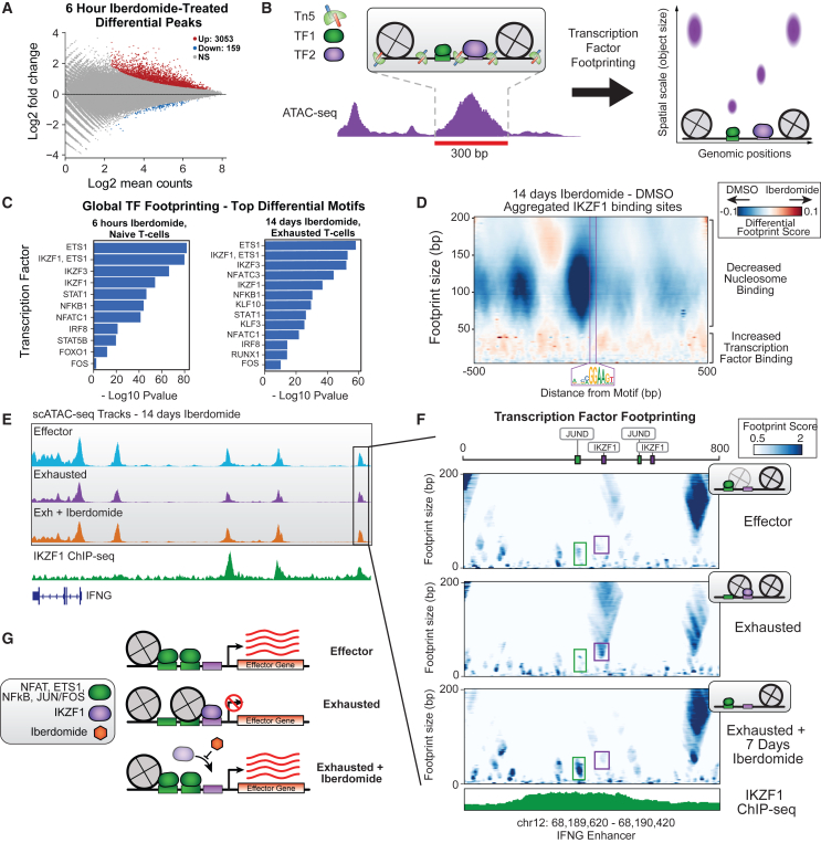
In cancer, chronic antigen stimulation drives effector T cells to exhaustion, limiting the efficacy of T cell therapies. Recent studies have demonstrated that epigenetic rewiring governs the transition of T cells from effector to exhausted states and makes a subset of exhausted T cells non-responsive to PD1 checkpoint blockade. Here, we describe an antigen-specific assay for T cell exhaustion that generates T cells phenotypically and transcriptionally similar to those found in human tumors. We perform a screen of human epigenetic regulators, identifying IKZF1 as a driver of T cell exhaustion. We determine that the IKZF1 degrader iberdomide prevents exhaustion by blocking chromatin remodeling at T cell effector enhancers and preserving the binding of AP-1, NF-κB, and NFAT. Thus, our study uncovers a role for IKZF1 as a driver of T cell exhaustion through epigenetic modulation, providing a rationale for the use of iberdomide in solid tumors to prevent T cell exhaustion.


Conflict of interest statement

Declaration of interests J.D.B. holds patents related to ATAC-seq and is an SAB member of Camp4 and seqWell. G.B., E.J., A.G., I.K.R., A.S.D., L.P., J.K., M.G.O., S.C., and D.A.M. are current or former employees of AstraZeneca. V.B.-W. is an employee of Revvity. S.L.T. was formerly an employee of Revvity.

Figures

None
Graphical abstract
Figure 1
Figure 1
A human antigen-specific model for T cell exhaustion (A) A schematic of the exhaustion assay using peptide stimulation. Figure was created using BioRender.com. (B) Tumor: T cell co-culture showing loss of cytotoxic ability of day 14 Tex. (C) Cytokine analysis for IFNγ (left, p = 0.1079), IL-2 (center, p = 0.068), and TNF-α (right, p = 0.1134) of culture supernatants measured by MSD. (D) Distribution of day 7 Teff cells in RNA UMAP. (E) Marker gene expression on UMAP. (F) Gene expression signatures of effector or exhausted T cells from current dataset or the study by Zheng et al., 2021. (G) Composition of effector or exhausted T cells by antigen stimulation time. (H) Gene expression for cells sorted by combined exhausted score. (I) ATAC-seq tracks aggregating scATAC-seq profiles from top 5% of most exhausted or most effector cells (XCL2 p = 1.2∗10−48, CRTAM p = 1.7∗10−23). (J) Differential accessibility at domains of regulatory chromatin (DORCs) associated with specific genes. Significance thresholds are absolute value log2FC > 0.1 and p < 0.01. (K) Differential TF motif activity calculated using chromVAR scores. Significance thresholds are absolute value log2FC > 0.1 and p < 0.01.
Figure 2
Figure 2
A CRISPR screen in exhausted T cells identifies IKZF1 as a driver of T cell exhaustion Iberdomide, a clinical IKZF1 degrader, prevents T cell exhaustion. (A) Schematic of the CRISPR screen workflow using Immunocult reagent to generate exhausted T cells. (B) T cells lose their ability to proliferate and produce cytokines upon repeated chronic stimulation. p values are <0.0001 for IL-2 and 0.0119 for TNF-α. (C) Heatmap of top 13 hits from CRISPR screen with increased cytokine expression and viability shown. (D) Western blot showing IKZF1 degradation after 7 days of iberdomide treatment. (E) Iberdomide treatment of cells during chronic stimulation prevents them from losing (E) cytotoxic ability and (F) cytokine secretion activity. p values are 0.0003 for IFNG, 0.0706 for IL-2, and 0.0007 for TNF-α.
Figure 3
Figure 3
Iberdomide affects gene expression of key effector/regulator genes in Tex cells (A) RNA UMAP of iberdomide and DMSO-treated cells. (B) Differential RNA expression in iberdomide vs. DMSO cells at 14 days. (C) Cumulative enrichment analysis of iberdomide-treated cells with higher effector function. (D) ATAC-seq tracks aggregating scATAC-seq profiles from iberdomide vs. DMSO-treated exhausted cells. (E) MA plot of peaks with differential chromatin accessibility in scATAC-seq profiles after iberdomide treatment. (F) Differential TF motif accessibility in iberdomide vs. DMSO-treated exhausted cells (chromVAR).
Figure 4
Figure 4
TF footprinting analysis reveals loss of nucleosomes and increased AP-1, NF-κB, and NFAT binding at effector enhancers following iberdomide treatment (A) MA plot of peaks with differential chromatin accessibility in bulk ATAC profiles after 6-h iberdomide treatment. (B) Schematic of TF footprinting analysis. (C) Top motifs with differential binding scores across all peaks in bulk ATAC (left) or scATAC-seq (right). (D) Aggregated multiscale footprinting plots of peaks containing IKZF1/ETS1 motif filtered for the highest 25% in IKZF1 ChIP-seq signal. (E) ATAC-seq tracks aggregating scATAC-seq profiles from untreated effector, untreated exhausted, or iberdomide-treated exhausted cells. IKZF1 ChIP-seq tracks from Sun et al., 2022 in purple (bottom). (F) Multiscale TF footprinting in enhancer marked in (E) from effector, exhausted, or iberdomide-treated exhausted cells. Schematics of TF and nucleosome binding inferred from the plots shown top-right of each plot with the IKZF1 ChIP-seq track from Sun et al., 2022 (bottom). (G) Model of iberdomide mechanism of action in restoring effector function.

References

    1. Tang L., Zhang Y., Hu Y., Mei H. T Cell Exhaustion and CAR-T Immunotherapy in Hematological Malignancies. BioMed Res. Int. 2021;2021 - PMC - PubMed
    1. Gumber D., Wang L.D. Improving CAR-T immunotherapy: Overcoming the challenges of T cell exhaustion. EBioMedicine. 2022;77 - PMC - PubMed
    1. Poorebrahim M., Melief J., Pico de Coaña Y., L Wickström S., Cid-Arregui A., Kiessling R. Counteracting CAR T cell dysfunction. Oncogene. 2021;40:421–435. - PMC - PubMed
    1. Friedrich M.J., Neri P., Kehl N., Michel J., Steiger S., Kilian M., Leblay N., Maity R., Sankowski R., Lee H., et al. The pre-existing T cell landscape determines the response to bispecific T cell engagers in multiple myeloma patients. Cancer Cell. 2023;41:711–725.e6. - PubMed
    1. Ghoneim H.E., Fan Y., Moustaki A., Abdelsamed H.A., Dash P., Dogra P., Carter R., Awad W., Neale G., Thomas P.G., Youngblood B. De Novo Epigenetic Programs Inhibit PD-1 Blockade-Mediated T Cell Rejuvenation. Cell. 2017;170:142–157.e19. - PMC - PubMed

MeSH terms

LinkOut - more resources