. 2024 Nov 1;14(1):26313.
doi: 10.1038/s41598-024-76664-8.

A DNA base-specific sequence interposed between CRX and NRL contributes to RHODOPSIN expression

Affiliations

A DNA base-specific sequence interposed between CRX and NRL contributes to RHODOPSIN expression

Rosa Maritato et al. Sci Rep. .

Abstract

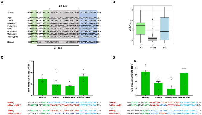
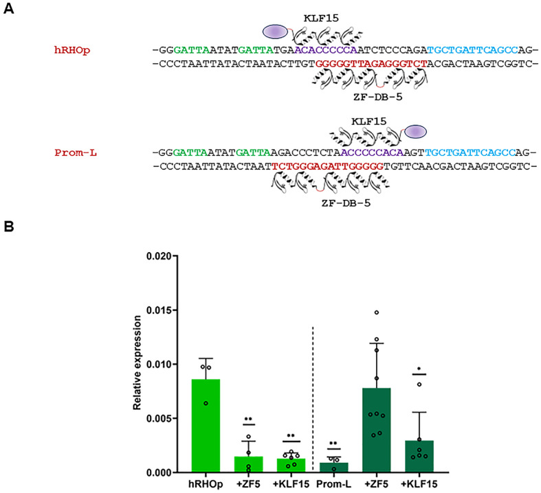
Gene expression emerges from DNA sequences through the interaction of transcription factors (TFs) with DNA cis-regulatory sequences. In eukaryotes, TFs bind to transcription factor binding sites (TFBSs) with differential affinities, enabling cell-specific gene expression. In this view, DNA enables TF binding along a continuum ranging from low to high affinity depending on its sequence composition; however, it is not known whether evolution has entailed a further level of entanglement between DNA-protein interaction. Here we found that the composition and length (22 bp) of the DNA sequence interposed between the CRX and NRL retinal TFs in the proximal promoter of RHODOPSIN (RHO) largely controls the expression levels of RHO. Mutagenesis of CRX-NRL DNA linking sequences (here termed "DNA-linker") results in uncorrelated gene expression variation. In contrast, mutual exchange of naturally occurring divergent human and mouse Rho cis-regulatory elements conferred similar yet species-specific Rho expression levels. Two orthogonal DNA-binding proteins targeted to the DNA-linker either activate or repress the expression of Rho depending on the DNA-linker orientation relative to the CRX and NRL binding sites. These results argue that, in this instance, DNA itself contributes to CRX and NRL activities through a code based on specific base sequences of a defined length, ultimately determining optimal RHO expression levels.


Conflict of interest statement

E.M.S., S.B. and E.M. are inventors on patents, “Artificial DNA-binding proteins and uses thereof”, WO2015075154A3, US20160289284A1. E.M.S., S.B. and E.M. are inventors on a pending patent “Ectopically expressed transcription factors and uses thereof”, PCT/EP2018/086782, Department of Translational Medical Sciences (DiSMeT) University of Naples "Federico II", Italy, which may encompass the findings. The remaining authors declare no competing interests.

Figures

Fig. 1
Fig. 1
Forty-nine bp containing CRX and NRL BSs control RHO expression in adult retina. (A), Proximal promoter region of the human RHO containing the Rho Regulatory Unit (hRRU, chr3:129,528,537 − 129,528,585). hRRU is composed of the BAT-1 and NRE elements (dashed grey boxes), CRX and NRL TFs binding sites (in green and light blue, respectively) and DNA-linker (in red). The “footprints” onto hRHOp of the synthetic zinc finger protein (drawing of the six zinc-finger array of ZF-DB below the negative strand, amaranth) and the TF KLF15 (drawing of KLF15 including the effector domain in violet above the positive strand). Below, schematic representation of gene reporter assay with of Adeno-associated virus (AAV8) vector carrying eGFP under the control of the RHO proximal promoter (hRHOp) injected sub-retina in adult mice (4 weeks old), on the right-handed panel immunofluorescence analysis of a cross section of a retina transduced with AAV8-hRHOp-eGFP (3 × 109 genome copies, GC), co-injected with AAV8-hGNAT1-mCherry (3 × 108 GC) 15 days post-injection. (B), Histogram, quantitative PCR analysis (qPCR), eGFP expression levels 15 days after subretinal administration of AAV8-eGFP (3 × 109 GC) co-delivered with AAV8-hGNAT1-mCherry (3 × 108 GC) to account variability associated with subretinal AAV8 vector delivery (eGFP expression levels were normalised to those of mCherry, Methods; one promoter variant per eye, three replicates). (C) qPCR upon removal of hRRU (Prom-Q) and doubling the hRRU (Prom-S), and immunofluorescence analysis on Prom-Q, and Prom-S co-injected with a AAV8 vector encoding for mCherry (eGFP expression levels were normalised to those of mCherry, Methods; three individual retinal replicates for each construct). RPE, retinal pigment epithelium; OS, outer segment; ONL, outer nuclear layer; INL, inner nuclear layer; GCL, ganglion cell layer. Statistical significance was computed using the One-Way Anova test (p-values ≤ 0.05), and post-hoc Dunnett’s multiple comparisons test (p-values ≤ 0.05).
Fig. 2
Fig. 2
Changes of the DNA sequence interposed between CRX and NRL lead to uncorrelated reporter gene expression. (A), Sequences, and histogram, qPCR analysis, eGFP expression levels 15 days after AAV8-eGFP in vivo retinal delivery in adult mice (Methods), showing the impact of change of the order, orientation, and spacing, of CRX and NRL BSs. (B), Sequences, and qPCR of nucleotide substitutions in the DNA-linker (C), Immunofluorescence analysis on AAV8-hRHOp, AAV8-Prom-N, AAV8-Prom-Me, AAV8-Prom-H, AAV8-Prom-E and AAV8-Prom-3A. RPE, retinal pigment epithelium; OS, outer segment; ONL, outer nuclear layer; INL, inner nuclear layer; GCL, ganglion cell layer. (D), Quantitative effects (qPCR) of DNA-linker sequence cloned in the 5’ and 3’ orientation (sequence, AAV8-Prom-L). Statistical significance was computed using the One-Way Anova test (p-values ≤ 0.05), and post-hoc Dunnett’s multiple comparisons test (p-values ≤ 0.05). Only in Fig. 2D we performed t-student test (p-values ≤ 0.0001).
Fig. 3
Fig. 3
Human and murine DNA-linkers diverge but lead to species-specific RHO reporter expression. (A), Multiple sequence alignment for representative mammalian species from UCSC track. The CRX and NRL binding sites are highlighted in green and blue, respectively. The DNA-linker is highlighted in grey. Conserved bases are indicated in bold. (B), Distribution of PhyloP score of DNA bases in the NRL (green box) and CRX binding sites (blue box) and in the DNA-linker of the human RRU. PhyloP score indicates the degree of conservation of a DNA base in vertebrates. (C), Sequences and qPCR analysis of mutual changes of hRRU (AAV8-mRhop-hRRU) and mRRU (AAV8-hRHOp-mRRU) insertion in murine and human Rho/RHO promoters, respectively. (D), Sequence and qPCR analysis of the human RHO promoter with 5 bp of central murine DNA-linker (ATGAT, AAV8-hRHOp-mAT) and mouse Rho promoter with 5 bp of central human DNA-linker (CCCCA, AAV8-mRhop-hCG). Statistical significance was computed using the One-Way Anova test (p-values ≤ 0.05), and post-hoc Dunnett’s multiple comparisons test (p-values ≤ 0.05).
Fig. 4
Fig. 4
Orthogonal DNA-linker binding proteins perturb reporter gene expression in vitro. (), Drawing of hRHOp and Prom-L promoters with the orthogonal protein ZF-DB-5 and KLF15 bound to the DNA-linker in the opposite direction while maintaining the same double strand side. (B), Quantitative effects (qPCR) of transfecting ZF-DB-5 or KLF15,, and Fig. 4A, in HEK-293 cell line with hRHOp (light green, left hand side, and panel B) or Prom-L (DNA-linker sequence cloned in the 3’-5’ direction, dark green, right hand side, and panel B). Statistical significance was computed using the One-Way Anova test (p-values ≤ 0.05), and post-hoc Dunnett’s multiple comparisons test (p-values ≤ 0.05).

References

    1. Jacob, F. & Monod, J. Genetic regulatory mechanisms in the synthesis of proteins. J. Mol. Biol.3, 318–356 (1961). - PubMed
    1. Gilbert, W. Müller-Hill, B. The lac operator is DNA. Proc. Natl. Acad. Sci. U S A. 58, 2415–2421 (1967). - PMC - PubMed
    1. Klug, A. Gene regulatory proteins and their interaction with DNA. Ann. N Y Acad. Sci.758, 143–160 (1995). - PubMed
    1. Pabo, C. O. & Sauer, R. T. Transcription factors: Structural families and principles of DNA recognition. Annu. Rev. Biochem.61, 1053–1095 (1992). - PubMed
    1. Rohs, R. et al. Origins of specificity in Protein-DNA recognition. Annu. Rev. Biochem.79, 233–269 (2010). - PMC - PubMed

LinkOut - more resources