. 2024 Jan-Dec:33:9636897241290367.
doi: 10.1177/09636897241290367.

Human Stem Cell-Derived Cardiomyocytes Integrate Into the Heart of Monkeys With Right Ventricular Pressure Overload

Affiliations

Human Stem Cell-Derived Cardiomyocytes Integrate Into the Heart of Monkeys With Right Ventricular Pressure Overload

Jodi Scholz et al. Cell Transplant. 2024 Jan-Dec.

Abstract

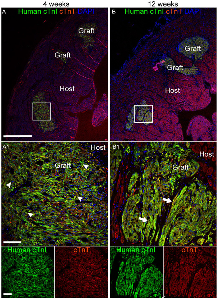
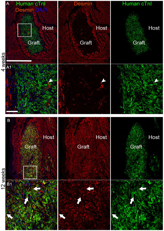
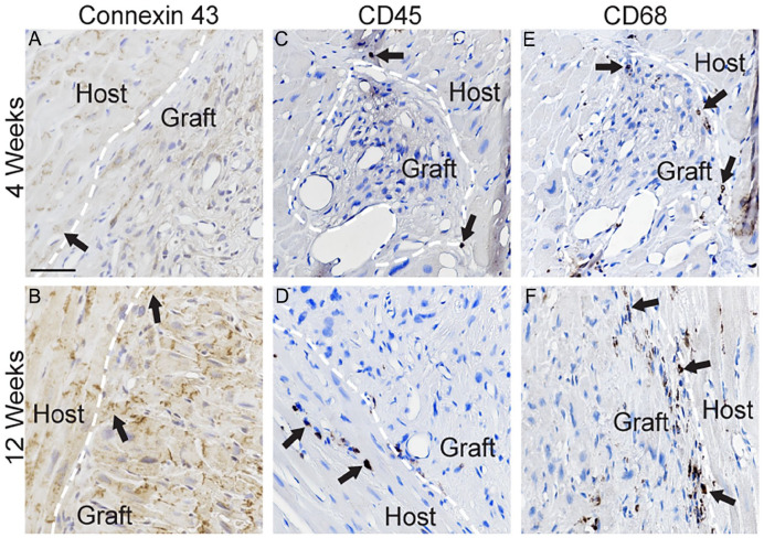
Cardiac ventricular pressure overload affects patients with congenital heart defects and can cause cardiac insufficiency. Grafts of stem cell-derived cardiomyocytes are proposed as a complementary treatment to surgical repair of the cardiac defect, aiming to support ventricular function. Here, we report successful engraftment of human induced pluripotent stem cell-derived cardiac lineage cells into the heart of immunosuppressed rhesus macaques with a novel surgical model of right ventricular pressure overload. The human troponin+ grafts were detected in low-dose (2 × 10⁶ cells/kg) and high-dose (10 × 106 cells/kg) treatment groups up to 12 weeks post-injection. Transplanted cells integrated and progressively matched the organization of the surrounding host myocardium. Ventricular tachycardia occurred in five out of 16 animals receiving cells, with episodes of incessant tachycardia observed in two animals; ventricular tachycardia events resolved within 19 days. Our results demonstrate that grafted cardiomyocytes mature and integrate into the myocardium of nonhuman primates modeling right ventricular pressure overload.

Keywords: cardiomyocytes; grafts; rhesus; right ventricular pressure overload; stem cells.


Conflict of interest statement

Declaration of Conflicting InterestsThe author(s) declared no potential conflicts of interest with respect to the research, authorship, and/or publication of this article.

Figures

Figure 1.
Figure 1.
Development and characterization of a nonhuman primate model of cardiac pressure overload. (A) Cartoon of cardiac anatomy and surgical targets. (B, C) To create right ventricular pressure overload, a left thoracotomy was performed to access the pulmonary artery (asterisk), which was encircled with umbilical tape and gradually tightened with ligating clips (white arrow). Intraoperative echocardiography was used to measure the pressure gradient across the band as it was tightened by the addition of clips until (D) the gradient reached ~15–23 mmHg (green arrow) and (E) mild-moderate tricuspid regurgitation was present. Postmortem analyses detected myofibril disarray (F, F1) (H&E staining) and (G, G1) fibrosis (Masson’s trichrome staining, blue coloration) (P5). Grafts of hiPSC-CLs (area demarcated by white dashed line) were recognized with (H, H1) H&E staining and (I, I1) Masson’s trichrome staining and confirmed by (J, J1) human cardiac troponin I (cTnI) immunostaining counterstained with hematoxylin (P7). (F1-J1) high magnification images of areas demarcated by black rectangles in (F-J). Scale bar: (F-J) 200 µm, (F1-J1) 50 µm.
Figure 2.
Figure 2.
Ventricular arrhythmia events detected after injection of hiPSC-CLs. (A) Experimental design evaluating cardiac effects of hiPSC-CLs in 28 monkeys. Representative electrocardiogram (ECG) tracings: (B) normal sinus rhythm, (C) supraventricular tachycardia, (D) premature ventricular contractions (black arrows), (E) ventricular couplet (black arrow), (F) intermittent, non-sustained ventricular tachycardia (VT), and (G) incessant VT. (H) Frequency and duration of all VT events in the five affected subjects. PAB, pulmonary artery banding; ICM, implantable cardiac monitor.
Figure 3.
Figure 3.
Grafts of human cardiac troponin I (cTnI)+ hiPSC-CLs across the right ventricular myocardium. (A) Representative images of the right ventricular myocardium of all hiPSC-CL injected animals. Cardiac tissue was immunostained for human cTnI (brown) and counterstained with hematoxylin (blue). (B) High-magnification image of area demarcated by black rectangle in HD12-1. Scale bar: (A) 1,000 µm, (B) 100 µm. (C) Heat map of human cTnI+ graft area (mm2) per subject across cardiac tissue levels. Each heart was sliced in 4-mm blocks from the apex to the base, and tissue from each block was immunolabeled for human cTnI to detect grafted cells across the entire right ventricle. The number of blocks depended on individual animal size; gray boxes indicate no right ventricle tissue at this level. White, empty boxes indicate no human cTnI+ graft detected. *, some human cTnI was detected, but graft size was not quantified due to lack of perfusion impacting immunolabeling. DNC, did not complete study.
Figure 4.
Figure 4.
hiPSC-CLs integrate into the right ventricular myocardium. (A, B) Representative images from high-dose 4-week (A, HD4-4) and high-dose 12-week (B, HD12-2) subjects showing immunofluorescence labeling of human cardiac troponin I (cTnI) (green) grafted cells integrating into the myocardium (red = rhesus and human cardiac troponin T (cTnT)) counterstained with the nuclear marker 4′,6-diamidino-2-phenylindole (DAPI; blue). Grafted cells exhibited increased organization and maturity from 4 (arrowheads—disorganized with pleomorphic morphology) to 12 (arrows—increased parallel organization of cells, spindle-like morphology, and visible cross-striations) weeks. (A1, B1) Higher magnification image of area demarcated by white squares in (A, B). Scale bar: (A, B) 1,000 µm, (A1, B1) 100 µm.
Figure 5.
Figure 5.
Desmin is abundantly present in grafted hiPSC-CLs at 12 weeks post-transplantation. (A, B) Representative images from high-dose 4-week (HD4-1) and high-dose 12-week (HD12-4) subjects showing immunofluorescence labeling of human cardiac troponin I (cTnI) (green) and desmin (red) counterstained with the nuclear marker 4′,6-diamidino-2-phenylindole (DAPI; blue). Desmin was rarely present in grafted cells at 4 weeks (white arrowhead). At 12 weeks, desmin was present in nearly all grafted cells, often displaying a striated pattern similar to cTnI (white arrows). (A1, B1) Higher magnification image of area demarcated by white squares in (A, B). Scale bar: (A, B) 1,000 µm, (A1, B1) 100 µm.
Figure 6.
Figure 6.
Cardiac grafts have similar vascularization and presence of dividing cells at 4 and 12 weeks post-cell delivery. (A–D) Representative images from high-dose 4-week (A, HD4-1; C, HD4-4) and high-dose 12-week (B, HD12-4; d, HD12-1) subjects showing immunofluorescence labeling of human cardiac troponin I (cTnI) (green) and either the endothelial marker CD31 (A, B = red; white arrows) or the dividing cell marker Ki67 (C, D = red; white arrows). Tissue was counterstained with the nuclear marker 4′,6-diamidino-2- phenylindole (DAPI; blue). Scale bar: 100 µm.
Figure 7.
Figure 7.
Expression of connexin 43 and minimal immune cell labeling in cardiac grafts. (A–F) Representative images from high-dose 4-week (A, C, E, HD4-4) and 12-week (B, HD12-1; D, F, HD12-4) subjects showing immunolabeling for the gap junction marker connexin 43 (A, B = brown; black arrows), immune cell marker CD45 (C, D = brown; black arrows), and macrophage marker CD68 (E, F = brown; black arrows). Tissue was counterstained with hematoxylin (blue). White dashed line is drawn at border of grafted cells. Scale bar: 100 µm.

References

    1. Garcia AM, Beatty JT, Nakano SJ. Heart failure in single right ventricle congenital heart disease: physiological and molecular considerations. Am J Physiol Heart Circ Physiol. 2020;318(4):H947–65. - PMC - PubMed
    1. Khairy P, Poirier N, Mercier LA. Univentricular heart. Circulation. 2007;115(6):800–12. - PubMed
    1. Lewis M, Rosenbaum M. The miracle baby grows up: hypoplastic left heart syndrome in the adult. Curr Cardiol Rep. 2017;19(8):74. - PubMed
    1. Niebler RA, Shah TK, Mitchell ME, Woods RK, Zangwill SD, Tweddell JS, Berger S, Ghanayem NS. Ventricular assist device in single-ventricle heart disease and a superior cavopulmonary anastomosis. Artif Organs. 2016;40(2):180–4. - PubMed
    1. Chu MWA, Gersch KA, Rodriguez E, Nifong LW, Chitwood WR. Robotic “haircut” mitral valve repair: posterior leaflet-plasty. Ann Thorac Surg. 2008;85(4):1460–62. - PubMed

LinkOut - more resources