. 2024 Nov 2;74(1):6.
doi: 10.1007/s00262-024-03860-w.

Three-dimensional dynamics of mesothelin-targeted CAR.CIK lymphocytes against ovarian cancer peritoneal carcinomatosis

Affiliations

Three-dimensional dynamics of mesothelin-targeted CAR.CIK lymphocytes against ovarian cancer peritoneal carcinomatosis

Federica Galvagno et al. Cancer Immunol Immunother. .

Abstract

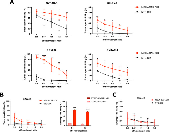
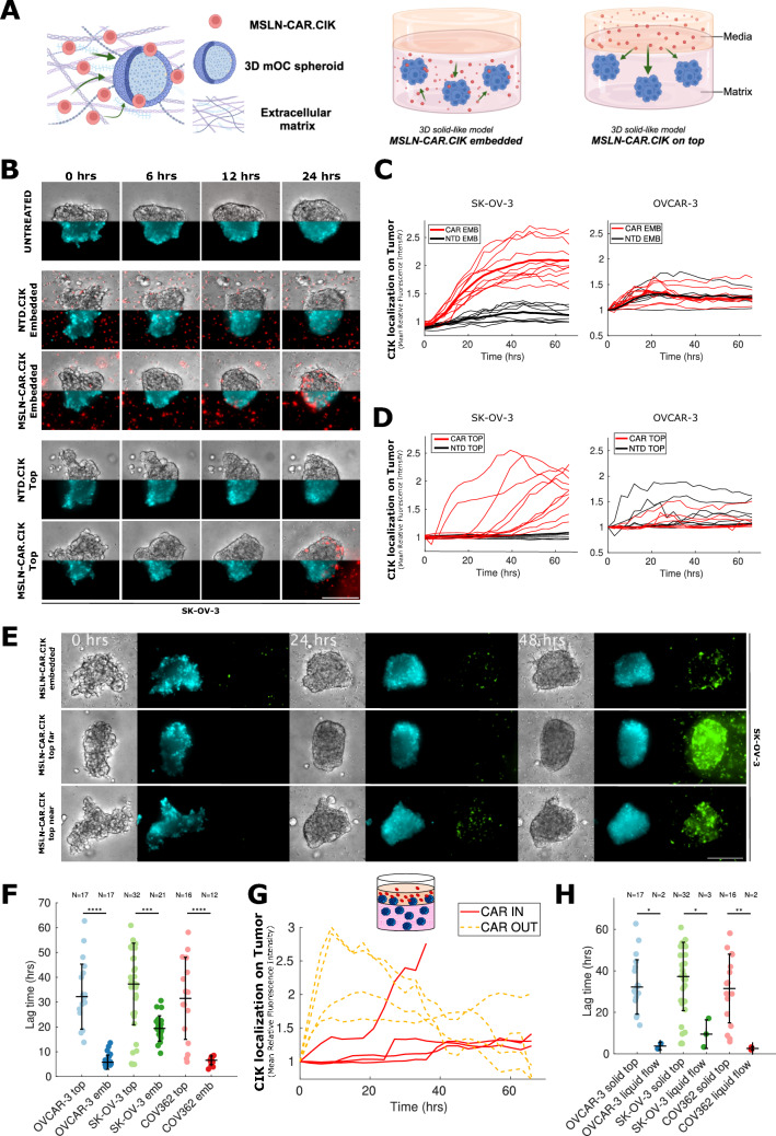
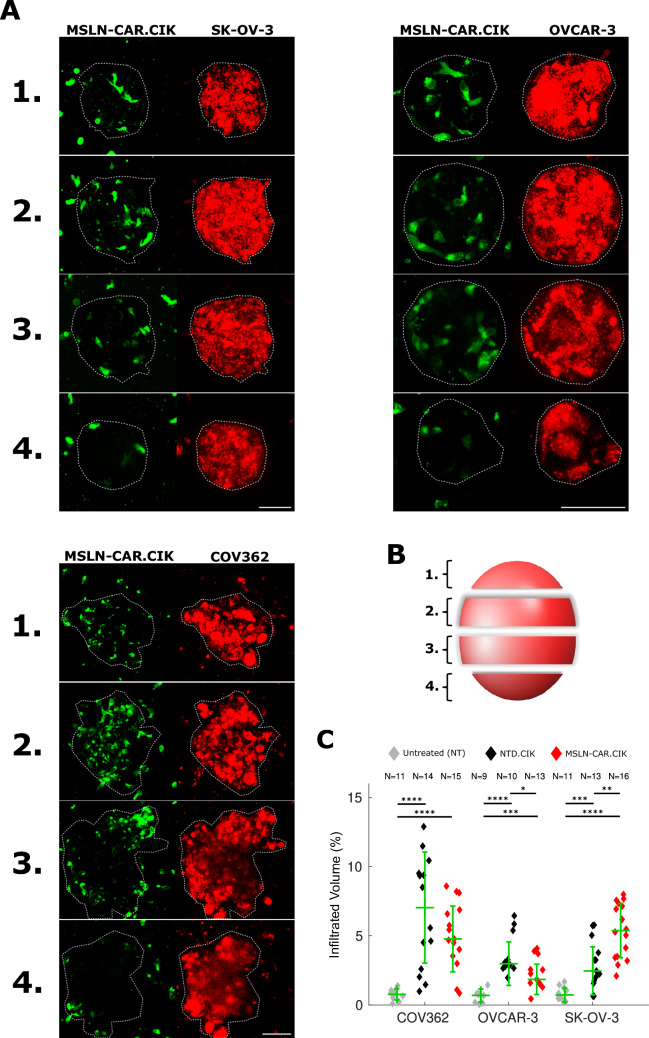
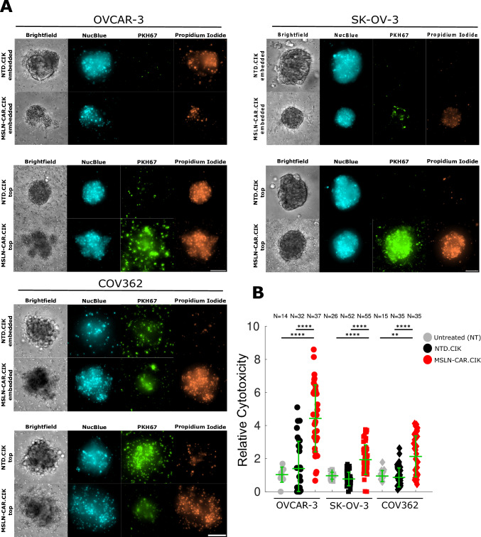
Intraperitoneal cellular immunotherapy with CAR-redirected lymphocytes is an intriguing approach to target peritoneal carcinomatosis (PC) from ovarian cancer (OC), which is currently evaluated in clinical trials. PC displays a composite structure with floating tumor cells within ascites and solid-like masses invading the peritoneum. Therefore, a comprehensive experimental model is crucial to optimize CAR-cell therapies in such a peculiar environment. Here, we explored the activity of cytokine-induced killer lymphocytes (CIK), redirected by CAR against mesothelin (MSLN-CAR.CIK), within reductionistic 3D models resembling the structural complexity of both liquid and solid components of PC. MSLN-CAR.CIK effectively killed and were functionally efficient against OC targets. In a "floating-like" 3D context with floating OC spheroids, both tumor localization and killing by MSLN-CAR.CIK were significantly boosted by fluid flow. In a "solid-like" context, MSLN-CAR.CIK were recruited through the extracellular matrix on embedded tumor aggregates, with variable kinetics depending on the effector-target distance. Furthermore, MSLN-CAR.CIK penetrated the inner levels of OC spheroids exerting effective tumor killing. Our findings provide currently unknown therapeutically relevant information on intraperitoneal approaches with CAR.CIK, supporting further developments and improvements for clinical studies in the context of locoregional cell therapy approaches for patients with PC from OC.

Keywords: 3D models; Cellular immunotherapy; Chimeric antigen receptor; Ovarian cancer; Peritoneal carcinomatosis.


Conflict of interest statement

The authors declare no potential conflicts of interest.

Figures

Fig. 1
Fig. 1
MSLN-CAR.CIK effectively and specifically kill mOC cells in 2D. A MSLN-CAR.CIK killed mOC cell lines more specifically than NTD.CIK. Tumor cell-specific cytotoxicity values from > 3 biological replicates are reported (mean ± SD). B MSLN-CAR.CIK and NTD.CIK similarly killed the MSLN low mOC cell line OAW42 (n = 4) (mean ± SD) (left). MSLN-CAR.CIK killing of mOC cells depends on MSLN expression levels at therapeutically relevant E/T ratios (n = 3/4) (mean ± SD) (OVCAR-3 chosen as MSLN high cell line, while OAW42 as MSLN low) (right). C MSLN-CAR.CIK and NTD.CIK showed similar cytotoxic activity against colorectal cancer cell line Caco-2 not expressing MSLN (n = 3) (mean ± SD). (*P ≤ 0.05; **P ≤ 0.01; ***P ≤ 0.001; ****P ≤ 0.0001)
Fig. 2
Fig. 2
MSLN-CAR.CIK are more stably localized on the target when coupled with fluid flow in floating-like cultures. A Sketch of the reductionistic experimental floating 3D context (Created in BioRender.com). B-C MSLN-CAR.CIK cells were monitored by time-lapse imaging with (B) and without (C) flow, compared to NTD.CIK. Representative time-lapse microscopy images refer to snapshots at indicated timepoints for brightfield and fluorescence, the latter corresponding to lymphocytes only. Scale bar: 200 μm. DE Measure of the intensity of fluorescence signal in the regions of the image corresponding to the spheroids. The plot shows the localization of lymphocytes on mOC spheroids in the presence (D) or absence (E) of a fluid flow at different times after the addition of MSLN-CAR.CIK. Each line represents the mean of many single spheroids (CAR flow N = 3; NTD flow N = 3; CAR no flow N = 5; NTD no flow N = 3), while shades represent SD. F Quantitative analysis obtained from localization data: localization time extracted by fitting the localization time sequences as fluorescent signal localized onto the tumor spheroids for MSLN-CAR.CIK vs NTD.CIK in the presence or absence of a flow. In floating cultures, mean and dispersions were (3.20 ± 1.02) h for MSLN-CAR.CIK (N = 5 spheroids), compared with NTD.CIK (4.50 ± 2.72) h (N = 7 spheroids). Localization time in static cultures had mean and dispersions of (13.05 ± 2.04) h for MSLN-CAR.CIK (N = 4 spheroids), (12.85 ± 5.09) h for NTD.CIK (N = 6 spheroids). GH Representative images of mOC spheroids labeled with NucBlue and cultured with lymphocytes in the presence (G) or absence (H) of flow. Red arrows indicate small and fragmented nuclei, while white arrows indicate intact nuclei. Scale bar: 200 μm. I MSLN-CAR.CIK target more effectively 3D mOC spheroid in the presence of forced flow. LDH release from spheroids cultured in the presence or absence of fluid flow (n = 4) (mean ± SD). (*P ≤ 0.05; **P ≤ 0.01; ***P ≤ 0.001; ****P ≤ 0.0001)
Fig. 3
Fig. 3
MSLN-CAR.CIK migrate and are recruited on mOC tumor spheroids. A Sketch of the reductionistic experimental solid 3D contexts (Created in BioRender.com). B MSLN-CAR.CIK and NTD.CIK were monitored by time-lapse imaging when embedded in the hydrogel (upper three rows) and when seeded on top (lower two rows) against mOC spheroids. Representative microscope images refer to snapshots at indicated times where the top half of the image is the brightfield channel while the lower part corresponds to tumors labeled with NucBlue, and for both the additional fluorescent channel corresponds to lymphocytes. Scale bar: 200 μm. CD Intensity of fluorescence signal in the regions of the image corresponding to the spheroids. The plot shows the recruitment of MSLN-CAR.CIK or NTD.CIK on mOC spheroids when embedded C or seeded on top of the matrix (D), as a function of time. Each thin line represents a single spheroid, while the ticker lines represent the median. E Comparison of MSLN-CAR.CIK monitored by time-lapse imaging when embedded in the hydrogel with respect to when seeded on top of it, targeting mOC spheroids embedded at different distances from the liquid–matrix interface. mOC spheroids are labeled with NucBlue (cyan) and CAR lymphocytes labeled with PKH67 (green). Grayscale images correspond to brightfield images. Each row shows a mOC spheroid either co-embedded with MSLN-CAR.CIK (upper row) or alone (middle–lower rows), in representative snapshots at indicated times for each channel. Scale bar: 200 μm. F Lag time of recruitment on tumor spheroids for MSLN-CAR.CIK embedded or seeded on top of the hydrogel for each considered mOC cell line. For example, OVCAR-3 cell line challenged with MSLN-CAR.CIK, results in a lag time (31.50 ± 16.53) h when embedded (N = 17 spheroids), versus (6.55 ± 1.71) h when on top (N = 17 spheroids). G The plot shows the recruitment of MSLN-CAR.CIK when seeded on top of the hydrogel against mOC spheroids seeded both on top of as well (CAR OUT) or completely embedded in (CAR IN). H Recruitment time of MSLN-CAR.CIK when seeded on top of the hydrogel in a 3D solid-like model or in a 3D floating-like model under a forced fluid flow for each considered mOC cell line. For example, OVCAR-3 cell line challenged with MSLN-CAR.CIK, results in a lag time (31.50 ± 16.53) h when on top of the hydrogel (N = 17 spheroids), versus (2.60 ± 0.85) h in a forced flow (N = 2 spheroids). (*P ≤ 0.05; **P ≤ 0.01; ***P ≤ 0.001; ****P ≤ 0.0001)
Fig. 4
Fig. 4
MSLN-CAR.CIK infiltrate the tumor target in a 3D solid-like biological setting. A Maximum intensity projections of four grouped confocal sections of mOC spheroids challenged with MSLN-CAR.CIK, both embedded in 3D hydrogel after 16 h of coculture. (MSLN-CAR.CIK: green—PKH67; mOC spheroid: red—PKH26). Scale bar: 50 μm. B Sketch of the mOC spheroid sectioning done to generate four different maximum intensity projections along the z-plane of each spheroid. C Fraction of mOC spheroid volume occupied by MSLN-CAR.CIK or NTD.CIK, obtained by quantifying the segmented signal within the spheroid volume. (*P ≤ 0.05; **P ≤ 0.01; ***P ≤ 0.001; ****P ≤ 0.0001)
Fig. 5
Fig. 5
MSLN-CAR.CIK are cytotoxic against the solid component of PC in 3D solid-like biological settings. A Comparison of MSLN-CAR.CIK- and NTD.CIK-treated mOC spheroids lapsed after 72 h of coculture, when CAR lymphocytes are embedded in the hydrogel with respect to when seeded on top of it. mOC spheroids are labeled with NucBlue (cyan), CIK with PKH67 (green), and the killing effect is shown by propidium iodide labeling (orange). Grayscale images correspond to brightfield images. Scale bar: 100 μm. B Cytotoxic activity on single mOC spheroids either untreated (gray), cocultured with MSLN-CAR.CIK (red) or with NTD.CIK (black) in both 3D solid-like models, all related to untreated condition. Green bars correspond to median and SD. (**P ≤ 0.01; ****P ≤ 0.0001)

References

    1. van Baal JOAM, van Noorden CJF, Nieuwland R, Van de Vijver KK, Sturk A, van Driel WJ et al (2017) Development of peritoneal carcinomatosis in epithelial ovarian cancer: a review. J Histochem Cytochem 66(2):67–83 - PMC - PubMed
    1. Burg L, Timmermans M, van der Aa M, Boll D, Rovers K, de Hingh I et al (2020) Incidence and predictors of peritoneal metastases of gynecological origin: a population-based study in the Netherlands. J Gynecol Oncol. 10.3802/jgo.2020.31.e58 - PMC - PubMed
    1. Matulonis UA, Sood AK, Fallowfield L, Howitt BE, Sehouli J, Karlan BY (2016) Ovarian cancer. Nat Rev Dis Primers 2(1):16061 - PMC - PubMed
    1. Breusa S, Zilio S, Catania G, Bakrin N, Kryza D, Lollo G (2023) Localized chemotherapy approaches and advanced drug delivery strategies: a step forward in the treatment of peritoneal carcinomatosis from ovarian cancer. Front Oncol. 10.3389/fonc.2023.1125868 - PMC - PubMed
    1. Cortés-Guiral D, Hübner M, Alyami M, Bhatt A, Ceelen W, Glehen O et al (2021) Primary and metastatic peritoneal surface malignancies. Nat Rev Dis Primers 7(1):91 - PubMed

LinkOut - more resources