. 2025 Feb;24(2):e14387.
doi: 10.1111/acel.14387. Epub 2024 Nov 2.

TMEM16F regulates pathologic α-synuclein secretion and spread in cellular and mouse models of Parkinson's disease

Affiliations

TMEM16F regulates pathologic α-synuclein secretion and spread in cellular and mouse models of Parkinson's disease

Stav Cohen-Adiv et al. Aging Cell. 2025 Feb.

Abstract

One of the main hallmarks of Parkinson's disease (PD) pathology is the spread of the aggregate-prone protein α-synuclein (α-syn), which can be detected in the plasma and cerebrospinal fluid of patients as well as in the extracellular environment of neuronal cells. The secreted α-syn can exhibit "prion-like" behavior and transmission to naïve cells can promote conformational changes and pathology. The precise role of plasma membrane proteins in the pathologic process of α-syn is yet to be fully resolved. The TMEM16 family of lipid scramblases and ion channels has been recently associated with cancer and infectious diseases but is less known for its role in aging-related diseases. To elucidate the role of TMEM16F in α-syn spread, we transduced neurons derived from TMEM16F knockout mice with a reporter system that enables the distinction between donor and recipient neurons of pathologic α-synA53T. We found that the spread of α-synA53T was reduced in neurons derived from TMEM16F-knockout mice. These findings were recapitulated in vivo in a mouse model of PD, where attenuated α-synA53T spread was observed when TMEM16F was ablated. Moreover, we identified a single nucleotide polymorphism in TMEM16F of Ashkenazi Jewish PD patients resulting in a missense Ala703Ser mutation with enhanced lipid scramblase activity. This mutation is associated with altered regulation of α-synA53T extracellular secretion in cellular models of PD. Our study highlights TMEM16F as a novel regulator of α-syn spread and as a potential therapeutic target in synucleinopathies.

Keywords: aggregate‐prone proteins; aging‐related diseases; extracellular secretion; lipid translocases; proteostasis.


Conflict of interest statement

The authors report no competing interests.

Figures
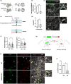
FIGURE 1
FIGURE 1
Reduced α‐synucleinA53T spread in primary neurons derived from TMEM16F knockout mice. (a) Heterozygous mice for the TMEM16F knockout (KO) allele (C57BL/6‐Ano6 Gt(EUCJ0166e09)Hmgu /DgiJ mice) were crossed and primary cortical neurons were cultured from brains of E17 WT and KO embryos. (b) qRT‐PCR quantification of TMEM16F mRNA levels relative to GAPDH in the primary neurons (n = 3 independent cultures). (c) Representation of TMEM16F activity assay: Intracellular calcium‐dependent phospholipid scrambling of TMEM16F increases transbilayer movement of phosphatidylserine (PS) from the inner to the out leaflet, which can be detected by recombinant FITC‐Annexin V binding. (d, e) Analysis of extracellular PS exposure in ionomycin‐treated (250 nM for 10 min) TMEM16F WT and KO primary neurons by FITC‐Annexin V binding. Confocal images are presented for FITC fluorescence (colored green) and neuronal morphology marker NFH (colored gray). Scale bar 20 μm (insets scale 10 μm). FITC intensity in different image fields was obtained and normalized to control WT neurons. n = 3 independent cultures. (f) Analysis of neurite length and cell body area in the NFH‐stained TMEM16F WT and KO neurons. Results are average of the morphological parameters in different image fields in n = 3 independent cultures. (g) Diagram of the AAV system encoding eGFP‐P2A‐α‐synA53T‐HA used to investigate neuronal α‐syn spread. (h, i) TMEM16F WT and KO neurons were transduced with the AAV system and spread was analyzed after one‐week post infection. (h) NFH staining (colored gray), eGFP (colored green), α‐synA53T‐HA (HA staining, colored red). Images are shown for the transduced donor neurons and acceptor neurons (scale bar 20 μm). Acceptor neurons are marked with arrows and are shown in insets (inset scale bar 10 μm). (i) Approximately 200 neuronal cell bodies were quantified per experiment. The results of spread were normalized to control neurons and calculated as described in the Methods. n = 3 independent cultures. 2‐tailed t‐test *p < 0.05, **p < 0.01.
FIGURE 2
FIGURE 2
TMEM16F knockdown diminishes α‐synucleinA53T spread in vivo. (a) Diagram of the in vivo experimental design of two‐step AAV injections for silencing TMEM16F using two independent shRNAs followed by expression of the α‐synA53T spreading system. (b) Primary cortical neurons were transduced with AAV encoding for control scrambled shRNA or for one of two different TMEM16F shRNAs (TMEM16F targeting sh1 or sh2) as well as encoding for mCherry reporter. Images show robust expression of the shRNAs (mCherry, colored red). Scale bar 10 μm. (c) Representative mosaic of mice injected with AAV encoding for control scrambled shRNA, TMEM16F shRNA 1, or TMEM16F shRNA 2. Scale bar 1 mm. (d) Insets show the view of recipient neurons (mCherry+ colored red/ eGFP /α‐syn A53T‐HA+, colored gray) at the contralateral cerebral cortex. Scale bar 50 μm (e) Quantification of brain section sizes of mice used for analysis. (f) Whole brain (contralateral and ipsilateral side) quantification of mCherry+/ eGFP / α‐syn A53T‐HA+ recipient cells in mice injected with scrambled shRNA or TMEM16F shRNAs. (g) Quantification of mCherry+/ eGFP / α‐syn A53T‐HA+ recipient cells of whole brain divided to cortical and subcortical regions in mice injected with scrambled shRNA or TMEM16F shRNAs. n = 3–5 mice per group. 2‐tailed t‐test. ns, nonsignificant, *p < 0.05, **p < 0.01.
FIGURE 3
FIGURE 3
Variations in the TMEM16F gene observed in the Ashkenazi Jewish PD/ DLB patients. There are five TMEM16F splice variants (SV1, 2, 3, 5, 6) in humans. Eight single nucleotide variants (SNVs) were observed in whole‐genome‐sequencing of 250 Ashkenazi Jewish (AJ)‐PDs/DLBs. Light blue domains are predicted to be TMEM16F transmembrane domains. Dark blue domains are topological domains. Cyt, cytoplasmic; EC, extracellular. Numbers in parentheses are Phred‐CADD scores for functional prediction.
FIGURE 4
FIGURE 4
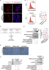
Functional perturbations in TMEM16F regulate the extracellular secretion of α‐synucleinA53T. (a) HEK293T cells were transduced with lentiviruses expressing mCherry‐TMEM16F WT or mCherry‐TMEM16F A703S and were analyzed for TMEM16F cellular expression by imaging. Scale bar 10 μm. (b) Flow cytometry analysis of Annexin V binding to extracellular PS in WT and A703S mutant TMEM16F stably‐expressing cells that were pre‐treated with ionomycin. Representative histograms are presented for Annexin V binding (FITC signal) in the TMEM16F‐expressing cells (mCherry signal). Results were normalized to control cells (n = 6 experiments). (c, d) Control HEK293T cells were transduced with AAV encoding the mCherry reporter (no TMEM16F). The mCherry‐TMEM16F stably‐expressing cells and the mCherry control cells were co‐transduced with lentiviruses encoding α‐synA53T‐HA. The levels of α‐synA53T in the intracellular (cell lysate) and extracellular (cell media) fractions were analyzed. Results are normalized to WT TMEM16F cells. n = 3 experiments, 2‐tailed t‐test *p < 0.05, **p < 0.01. (e) Images of the morphology of the stable cell lines. Scale bar 50 μm. (f) Extracellular vesicles were purified from cell media of stably‐expressing cells (control cells: MCherry + α‐synA53T‐HA, WT cells: MCherry‐TMEM16F + α‐synA53T‐HA, and mutant cells: MCherry‐TMEM16F A703S + α‐synA53T‐HA). The vesicles were analyzed for various membrane protein markers and for α‐synA53T. Representative blots are presented (n = 3 experiments).
FIGURE 5
FIGURE 5
PFF modeling of TMEM16F‐regulated spread of α‐synucleinA53T in human cell lines and iPSC‐derived dopaminergic neurons. (a) Schematic representation of experimental conditions analyzed in b–d. Different stably expressing donor HEK293T cells (control cells: MCherry + α‐synA53T‐HA, WT cells: MCherry‐TMEM16F + α‐synA53T‐HA, and mutant cells: MCherry‐TMEM16F A703S + α‐synA53T‐HA) were incubated with buffer or recombinant α‐synA53T PFF (1 μg/mL). After 4 days, the number of cells with aggregates was analyzed by quantifying aggregation puncta of α‐synA53T (HA staining). (b) Images of the donor cells (mCherry in red and HA staining in green) after 4 days with or without PFF treatment. Scale bar 10 μm. The quantification of percentage of cells with aggregation puncta is presented in different image fields in n = 3 experiments (900–1800 cells were analyzed for each stable cell line). (c, d) Wild type HEK293T acceptor cells were incubated for 2 days with conditioned media from the PFF‐treated donor cell lines or with media from buffer‐treated donor cell lines (no PFF), and were analyzed for α‐synA53T transfer. (c) Images of acceptors cells stained for α‐synA53T‐HA (HA staining colored green) or probed with proteostat dye (colored gray) together with nuclei staining (colored blue). Scale bar 10 μm. (d) The quantification of percentage of cells with proteostat‐stained aggregates or positive to HA staining is presented in different image fields in n = 3 experiments (450–900 cells were analyzed for each condition). (e) Human iPSC‐derived dopaminergic neurons were incubated with extracellular vesicles (EVs) purified from conditioned media of the PFF‐treated donor cell lines. After 1 week, the neurons were stained for β‐tubulin III (DAPI used for nuclei staining), and analyzed for mean single neurite length and the mean total length of the complete neurite tree per neuron (50 neurons were analyzed for each condition). Image scale bar 10 μm. The results are from two independent iPSC differentiations. One‐way ANOVA (b, d, e). ns, nonsignificant, *p < 0.05, **p < 0.01, ****p < 0.0001.
FIGURE 6
FIGURE 6
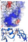
TMEM16F A703S structural model correlates calcium coordination with scrambling activity. (a) A structural model of the human TMEM16F containing the A703S was generated using AlphaFold2 and Phyre2. The structures of the mouse TMEM16F with Ca2+ (PDB 6P46) served as templates to model the Ca2+ binding residues within transmembrane 6 (TM6), TM7, and TM8. The positive patch involved in lipid association (yellow circled) comprising R635 located at the carboxy terminus of TM6, and R270, H274, and R276 located at the pre‐TM1 elbow. (b) A703 is located close to the negatively charged Ca2+ binding pocket and interacts with W272 within the pre‐TM1 elbow. The substitution of alanine 703 with serine pushes W272 towards the positive patch.

References

    1. Alvarez‐Erviti, L. , Seow, Y. , Schapira, A. H. , Gardiner, C. , Sargent, I. L. , Wood, M. J. , & Cooper, J. M. (2011). Lysosomal dysfunction increases exosome‐mediated alpha‐synuclein release and transmission. Neurobiology of Disease, 42, 360–367. - PMC - PubMed
    1. Anderson, J. P. , Walker, D. E. , Goldstein, J. M. , de Laat, R. , Banducci, K. , Caccavello, R. J. , Barbour, R. , Huang, J. , Kling, K. , Lee, M. , Diep, L. , Keim, P. S. , Shen, X. , Chataway, T. , Schlossmacher, M. G. , Seubert, P. , Schenk, D. , Sinha, S. , Gai, W. P. , & Chilcote, T. J. (2006). Phosphorylation of Ser‐129 is the dominant pathological modification of alpha‐synuclein in familial and sporadic Lewy body disease. The Journal of Biological Chemistry, 281, 29739–29752. - PubMed
    1. Arend, R. C. , Londono‐Joshi, A. I. , Gangrade, A. , Katre, A. A. , Kurpad, C. , Li, Y. , Samant, R. S. , Li, P. K. , Landen, C. N. , Yang, E. S. , Hidalgo, B. , Alvarez, R. D. , Straughn, J. M. , Forero, A. , & Buchsbaum, D. J. (2016). Niclosamide and its analogs are potent inhibitors of Wnt/beta‐catenin, mTOR and STAT3 signaling in ovarian cancer. Oncotarget, 7, 86803–86815. - PMC - PubMed
    1. Braak, H. , Ghebremedhin, E. , Rub, U. , Bratzke, H. , & Del Tredici, K. (2004). Stages in the development of Parkinson's disease‐related pathology. Cell and Tissue Research, 318, 121–134. - PubMed
    1. Braga, L. , Ali, H. , Secco, I. , Chiavacci, E. , Neves, G. , Goldhill, D. , Penn, R. , Jimenez‐Guardeno, J. M. , Ortega‐Prieto, A. M. , Bussani, R. , Cannatà, A. , Rizzari, G. , Collesi, C. , Schneider, E. , Arosio, D. , Shah, A. M. , Barclay, W. S. , Malim, M. H. , Burrone, J. , & Giacca, M. (2021). Drugs that inhibit TMEM16 proteins block SARS‐CoV‐2 spike‐induced syncytia. Nature, 594, 88–93. - PMC - PubMed

LinkOut - more resources