

Mapping grey matter and cortical thickness alterations associated with subjective cognitive decline and mild cognitive impairment among rural-dwelling older adults in China: A population-based study
- PMID: 39488196
- PMCID: PMC11566878
- DOI: 10.1016/j.nicl.2024.103691
Mapping grey matter and cortical thickness alterations associated with subjective cognitive decline and mild cognitive impairment among rural-dwelling older adults in China: A population-based study
Abstract
Background: The structural brain alterations for subjective cognitive decline (SCD) and mild cognitive impairment (MCI) are poorly defined. We sought to characterize grey matter volume (GMV) and cortical thickness associated with SCD and MCI among rural-dwelling older adults in China.
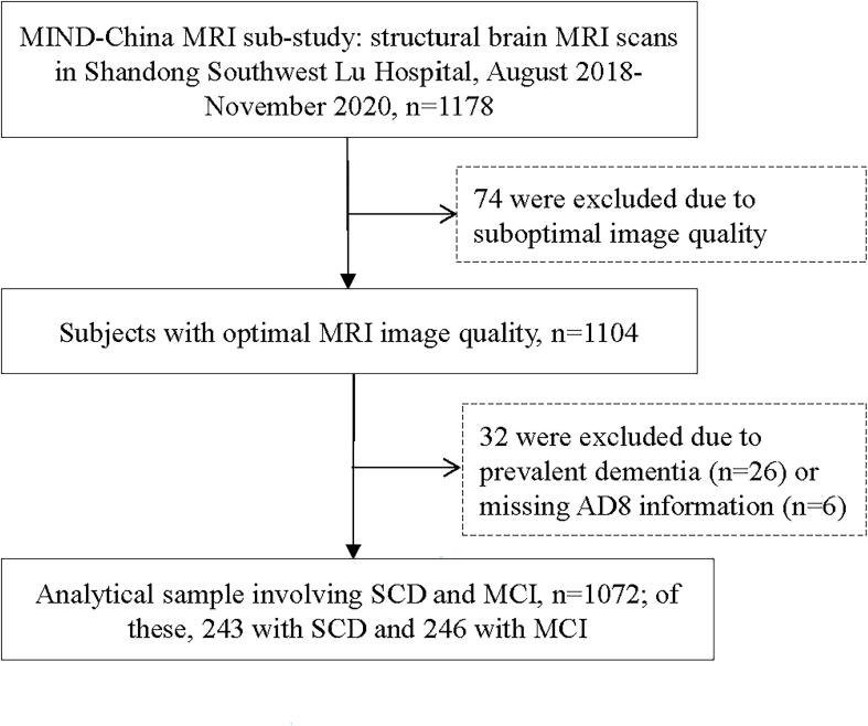
Methods: This population-based cross-sectional study included 1072 dementia-free participants from the brain MRI sub-study of MIND-China (2018-2020). We defined MCI following the Petersen's criteria, and SCD as the self-rated Ascertain Dementia 8-item Questionnaire score ≥ 2. Data were analyzed using voxel-based morphometry (VBM), surface-based morphometry analysis (SBM), and logistic regression models.
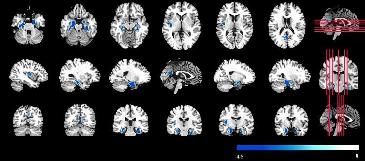
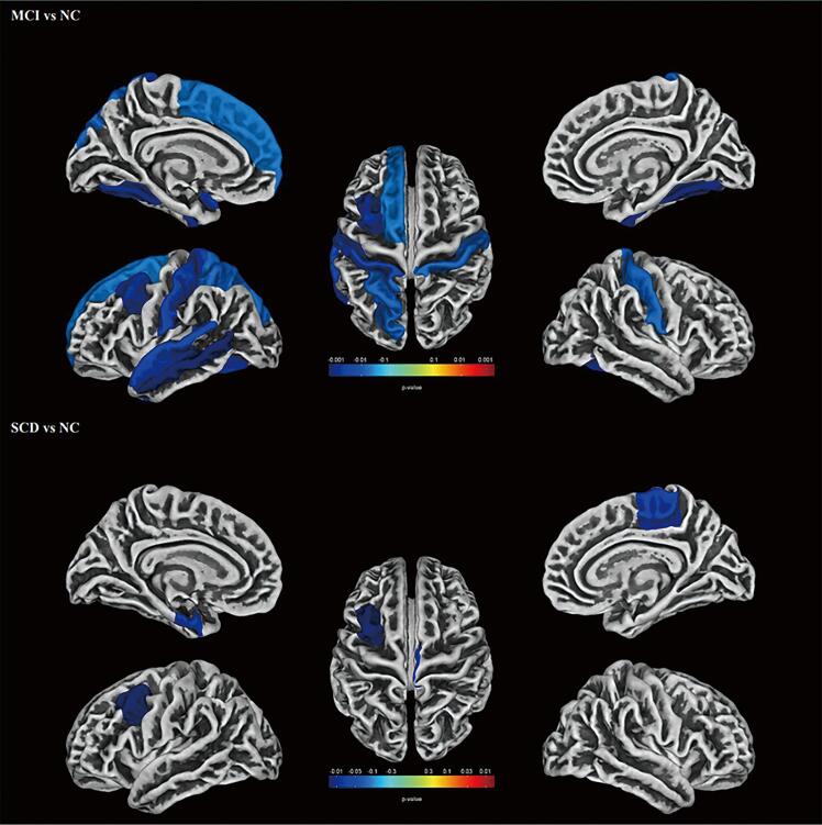
Results: SCD was defined in 243 persons and MCI in 246 individuals. The VBM analysis showed that MCI (vs. normal cognition) was significantly associated with reduced GMV in brain regions such as the bilateral parahippocampus, bilateral hippocampus, and bilateral fusiform (P < 0.05), but SCD exhibited no significant differences with normal cognition in GMV (P > 0.05). The ROI-wise SBM analysis revealed that SCD was significantly associated with cortical thinning in the right paracentral sulcus, left caudal middle frontal gyrus, and left entorhinal cortex (P < 0.05) and that MCI was significantly associated with cortical thinning in the left temporal lobe, left frontal lobe, bilateral parietal lobe and bilateral fusiform (P < 0.05).
Conclusions: The brain regions with reduced GMV or cortical thickness in older adults gradually expand from normal cognition through SCD to MCI, suggesting that characterizing structural brain alterations may help define the cognitive spectrum at the pre-dementia phase. These findings have potential implications for understanding the neuropathological process of cognitive deterioration in aging.
Keywords: Cortical thickness; Magnetic resonance imaging; Mild cognitive impairment; Population-based study; Regional brain volume; Subjective cognitive decline.
Copyright © 2024 The Authors. Published by Elsevier Inc. All rights reserved.
Figures
References
-
- Anticevic A., Dierker D.L., Gillespie S.K., Repovs G., Csernansky J.G., Van Essen D.C., Barch D.M. Comparing surface-based and volume-based analyses of functional neuroimaging data in patients with schizophrenia. Neuroimage. 2008;41(3):835–848. doi: 10.1016/j.neuroimage.2008.02.052. - DOI - PMC - PubMed
MeSH terms
LinkOut - more resources
Full Text Sources
Medical
