. 2024 Dec 10;57(12):2914-2927.e7.
doi: 10.1016/j.immuni.2024.10.001. Epub 2024 Nov 1.

Isolation and escape mapping of broadly neutralizing antibodies against emerging delta-coronaviruses

Affiliations

Isolation and escape mapping of broadly neutralizing antibodies against emerging delta-coronaviruses

Megi Rexhepaj et al. Immunity. .

Abstract

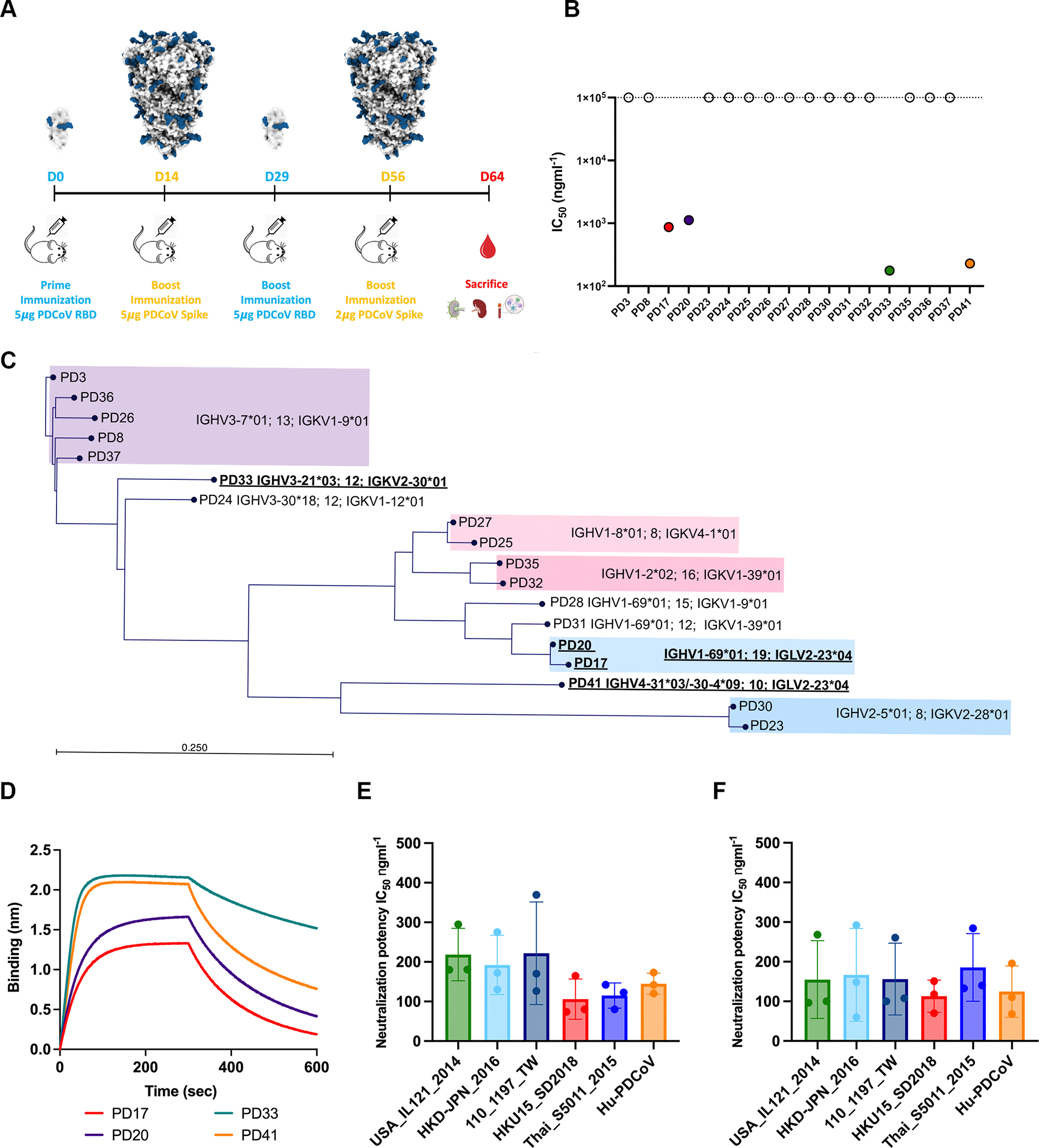
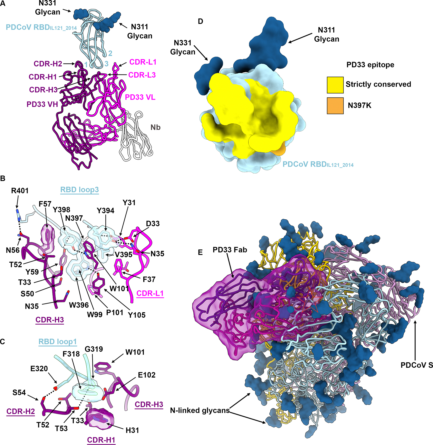
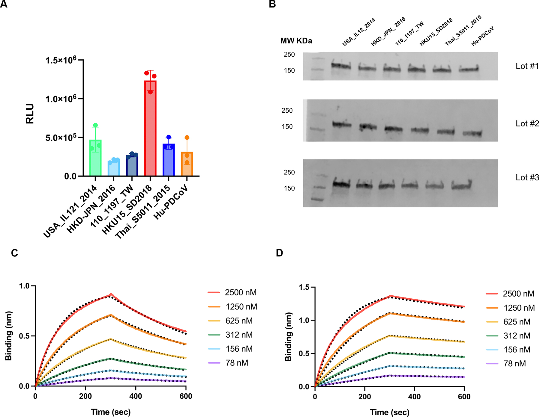
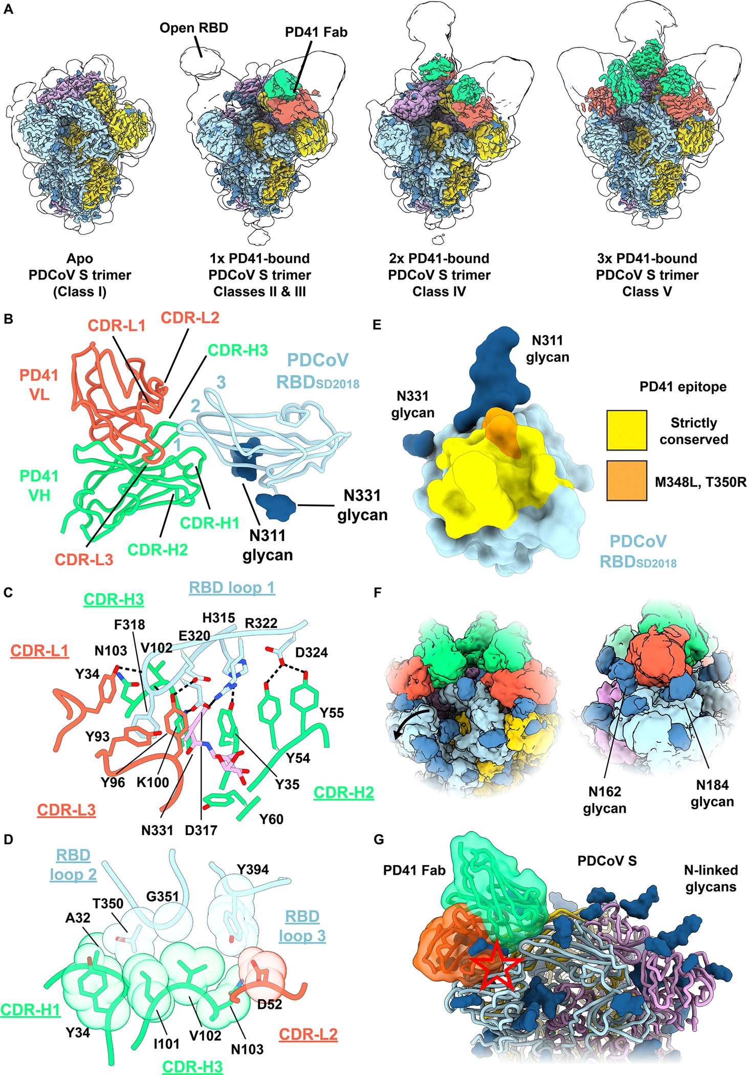
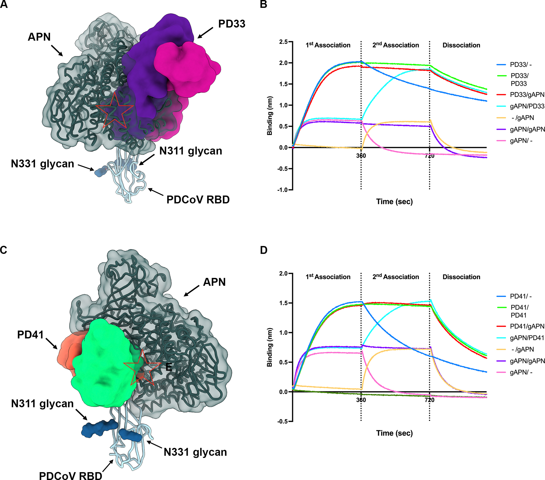
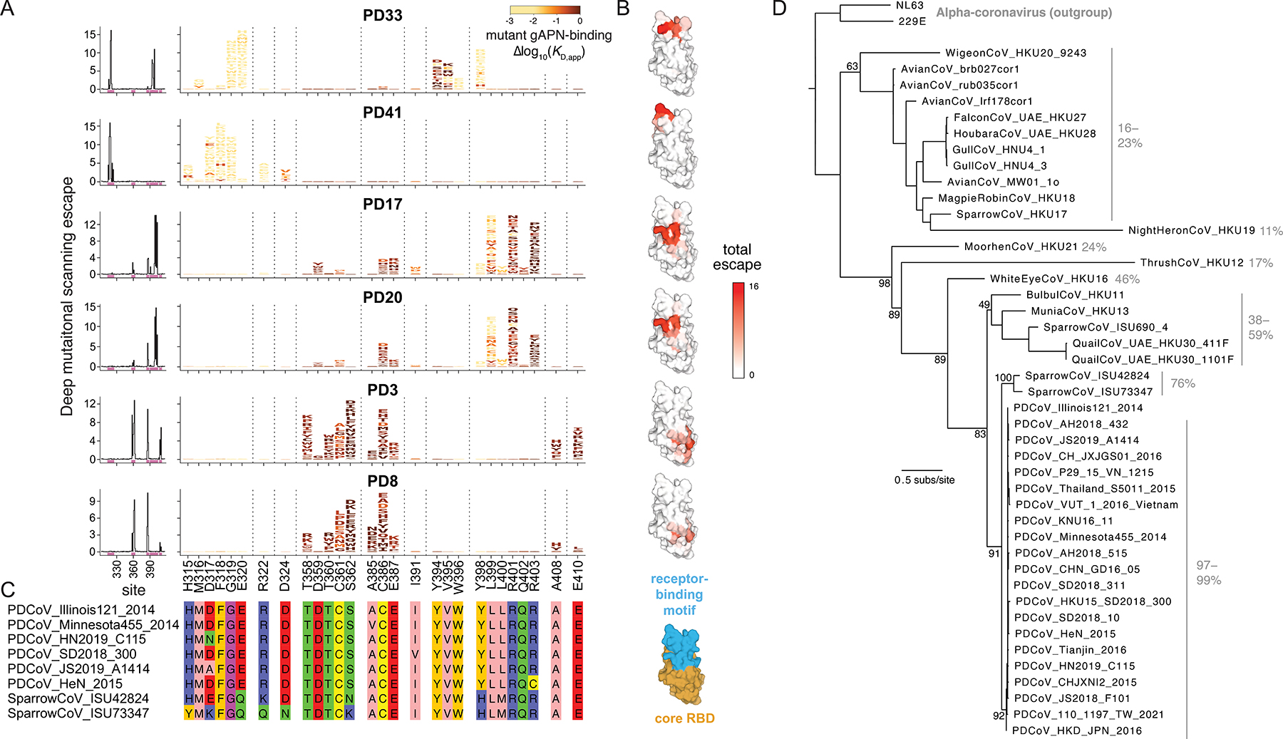
Porcine delta-coronavirus (PDCoV) spillovers were recently detected in febrile children, underscoring the recurrent zoonoses of divergent CoVs. To date, no vaccines or specific therapeutics are approved for use in humans against PDCoV. To prepare for possible future PDCoV epidemics, we isolated PDCoV spike (S)-directed monoclonal antibodies (mAbs) from humanized mice and found that two, designated PD33 and PD41, broadly neutralized a panel of PDCoV variants. Cryoelectron microscopy (cryo-EM) structures of PD33 and PD41 in complex with the S receptor-binding domain (RBD) and ectodomain trimer revealed the epitopes recognized by these mAbs, rationalizing their broad inhibitory activity. We show that both mAbs competitively interfere with host aminopeptidase N binding to neutralize PDCoV and used deep-mutational scanning epitope mapping to associate RBD antigenic sites with mAb-mediated neutralization potency. Our results indicate a PD33-PD41 mAb cocktail may heighten the barrier to escape. PD33 and PD41 are candidates for clinical advancement against future PDCoV outbreaks.

Keywords: PDCoV; cryo-EM structures; deep mutational scanning; neutralizing antibodies; porcine deltacoronavirus; spike glycoprotein; zoonosis.


Conflict of interest statement

Declaration of interests D.V. is named as inventor on patents for CoV nanoparticle vaccines filed by the University of Washington. D.C., L.P., A.D., K.C., C.S., A.B., and F.B. are employees of Vir Biotechnology and may hold shares in Vir Biotechnology.

Figures

Figure 1:
Figure 1:. Discovery and characterization of broadly neutralizing PDCoV monoclonal antibodies (mAbs).
A, Study design for immunization of ATX mice with PDCoVIL121_2014 RBD and S. B, Screening of isolated mAbs for neutralization of VSV pseudotyped with PDCoVIL121_2014 S using HEK293T cells transfected with the galline APN (gAPN) receptor. Each point represents the mean of technical triplicates. C, Phylogenetic tree of the panel of mAbs isolated in this study showing germline gene usage. D, BLI analysis of PD17, PD20, PD33, and PD41 Fabs binding to the PDCoVIL121_2014 RBD immobilized at the surface of Ni-NTA biosensors. E-F, PD33 (E) and PD41 (F) mAb-mediated neutralization of VSV pseudotyped with PDCoV S variants using HEK293T target cells transfected with human APN (hAPN). The bars correspond to mean titers with SD and each data point is one out of 3 biological replicates, each one using a different batch of pseudovirus. See also Figures S1, S2 and S3.
Figure 2:
Figure 2:. Broad neutralization by PD33 is conferred by binding to a conserved epitope
A, Ribbon diagram of the PD33 Fab-bound PDCoVIL121_2014 RBD cryoEM structure at 3.0 Å resolution. PD33 VH and VL are respectively rendered in purple and pink whereas the PDCoV RBD is colored cyan. The kappa light chain nanobody (Nb) used for assisting structural determination is shown in white. The PDCoV receptor-binding loops are annotated 1–3 and the PD33 complementary determining regions (CDR) for light and heavy chains are annotated CDR-L1, 2, 3 and CHR-H1, 2, 3. N-linked glycans are rendered as blue spheres. B-C, Zoomed-in views of the interface between PDCoV RBDIL121_2014 loop 3 (B) or loop 1 (C) and the PD33 Fab. Selected hydrogen bonds and salt bridges are shown as black dashed lines. A few side chains are shown in surface representation to highlight shape complementarity. D, PD33 epitope conservation across the panel of PDCoV S variants analyzed for neutralization. Yellow indicates strict residue conservation whereas orange shows the position of the N397K substitution present in SSD2018/300. E, Superimposition of the PD33-bound PDCoV RBD structure with the PDCoV S ectodomain trimer structure (PDB 6BFU) showing that PD33 could not bind to a closed S trimer due to masking of the receptor-binding loops and resulting steric clashes (red star). See also Figure S4.
Figure 3.
Figure 3.. The PDCoVSD2018 RBD utilizes human APN most efficiently among PDCoV variants
A, Entry of PDCoV S VSV variants into HEK293T target cells transiently transfected with human APN. Bars represent mean with SD and each data point was obtained from an independently produced batch of pseudovirus. B, Western blot quantification of PDCoV S incorporation in VSV pseudotypes for each of the three biological replicates (lots #1–3) for each variant used in panel A. C-D, BLI analysis of hAPN-Fc binding to the PDCoVIL121_2014 RBD (left) or to the PDCoVSD_2018 RBD (right) immobilized onto Ni-NTA tips. Fits to the data are shown as black dotted lines and were used to determine apparent binding affinities (KD,app) due to the avid binding of dimeric APN.
Figure 4.
Figure 4.. Broad neutralization by PD41 is conferred by binding to a conserved epitope.
A, Surface renderings of the cryoEM maps of the PDCoVSD2018 S trimer (gold, cyan and pink) without and with one, two or three bound PD41 Fabs (green and orange for heavy and light chains, respectively). N-linked glycans are rendered as blue spheres. The semi-transparent surfaces correspond to the same maps at lower contour for visualization of weak densities. B, Ribbon diagram of the PD41 Fab-bound PDCoVSD2018 RBD cryoEM structure at 2.8Å resolution obtained through local refinement. The PDCoV receptor-binding loops are annotated 1–3 and the PD41 complementary determining regions (CDR) for light and heavy chains are annotated CDR-L1, 2, 3 and CHR-H1, 2, 3. C-D, Zoomed-in views of the interface between PDCoVSD2018 RBD loop 1 (C) and loops 2 and 3 (D) and the PD41 Fab. Selected hydrogen bonds and salt bridges are shown as black dashed lines. A few side chains are shown with Van der Waals spheres to highlight shape complementarity. E, PD41 epitope conservation across the panel of PDCoV S variants analyzed for neutralization. Yellow indicates strict residue conservation whereas orange shows the positions of the M348L and T350R substitutions present in SThailand/S5011/2015. F, Two views of the cryoEM map of class V depicting the NTD displacement and glycan accommodation that relieve clashes that would occur with the canonical closed (apo) PDCoV S trimer conformation. G, Superimposition of the PD41-bound PDCoV RBD structure with the PDCoV S ectodomain trimer structure (PDB 6BFU) showing that PD41 could not bind to a closed S trimer without structural changes due to masking of the receptor-binding loops and resulting steric clashes (red star). See also Figure S5.
Figure 5:
Figure 5:. PD33 and PD41 competitively inhibit receptor engagement by PDCoV.
A, PD33 Fab (purple and magenta surfaces for the heavy and light chains, respectively) and human APN (dark green) recognize overlapping sites on the PDCoV RBD (cyan ribbon). N-linked glycans are rendered as blue surfaces. The red star indicates steric clashes. B, BLI analysis of Fab PD33 and galline APN (gAPN) binding to the PDCoV RBD immobilized at the surface of BLI biosensors in various competitive or non-competitive conditions. C, PD41 Fab (purple and magenta surfaces for the heavy and light chains, respectively) and APN (dark green) recognize overlapping sites on the PDCoV RBD (cyan ribbon). N-linked glycans are rendered as blue surfaces. The red star indicates steric clashes. D, BLI analysis of Fab PD41 and galline APN (gAPN) binding to the PDCoV RBD immobilized at the surface of BLI biosensors in various competitive or non-competitive conditions. See also Figure S6.
Figure 6.
Figure 6.. Prospective mapping of PDCoV mAb escape mutants by deep mutational scanning.
A, Maps of escape from PDCoV RBD-directed mAbs. (Left) Lineplots showing the total extent of mutational escape from binding of each mAb for mutations at each site in the RBD. (Right) Logoplots showing the magnitude of escape from mAb binding promoted by individual mutations for sites of strong escape (pink bars in lineplot). Full-height letters correspond to 10-fold or greater loss of mAb binding in the DMS assay. Mutations are colored according to their orthogonally measured impacts on gAPN-binding avidity, with increasingly light yellow reflecting increasingly deleterious impacts on receptor binding. B, Sitewise escape (y-axis in lineplots in panel A) mapped on the PDCoV RBD structure (PDB 6BFU). Key at right delineates the receptor-binding loops (blue) versus core RBD with matched RBD orientation. C, Alignment of PDCoV S sequence isolates at sites illustrated in the logoplots in panel A. D, Phylogeny of the deltacoronavirus genus inferred from RBD amino acid sequence. Gray annotations indicate the range of sequence identity to PDCoVIL121_2014 of each deltacoronavirus RBD amino acid sequence. See also Figure S7.

Update of

Similar articles

References

    1. Hamre D, and Procknow JJ (1966). A new virus isolated from the human respiratory tract. Proc. Soc. Exp. Biol. Med. 121, 190–193. - PubMed
    1. van der Hoek L, Pyrc K, Jebbink MF, Vermeulen-Oost W, Berkhout RJM, Wolthers KC, Wertheim-van Dillen PME, Kaandorp J, Spaargaren J, and Berkhout B (2004). Identification of a new human coronavirus. Nat. Med. 10, 368–373. - PMC - PubMed
    1. Woo PCY, Lau SKP, Chu C-M, Chan K-H, Tsoi H-W, Huang Y, Wong BHL, Poon RWS, Cai JJ, Luk W-K, et al. (2005). Characterization and complete genome sequence of a novel coronavirus, coronavirus HKU1, from patients with pneumonia. J. Virol. 79, 884–895. - PMC - PubMed
    1. Vijgen L, Keyaerts E, Moes E, Thoelen I, Wollants E, Lemey P, Vandamme AM, and Van Ranst M (2005). Complete genomic sequence of human coronavirus OC43: molecular clock analysis suggests a relatively recent zoonotic coronavirus transmission event. J. Virol. 79, 1595–1604. - PMC - PubMed
    1. Drosten C, Gunther S, Preiser W, van der Werf S, Brodt HR, Becker S, Rabenau H, Panning M, Kolesnikova L, Fouchier RA, et al. (2003). Identification of a novel coronavirus in patients with severe acute respiratory syndrome. N. Engl. J. Med. 348, 1967–1976. - PubMed

Publication types

MeSH terms

LinkOut - more resources