. 2024 Nov 2;15(1):9475.
doi: 10.1038/s41467-024-53750-z.

CLSTN3B promotes lipid droplet maturation and lipid storage in mouse adipocytes

Affiliations

CLSTN3B promotes lipid droplet maturation and lipid storage in mouse adipocytes

Chuanhai Zhang et al. Nat Commun. .

Abstract

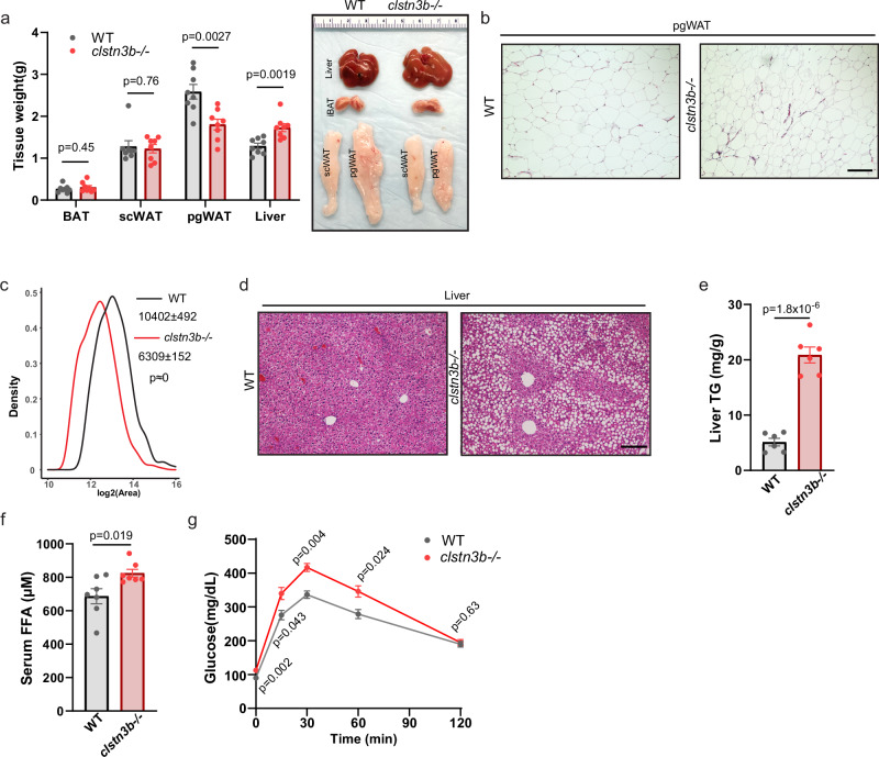
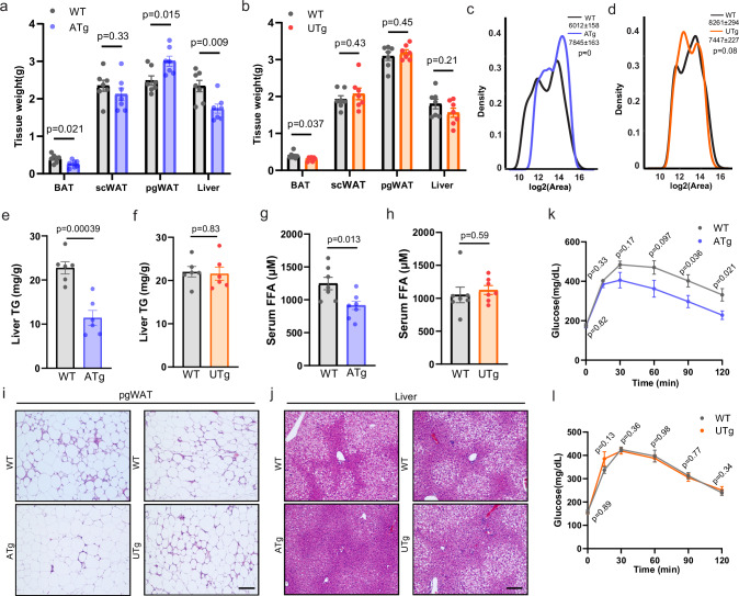
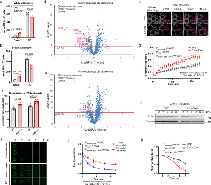
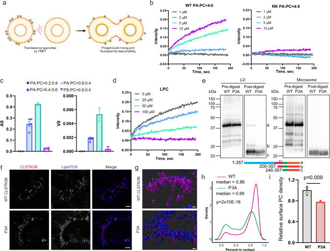
Interorganelle contacts facilitate material exchanges and sustain the structural and functional integrity of organelles. Lipid droplets (LDs) of adipocytes are responsible for energy storage and mobilization responding to body needs. LD biogenesis defects compromise the lipid-storing capacity of adipocytes, resulting in ectopic lipid deposition and metabolic disorders, yet how the uniquely large LDs in adipocytes attain structural and functional maturation is incompletely understood. Here we show that the mammalian adipocyte-specific protein CLSTN3B is crucial for adipocyte LD maturation. CLSTN3B employs an arginine-rich segment to promote extensive contact and hemifusion-like structure formation between the endoplasmic reticulum (ER) and LD, allowing ER-to-LD phospholipid diffusion during LD expansion. CLSTN3B ablation results in reduced LD surface phospholipid density, increased turnover of LD-surface proteins, and impaired LD functions. Our results establish the central role of CLSTN3B in the adipocyte-specific LD maturation pathway that enhances lipid storage and maintenance of metabolic health under caloric overload in mice of both sexes.


Conflict of interest statement

The authors declare no competing interests.

Figures

Fig. 1
Fig. 1. CLSTN3B deficiency impairs lipid storage in the perigonadal white adipose tissue.
Tissue weights (n = 8) and gross appearances (a), pgWAT histology (b), pgWAT adipocyte size distributions (c), liver histology (d), liver TG contents (n = 6) (e), serum FFA (n = 7) (f), and glucose tolerance test (n = 7) (g) of WT and clstn3b−/− mice on HFD at thermoneutrality. Scale bar: 100 μm. Data are mean ± s.e.m. Statistical significance was calculated by unpaired Student’s two-sided t test for (a, c, e, f), and Two-way Repeated Measurement ANOVA for (g). Source data are provided as a Source Data file.
Fig. 2
Fig. 2. White but not brown adipocyte-derived CLSTN3B promotes lipid storage in the perigonadal white adipose tissue and suppresses ectopic lipid deposition.
Tissue weights and gross appearances (a), pgWAT, liver and BAT histology (b), and serum FFA (c) of WT mice with sham surgery, clstn3b−/−, or clstn3b−/−; adq-cre mice receiving AAV-DIO-clstn3b injection into the iBAT maintained on HFD at thermoneutrality (n = 6). Tissue weights and gross appearances of pgWAT and liver (d, e), serum FFA (f, g), pgWAT and liver histology (h, i) of adq-cre clstn3bfl/fl (n = 5), ucp1-cre clstn3bfl/fl (n = 6) mice and cre-negative littermates on HFD at thermoneutrality. Scale bar: 100 μm. Data are mean ± s.e.m. Statistical significance was calculated by one-way ANOVA with Tukey’s post hoc test for (a), and unpaired Student’s two-sided t test for (cg). Source data are provided as a Source Data file.
Fig. 3
Fig. 3. Transgenic overexpression CLSTN3B in white adipocyte enhances lipid storage in perigonadal white adipose tissue.
Tissue weights (n = 7) (a, b), pgWAT adipocyte size distributions (c, d), liver TG contents (n = 6) (e, f), serum FFA (n = 7) (g, h), pgWAT and liver histology (i, j), and glucose tolerance test (k, l) of adq-cre clstn3b transgenic mice (n = 7), ucp1-cre clstn3b transgenic mice (n = 7) and WT littermates on HFD at thermoneutrality. Scale bar: 100 μm. Data are mean ± s.e.m. Statistical significance was calculated by unpaired Student’s two-sided t test for (a, b, eh), two-sided Wilcoxon text test for (c, d), and Two-way Repeated Measurement ANOVA for (k, l). Source data are provided as a Source Data file.
Fig. 4
Fig. 4. CLSTN3B enhances LD phospholipid and protein levels.
Lipolysis assay of WT and clstn3b−/− white (a) and brown (b) adipocytes (n = 4 biological replicates). c Quantitation of area per phospholipid molecule on brown and white adipocyte LD isolated from WT and clstn3b−/− mice (n = 4). Proteomics analysis of brown (d) and white (e) adipocyte LDs isolated from WT and clstn3b−/− mice (n = 4). Representative FRAP images (f) and quantitative analysis (n = 10 LDs) (g) of PLIN1-mcherry turnover rate in WT and clstn3b−/− brown adipocytes. Representative images (h) and quantitative analysis (i) of fluorescent PLIN1 peptide coating of neat or PC-coated triolein droplets in the presence of excess unlabeled PLIN1 peptide (n = 8–18 LDs for each condition). Representative Western blot (j) and quantitative analysis (k) of PLIN1 half-life by cycloheximide chase in WT and clstn3b−/− brown adipocytes (n = 3 biological replicates). Scale bar: 1 μm in (f, h). Data are mean ± s.e.m. Statistical significance was calculated by unpaired Student’s two-sided t test for (ae), and Two-way Repeated Measurement ANOVA for (g, i, k). No adjustment was made for multiple comparisons for (d, e). Source data are provided as a Source Data file.
Fig. 5
Fig. 5. The arginine-rich region of CLSTN3B promotes extensive ER/LD contact.
a Ribbon diagram of AlphaFold predicted structure of CLSTN3B and the distribution of arginine (bold) and proline residues (bold italic) in the arginine-rich region of CLSTN3B (170–246). b Illustration of arginine/lysine phosphate hydrogen bonding. Fluorescence microscopic images (c), 3D reconstruction (d), quantitative analysis of ER/LD contact extent (e), and electron microscopic images (f) of WT CLSTN3B and RK mutant-mediated ER/LD contact in HEK293 cells. Scale bar: 5 μm in (c); 3 μm in (d); 200 nm in (f). Similar results have been obtained from at least 3 repeats for (c, f). Statistical significance was calculated by two-sided Wilcoxon test for (e). Source data are provided as a Source Data file.
Fig. 6
Fig. 6. The arginine-rich region of CLSTN3B facilitates ER-to-LD phospholipid diffusion.
a Fluorometric phospholipid quantitation of LDs isolated from empty vector, WT CLSTN3B or RK mutant-expressing HEK293 cells (n = 3 biological replicates). Schematics (b), representative images (c, e), and quantitation (d, f) of click-chemistry based ER-to-LD phospholipid transfer assay using fixed cell (n = 11) (c, d) or isolated LD (n = 13–20 LDs for each condition) (e, f). Scale bar: 1 μm. Schematics (g), representative images (h), and quantitation (i) of FRAP analysis of phospholipid exchange between ER and LD using copper-free click-chemistry labeled PC (n = 8–10 LDs for each condition). Scale bar: 1 μm. Schematics (j), representative images (k), and quantitation (l) of click-chemistry based semi-reconstituted phospholipid transfer assay. Scale bar: 2 μm. Data are mean ± s.e.m. Statistical significance was calculated by one-way ANOVA with Tukey’s post hoc test for (a, f, i, l), and unpaired Student’s two-sided t test for (d). Source data are provided as a Source Data file. b, g, and j were created in BioRender.
Fig. 7
Fig. 7. CLSTN3B promotes hemifusion-like structure formation.
Schematics (a) of FRET-based detection of liposome lipid mixing induced by a CLSTN3B-derived R-rich peptide or a RK mutant peptide (b), the dependence on phospholipid composition for lipid mixing (n = 2–5 independent replicates) (c), and the effect of LPC on lipid mixing (d). e Western blot analysis of WT or P3A CLSTN3B fragments generated from trypsin digestion of LDs from HEK293 cells. The positions of fragments with defined lengths are shown as references (see also Extended Data Fig. 8e). Arrows denote positions of mutated prolines. Fluorescence microscopic images (f), 3D reconstruction (g), quantitative analysis of ER/LD contact extent (h), and measurement of LD surface PC levels (n = 3 biological replicates) (i) in WT CLSTN3B and P3A mutant-expressing HEK293 cells. Scale bar: 2 μm. Similar results have been obtained from at least 3 repeats for (f). Data are mean ± s.e.m. Statistical significance was calculated by two-sided Wilcoxon test for (h), and unpaired Student’s two-sided t test for (i). Source data are provided as a Source Data file. a was created in BioRender.
Fig. 8
Fig. 8. CLSTN3B promotes LD maturation.
a Fluorescence microscopic analysis of CLSTN3B and KDEL localization in primary adipocytes. Scale bar: 5 μm. b Relationship between LD-associated CLSTN3B intensity and LD size. c Fluorescence microscopic analysis of CLSTN3B localization in primary brown adipocytes over a NE-treatment time course. Scale bar: 5 μm. Quantitation of LD-associated CLSTN3B signal intensity (d) and LD size (e) in (c). Fluorescence microscopic images (f) and quantitative analysis (g) of LD sizes in HEK293 cells transfected with empty vector (EV), WT CLSTN3B, RK CLSTN3B, or P3A CLSTN3B at indicated time points after OA treatment (n = 200–2000 LDs for each condition). Scale bar: 5 μm. h Model of CLSTN3B action at the molecular, organellar, cellular and organismal levels. Similar results have been obtained from at least 3 repeats for (a, c). Data are mean ± s.e.m. Statistical significance was calculated by one-way ANOVA with Tukey’s post hoc test for (g). Source data are provided as a Source Data file. h was created in BioRender.

Similar articles

Cited by

References

    1. Shulman, G. I. Ectopic fat in insulin resistance, dyslipidemia, and cardiometabolic disease. N. Engl. J. Med.371, 1131–1141 (2014). - PubMed
    1. Akazawa, S., Sun, F., Ito, M., Kawasaki, E. & Eguchi, K. Efficacy of troglitazone on body fat distribution in type 2 diabetes. Diab. Care23, 1067–1071 (2000). - PubMed
    1. Yki-Järvinen, H. Thiazolidinediones. N. Engl. J. Med.351, 1106–1118 (2004). - PubMed
    1. Lipscombe, L. L. et al. Thiazolidinediones and cardiovascular outcomes in older patients with diabetes. JAMA298, 2634–2643 (2007). - PubMed
    1. Greenberg, A. S. et al. The role of lipid droplets in metabolic disease in rodents and humans. J. Clin. Invest.121, 2102–2110 (2011). - PMC - PubMed

LinkOut - more resources