. 2024 Nov 2;15(1):9485.
doi: 10.1038/s41467-024-53187-4.

Molecular mechanism of parental H3/H4 recycling at a replication fork

Affiliations

Molecular mechanism of parental H3/H4 recycling at a replication fork

Fritz Nagae et al. Nat Commun. .

Abstract

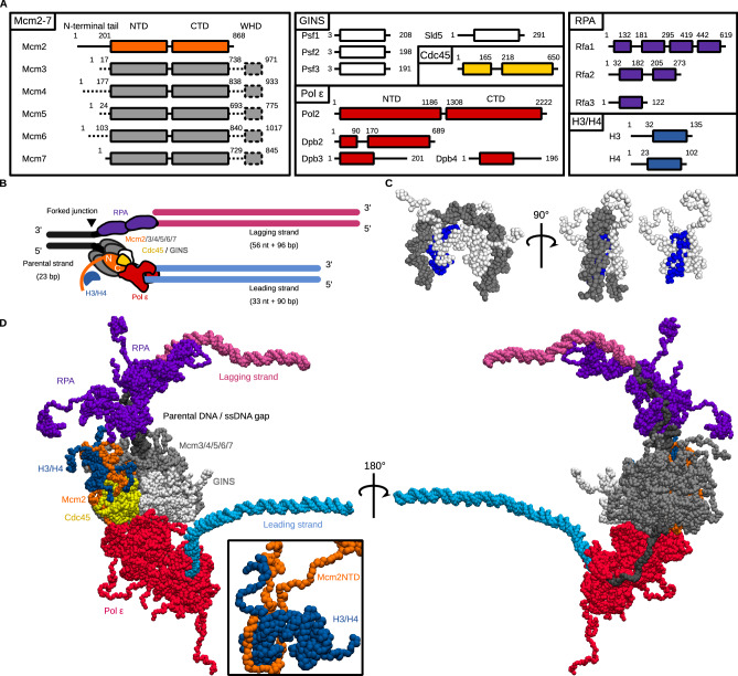
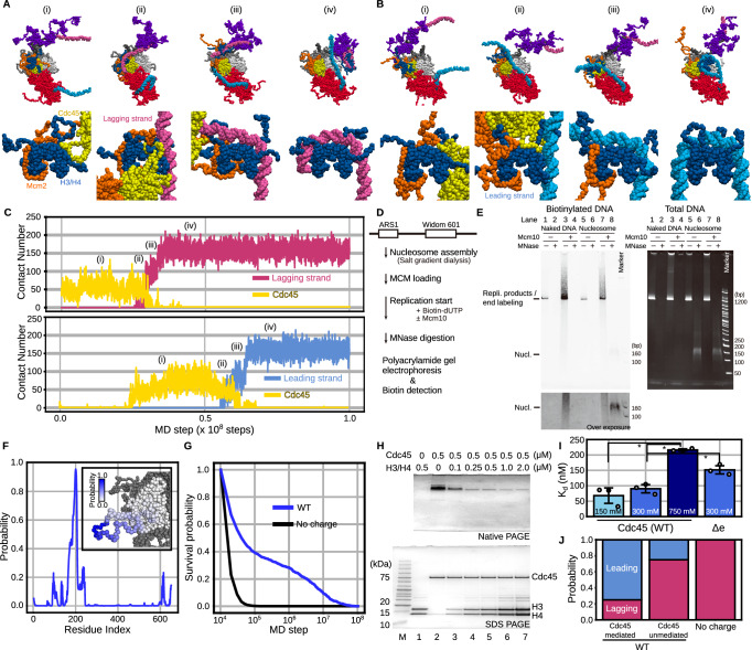
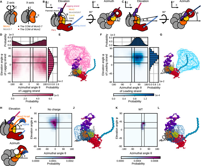
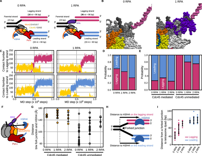
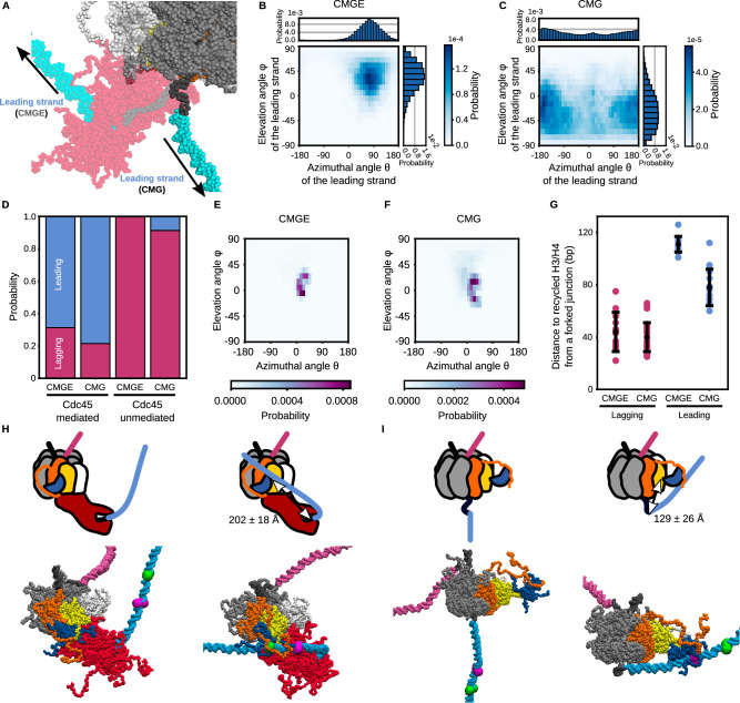
In chromatin replication, faithful recycling of histones from parental DNA to replicated strands is essential for maintaining epigenetic information across generations. A previous experiment has revealed that disrupting interactions between the N-terminal tail of Mcm2, a subunit in DNA replication machinery, and a histone H3/H4 tetramer perturb the recycling. However, the molecular pathways and the factors that regulate the ratio recycled to each strand and the destination location are yet to be revealed. Here, we performed molecular dynamics simulations of yeast DNA replication machinery, an H3/H4 tetramer, and replicated DNA strands. The simulations demonstrated that histones are recycled via Cdc45-mediated and unmediated pathways without histone chaperones, as our in vitro biochemical assays supported. Also, RPA binding regulated the ratio recycled to each strand, whereas DNA bending by Pol ε modulated the destination location. Together, the simulations provided testable hypotheses, which are vital for elucidating the molecular mechanisms of histone recycling.


Conflict of interest statement

The authors have no conflict of interest, financial or otherwise.

Figures

Fig. 1
Fig. 1. The initial structure for the coarse-grained molecular dynamics simulations of a replicated-DNA-engaged replisome binding to an H3/H4 tetramer.
A The domain composition of Mcm2-7, GINS, Cdc45, Pol ε, RPA, and the H3/H4 tetramer. The dotted lines represent the regions not included in the simulations. B Schematic illustration of the initial structures. The black, pink, and cyan lines represent the parental, lagging, and leading strands. The orange, gray, white, yellow, red, blue, and purple objects represent Mcm2, Mcm3/4/5/6/7, GINS, Cdc45, Pol ε, H3/H4, and RPA, respectively. C The structures of a tetrasome and an H3/H4 tetramer. The residues contacting Mcm2 in the crystal structure (PDB ID: 4UUZ) are colored blue. D The initial coarse-grained structures of the replicated-DNA-engaged replisome.
Fig. 2
Fig. 2. The H3/H4 tetramer was recycled to the replicated strands by the Mcm2 N-tail in the simulations of a replicated-DNA-engaged replisome. Source data are provided as a Source Data file.
A, B Representative snapshots of the simulation trajectories in which the H3/H4 tetramer was deposited on the lagging (A) and leading (B) strands. The top panels are the magnified version of the bottom ones. C Time trajectories of the number of residues in the H3/H4 tetramer contacting the lagging strand (pink in the top panel), the leading strand (cyan in the bottom panel), and Cdc45 (yellow) from the trajectories in (A) and (B). D Schematic of the in vitro DNA replication with nucleosome-assembled DNA. E Gel images of biotinylated DNA (left) and total DNA (right) before and after MNase digestion on native gels. We obtained the similar results from three independent replicates. F Probability of each residue in Cdc45 contacting the H3/H4 tetramer in the simulations. The inset figure is the probabilities represented by shades of blue on the Cdc45 structure. G Survival probabilities of the association between Cdc45 and the H3/H4 tetramer in the simulations in the presence (WT) or absence (No charge) of charges in the Cdc45 acidic loop. H Gel images of native PAGE (top) and SDS-PAGE (bottom) of the mixtures of Cdc45 and H3/H4 tetramer (lanes 1–7). ‘M’ denotes a marker (ATTO; 2332346). I The apparent dissociation constants between the H3/H4 tetramer and Cdc45 (WT in 150, 300, and 750 mM KCl and Δe (Cdc45Δe) in 300 mM KCl). The error bars represent the mean ± standard deviation from three replicates of each condition. The asterisks represent P < 0.05 by two-tailed Welch’s t-test (p-values between 150 mM and 750 mM, between 300 mM and 750 mM, and between WT and Δe are 0.013, 0.004, and 0.010, respectively). J Ratios of the replicated strands to which the H3/H4 tetramer was recycled.
Fig. 3
Fig. 3. Distributions of orientations of the replicated strands and the H3/H4 tetramer in the simulations of the replicated-DNA-engaged replisome. Source data are provided as a Source Data file.
A Definition of X- and Z-axes. B, C Schematic illustrations of the replicated-DNA-engaged replisome, which explains the definition of the elevation (φ) and azimuthal (θ) angles to describe the lagging (B) and leading (C) strand orientation. The color scheme is the same as in Fig. 1B. D, F 1D and 2D probability distributions of the lagging (D) and leading (F) strand orientation calculated from the simulation trajectories until recycling. E, G Iso-surfaces of the spatial probability density of the lagging (E) and leading (G) strand orientation. The iso-value was set to 0.0001 [Å−3]. H Schematic illustrations of the replicated-DNA-engaged replisome explaining the definition of the elevation (φ) and azimuthal (θ) angles to describe the H3/H4 tetramer orientation. I, K 2D distributions of the H3/H4 tetramer orientation calculated from trajectories of the simulations in the absence (I) and presence (K) of charges in the Cdc45 acidic loop. J, L Iso-surfaces of spatial probability distributions of the H3/H4 tetramer orientation in the simulation in the absence (J) and presence (L) of charges in the Cdc45 acidic loop. The iso-value was set to 0.0001 [Å−3]. D, F, I, K The black auxiliary lines, which encircle the populated area in (K), mark the azimuthal angles of 10° and 40° and the elevation angles of 10° and 40°.
Fig. 4
Fig. 4. The H3/H4 tetramer was deposited on the replicated strands by the Mcm2 N-tail in the simulations of the replicated-DNA-engaged replisome with zero or one RPA.
Source data are provided as a Source Data file. A Schematic illustration of the initial structures. The color scheme is the same as in Fig. 1B. B Initial structures with zero (left) and one (right) RPA molecule. C The number of residues in the H3/H4 tetramer contacting the lagging strand (pink), the leading strand (cyan), and Cdc45 (yellow) in the case of zero (left) and one (right) RPA molecule. D Ratios of the replicated strands on which the H3/H4 tetramer was deposited in the simulations with zero, one, and two RPA molecules. E The same as in (D), but trajectories were classified into the Cdc45-mediated and unmediated pathways. F Schematic illustration explaining the definition of the 3D distance from the fork junction to the H3/H4 tetramer. G Distances from the fork junction to the H3/H4 tetramer at the moment of recycling to the lagging strand via the Cdc45-mediated pathway (orange; n = 4, 8, and 4) and the Cdc45-unmediated pathway (gray; n = 13, 12, and 3) in the simulations with zero, one, and two RPA molecules. The error bars represent the mean ± standard deviation. H Schematic illustration explaining the definition of the distance from the fork junction to the destination location. I Distances from the fork junction to the position of the tetrasome dyad on the lagging (pink; n = 17, 20, and 7) and leading strand (cyan; n = 11, 18, and 13) in the simulations with zero, one, and two RPA molecules. The error bars represent the mean ± standard deviation.
Fig. 5
Fig. 5. The H3/H4 tetramer was deposited on the daughter strands by the Mcm2 N-tail in the simulations of the replicated-DNA-engaged replisome without Pol ε and RPA.
Source data are provided as a Source Data file. A The structure of CMG (opaque) superimposed to that of CMGE (CMG + Pol ε) (transparent). B, C 1D and 2D probability distributions of the leading strand orientation calculated from the simulations of CMGE and CMG. D Ratios of the replicated strands on which the H3/H4 tetramer was deposited in the simulations of CMGE and CMG. E, F 2D distributions of the H3/H4 tetramer orientation calculated from trajectories of the simulations of CMGE and CMG (G) Distances from the fork junction to the deposited positions on the lagging (pink; n = 17 and 24) and leading strand (cyan; n = 11 and 13) in the simulations of CMGE and CMG. The error bars represent the mean ± standard deviation. H, I Schematic illustrations and representative structures of CMGE (H) and CMG (I). The initial structure and the structure at the exact moment of the H3/H4 deposition are shown on the left and right, respectively. The color scheme is the same as in Fig. 1B, D. Additionally, the green and magenta spheres on the leading strand represent the average recycled position in the CMGE and CMG simulations, respectively.

Similar articles

Cited by

References

    1. Luger, K., Mäder, A. W., Richmond, R. K., Sargent, D. F. & Richmond, T. J. Crystal structure of the nucleosome core particle at 2.8 Å resolution. Nature389, 251–260 (1997). - PubMed
    1. Lai, W. K. M. & Pugh, B. F. Understanding nucleosome dynamics and their links to gene expression and DNA replication. Nat. Rev. Mol. Cell Biol.18, 548–562 (2017). - PMC - PubMed
    1. Venkatesh, S. & Workman, J. L. Histone exchange, chromatin structure and the regulation of transcription. Nat. Rev. Mol. Cell Biol.16, 178–189 (2015). - PubMed
    1. Wooten, M. et al. Asymmetric histone inheritance via strand-specific incorporation and biased replication fork movement. Nat. Struct. Mol. Biol.26, 732–743 (2019). - PMC - PubMed
    1. Urban, J. A., Ranjan, R. & Chen, X. Asymmetric histone inheritance: establishment, recognition, and execution. Annu. Rev. Genet.56, 113–143 (2022). - PMC - PubMed

Publication types

MeSH terms

LinkOut - more resources