. 2024 Nov 2;14(1):26418.
doi: 10.1038/s41598-024-78221-9.

The combination of venetoclax and quercetin exerts a cytotoxic effect on acute myeloid leukemia

Affiliations

The combination of venetoclax and quercetin exerts a cytotoxic effect on acute myeloid leukemia

Renshi Kawakatsu et al. Sci Rep. .

Abstract

Venetoclax is a BH3 mimetic that was recently approved for the treatment of acute myeloid leukemia (AML) treatment. However, the effect of venetoclax on AML remains limited, and a novel strategy is required. Here, we demonstrate for the first time that the cytotoxic effect of venetoclax drastically increased when by combined with the naturally occurring flavonoid quercetin. Combined treatment with venetoclax and quercetin caused most of AML KG-1 cells to exhibit a condensed morphology. Cell cycle analysis revealed that the combination strongly induced cell death. Caspase inhibitor blocked this cell death, and the combination induced poly (ADP-ribose) polymerase (PARP) cleavage, indicating that apoptosis was the primary mechanism. These effects were also observed in another AML cell line Kasumi-1 but not in chronic myeloid leukemia (CML) K562 cells. Public data analysis demonstrated that B-cell/CLL lymphoma 2 (Bcl-2) expression is increased in AML cells compared to other malignant tumors, and the survival and the growth of AML cell line depends on Bcl-2. We found that quercetin increased Bcl-2-associated X protein (Bax) expression in KG-1. Our study provides a novel function for quercetin and presents a promising strategy for AML treatment using venetoclax.


Conflict of interest statement

The authors declare no competing interests.

Figures
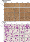
Fig. 1
Fig. 1
The combination of venetoclax and quercetin altered KG-1 cell morphology. KG-1 cells were treated with venetoclax and/or quercetin for 48 h at the indicated concentrations. (A) Cell morphology of KG-1 cells. A phase-contrast microscope was used. Red bars represent 50 μm. (B) Cell morphology of KG-1 cells stained with May-Gruenwald-Giemsa. Blue bars represent 50 μm.
Fig. 2
Fig. 2
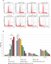
The combination of venetoclax and quercetin induced cell death. KG-1 cells were treated with venetoclax with or without quercetin, for 48 h at the indicated concentrations. (A,B) Cell cycle analysis of KG-1 cells stained with PI was examined using flow cytometry. Representative histograms (A) and a bar graph (B). The data was n = 3 ± S.D. **P < 0.01. Vene venetoclax, Que quercetin.
Fig. 3
Fig. 3
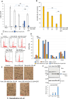
The combination of venetoclax and quercetin induced caspase-dependent apoptosis. KG-1 cells were treated with venetoclax with or without quercetin, for 48 h at the indicated concentrations. To inhibit caspase activation, Q-VD-OPh (QVD) was added. (A) Trypan blue dye exclusion assay of KG-1 cells was used to determine the number of live and dead cells. (B) CCK-8 assay of KG-1 cells. (C,D) Cell cycle analysis of KG-1 cells which were stained with PI was performed using flow cytometry. Representative histograms (C) and a bar graph (D). (E) Cell morphology of KG-1 cells under a phase-contrast microscope. Red bars represent 50 μm. (F) Western blotting of PARP. KG-1 cell lysates were extracted and western blotting was performed using PARP antibody (Upper panel) and β-actin antibody (Lower panel). The PARP-cleaved form produced by caspases is indicated by a blue arrow. Full-length blots are shown in Supplementary Fig. 5. Band intensities of cleaved PARP is shown below the blots. Control was set to 1.0. The data was n = 3 ± S.D. **P < 0.01. Vene venetoclax, Que quercetin.
Fig. 4
Fig. 4
The combination of venetoclax and quercetin induced apoptosis in Kasumi-1 cells. AML Kasumi-1 cells were treated with venetoclax in combination with or without quercetin for 48 h at the indicated concentrations. Q-VD-OPh (QVD) was added to inhibit caspase activation. (A) Cell morphology of Kasumi-1 cells under a phase-contrast microscope. Red bars represent 50 μm. (B,C) Cell cycle analysis of Kasumi-1 cells which were stained with PI was performed using flow cytometry. Representative histograms (B) and a bar graph (C). (D) Western blotting of PARP. Kasumi-1 cell lysates were extracted and western blotting was performed using PARP antibody (Upper panel) and β-actin antibody (Lower panel). Caspase activations produce the PARP-cleaved form. This cleaved form is indicated by blue arrow. Full-length blots are shown in Supplementary Fig. 5. Band intensities of cleaved PARP is shown below the blots. Control was set to 1.0. The data was n = 3 ± S.D. **P < 0.01. Vene venetoclax, Que quercetin.
Fig. 5
Fig. 5
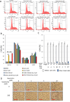
The combination of venetoclax and quercetin effected on chronic myeloid leukemia cells. CML K562 cells were treated with venetoclax with or without quercetin for 48 h at the indicated concentrations. (A,B) Cell cycle analysis of K562 cells stained with PI was performed using flow cytometry. Representative histograms (A) and a bar graph (B). (C) Trypan blue dye exclusion assay of K562 cells was used to determine the number of live and dead cells. (D) Cell morphology of K562 cells. Red bars show 50 μm. The data was n = 3 ± S.D. **P < 0.01. Vene venetoclax, Que quercetin.
Fig. 6
Fig. 6
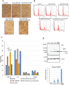
Quercetin up-regulated Bax expression. (A) Bcl-2 expression was analyzed by cBioPortal using the TCGA Pan Cancer Atlas Studies with 10,967 patient samples. Bcl-2 expression in leukemia was shown in a red box. (B) Bax, Bak and Bcl-2 expressions in KG-1 cells. KG-1 cells were treated with described concentration of quercetin for 48 h and cDNA were generated. RT-qPCR was performed using specific primers of each gene. (C) IAP family expressions in KG-1 cells. RT-qPCR of IAP family genes were performed as mentioned in B. The data was n = 3 ± S.D. *P < 0.05. **P < 0.01. Que quercetin.

Similar articles

Cited by

References

    1. Döhner, H. et al. Diagnosis and management of AML in adults. 2022 recommendations from an international expert panel on behalf of the ELN. Blood22, 1345–1377 (2022). - PubMed
    1. Siegel, R. L., Miller, K. D., Wagle, N. S. & Jemal, A. Cancer statistics, 2023. CA Cancer J. Clin.73, 17–48 (2023). - PubMed
    1. Short, N. J., Rytting, M. E. & Cortes, J. E. Acute myeloid leukaemia. Lancet392, 593–606 (2018). - PMC - PubMed
    1. Jahn, E. et al. Clinical impact of the genomic landscape and leukemogenic trajectories in non-intensively treated elderly acute myeloid leukemia patients. Leukemia37, 2187–2196 (2023). - PMC - PubMed
    1. Cancer Genome Atlas Research Network et al. Genomic and epigenomic landscapes of adult de novo acute myeloid leukemia. N. Engl. J. Med.368, 2059–2074 (2013). - PMC - PubMed

MeSH terms