. 2025 Jan:51:102164.
doi: 10.1016/j.tranon.2024.102164. Epub 2024 Nov 2.

TRIM47 is a prognostic biomarker for gallbladder cancer and promotes tumor progression through regulating K63-linked ubiquitination of PARP1

Affiliations

TRIM47 is a prognostic biomarker for gallbladder cancer and promotes tumor progression through regulating K63-linked ubiquitination of PARP1

Ming Xu et al. Transl Oncol. 2025 Jan.

Abstract

Background: Gallbladder cancer (GBC) is one of the most lethal malignancies worldwide with an extremely poor prognosis. Previous studies have suggested that tripartite motif containing 47 (TRIM47) is involved in the progression of numerous cancers. However, the molecular mechanism and function of TRIM47 in GBC remain unclear.

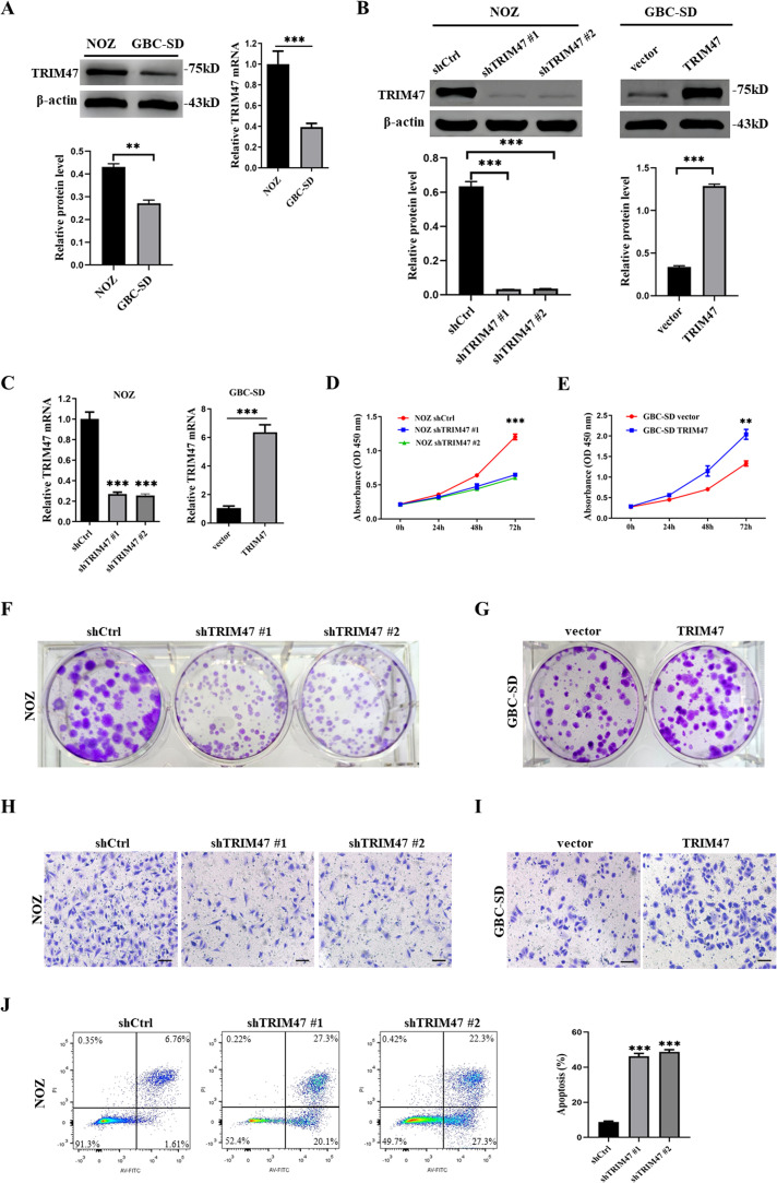
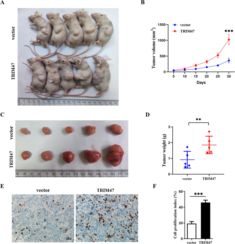
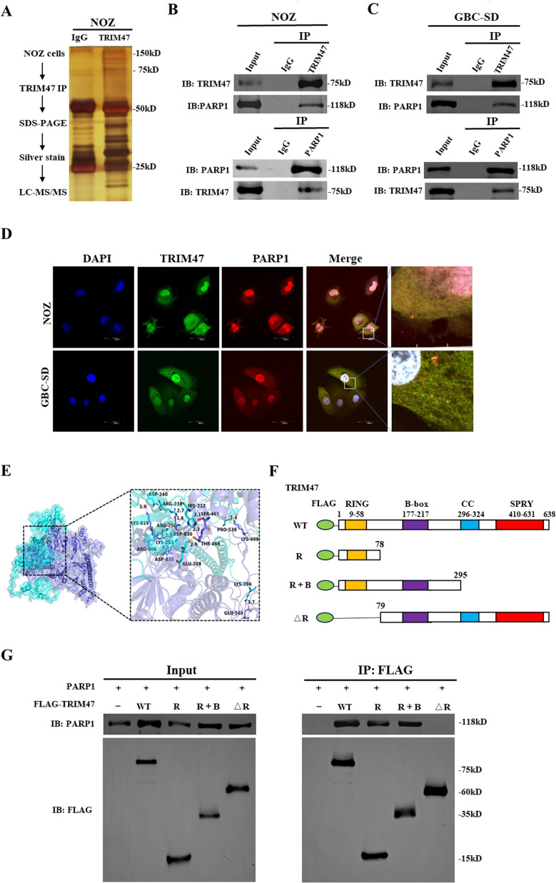
Methods: The clinical significance of TRIM47 was evaluated using immunohistochemistry. Functional assays were performed in vitro and in vivo to determine the role of TRIM47 in GBC. Mass spectrometric analysis, western blotting, and immunoprecipitation assays were performed to investigate the molecular mechanisms involved.

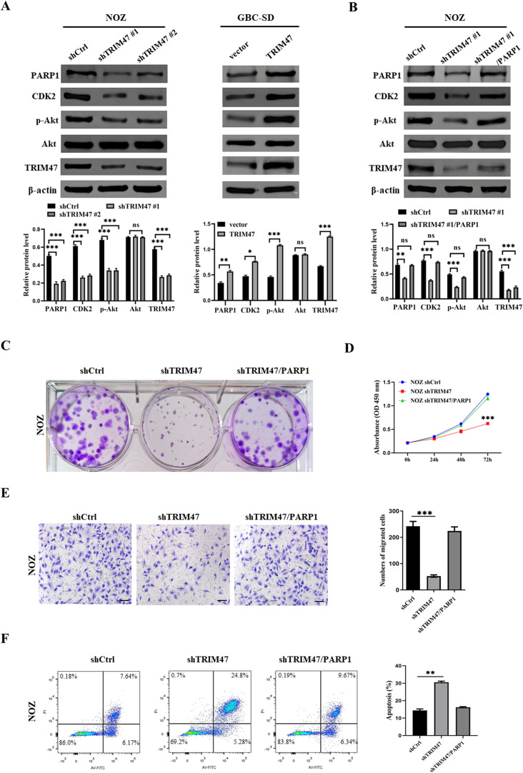
Results: In this study, TRIM47 was upregulated in GBC tissues and associated with shorter overall survival rates and TRIM47 was involved in GBC cell proliferation, migration, and apoptosis. Mechanistically, TRIM47 interacts with PARP1 and mediates the K63-linked polyubiquitination of PARP1, thereby stabilizing its expression. Furthermore, TRIM47 activated the AKT signaling pathway via PARP1.

Conclusion: The present study revealed that TRIM47 contributes to the progression of GBC and is therefore an important biomarker for predicting the prognosis of GBC and for therapeutic intervention.

Keywords: Gallbladder cancer; PARP1; Prognosis; TRIM47; Ubiquitination.


Conflict of interest statement

Declaration of competing interest The authors declare that they have no known competing financial interests or personal relationships that could have appeared to influence the work reported in this paper.

Figures

Image, graphical abstract
Graphical abstract
Fig 1
Fig. 1
TRIM47 overexpression in GBC patients was associated with a poor prognosis. A TRIM47 expression levels in various types of human cancers utilizing the TIMER database. B The expression of TRIM47 in 12 pairs of GBC tissues and adjacent non-tumor tissues (ANT) was analyzed by qRT-PCR. C The expression of TRIM47 is upregulated in GBC tissues, as demonstrated by western blot analysis. D Representative IHC staining images of GBC tissues with negative, low, and high expression of TRIM47. E TRIM47 expression is correlated with overall patient survival when analyzed by Kaplan-Meier analysis. *P < 0.05, **P < 0.01, ***P < 0.001.
Fig 2
Fig. 2
TRIM47 promotes the progression of GBC cells in vitro. A qRT-PCR and western blot analysis of TRIM47 expression in two GBC cell lines. B, C The expression of TRIM47 was detected by western blot and qRT-PCR in TRIM47-depleting NOZ cells and TRIM47-overexpressing GBC-SD cells. D, E CCK-8 assays were performed to detect cell growth upon TRIM47 knockdown or overexpression. F, G An assay assessing the colony formation ability of GBC cells was carried out following shRNA knockdown and overexpression of TRIM47. H, I Transwell assays were used to detect the effect of TRIM47 on GBC cell migration. J Apoptosis of NOZ cell lines with TRIM47 knockdown was analyzed by flow cytometry. **P < 0.01, ***P < 0.001.
Fig 3
Fig. 3
Tumor growth is promoted by overexpression of TRIM47 in vivo. A Tumor-bearing nude mice in a representative image. B Tumor volumes were measured every 5 days for 30 days using calipers. C Xenografts transfected with vector or TRIM47 were photographed at the end of the experiment. D Comparison of tumor weight between TRIM47 group and vector group. E The proliferation of cells in xenografts was measured using the Ki-67 stain. F Ki-67-positive nuclear cells were counted to determine the cell proliferation index. **P < 0.01, ***P < 0.001.
Fig 4
Fig. 4
TRIM47 binds to PARP1 with the RING domain. A Silver staining of SDS-PAGE gel after IgG or TRIM47 pulldown. B, C Protein interactions between PARP1 and TRIM47 were confirmed by co-IP and IB analysis of NOZ or GBC-SD cell extracts. D Confocal fluorescence microscope was performed to observe the location of TRIM47 and PARP1 in NOZ cells or GBC-SD cells. E Interactions between TRIM47 and PARP1 were analyzed using molecular docking. F Diagram of FLAG-tagged TRIM47 variants: full-length WT, RING domain, R + B (RING + B-box domains) and ΔR. G FLAG co-immunoprecipitated with PARP1 in HEK293T cells transfected with PARP1 and FLAG-tagged TRIM47 variants.
Fig 5
Fig. 5
TRIM47 mediates the K63-linked polyubiquitination of PARP1. A The expression of TRIM47 and PARP1 was examined by western blotting in NOZ cells with TRIM47 knockdown or in GBC-SD cells with TRIM47 overexpression. B mRNA expression of PARP1 in GBC cells with TRIM47 knockdown and overexpression. C Effects of protease inhibitor MG132 (20μM, 7 h) on TRIM47-induced PARP1 expression. D, E NOZ cells stably expressing control shRNA or two independent TRIM47 shRNA were treated with 50 μg/ml CHX, collected at the indicated times, and then analyzed with antibodies against TRIM47 and PARP1. An analysis of PARP1 levels compared with β-actin is shown (E). F, G GBC-SD cells were transfected with TRIM47, treated with 50 μg/ml CHX, harvested at the indicated times, and then subjected to IB analysis with antibodies against TRIM47 and PARP1. Quantification of PARP1 protein levels relative to β-actin is shown (G). H, I GBC cells with TRIM47 knockdown and overexpression were co-transfected with the indicated HA-Ub, and cell lysates were subjected to IP with PARP1 antibody, followed by IB analysis with antibodies against HA and TRIM47. A 7 h treatment with 20 μM MG132 was performed before harvesting the cells. J HEK-293T cells were co-transfected with FLAG-TRIM47, PARP1 and the indicated HA-Ub WT, or HA-K48-Ub (Lys48-only), or HA-K63-Ub (Lys63-only) plasmids, and then the PARP1 ubiquitylation linkage was analyzed by IB analysis using anti-HA antibody. K Constructed plasmids for FLAG-TRIM47, PARP1, HA-Ub, and various ubiquitin mutants (K6R, K11R, K27R, K29R, K33R, K48R, K63R) were co-transfected into HEK-293T cells to evaluate the levels of ubiquitination. **P < 0.01.
Fig 6
Fig. 6
TRIM47 activates the AKT signaling pathway through PARP1. A PARP1, CDK2, p-Akt (Ser 473), Akt, TRIM47 and β-actin protein expressions were detected by western blot in NOZ cells with TRIM47 knockdown or in GBC-SD cells with TRIM47 overexpression. B PARP1, CDK2, p-Akt (Ser 473), Akt, TRIM47 and β-actin protein expressions were detected were detected after overexpressing PARP1 in TRIM47-knockdown NOZ cells. C The plate colony formation ability after overexpressing PARP1 in TRIM47-knockdown NOZ cells. D CCK-8 assays were performed to detect cell growth after overexpressing PARP1 in TRIM47-knockdown NOZ cells. E Transwell migration assays were used to determine the cell migration of TRIM47 and PARP1 on NOZ cells. F Apoptosis of overexpressing PARP1 in TRIM47-knockdown NOZ cells was analyzed by flow cytometry. **P < 0.01, ***P < 0.001.
Fig 7
Fig. 7
TRIM47 and PARP1 has an positive correlation in GBC patients’ tissues. A, B Patients with high expression of TRIM47 protein in 130 GBC patents were frequently associated with high expression of PARP1 protein. C PARP1 expression is correlated with overall patient survival when analyzed by Kaplan-Meier analysis. D Diagram illustrating TRIM47-mediated polyubiquitination of PARP1 and activation of the AKT pathway. ***P < 0.001.

References

    1. Sung H., Ferlay J., Siegel R.L., Laversanne M., Soerjomataram I., Jemal A., et al. Global cancer statistics 2020: GLOBOCAN estimates of incidence and mortality worldwide for 36 cancers in 185 countries. CA Cancer J. Clin. 2021;71(3):209–249. - PubMed
    1. Vuthaluru S., Sharma P., Chowdhury S., Are C. Global epidemiological trends and variations in the burden of gallbladder cancer. J. Surg. Oncol. 2023;128(6):980–988. - PubMed
    1. He Z., Zhong Y., Hu H., Li F. ZFP64 promotes Gallbladder cancer progression through recruiting HDAC1 to activate NOTCH1 signaling pathway. Cancers. 2023;15(18) - PMC - PubMed
    1. Ghosh I., Dey Ghosh R., Mukhopadhyay S. Identification of genes associated with gall bladder cell carcinogenesis: implications in targeted therapy of gall bladder cancer. World J. Gastrointest. Oncol. 2023;15(12):2053–2063. - PMC - PubMed
    1. Lv T.R., Hu H.J., Liu F., Ma W.J., Jin Y.W., Li F.Y. The significance of peri-neural invasion in patients with gallbladder carcinoma after curative surgery: a 10 year experience in China. Updates Surg. 2023;75(5):1123–1133. - PubMed

LinkOut - more resources