. 2024 Nov 3;14(1):26528.
doi: 10.1038/s41598-024-77985-4.

Dyslipidemia in Juvenile Dermatomyositis

Affiliations

Dyslipidemia in Juvenile Dermatomyositis

Amer Khojah et al. Sci Rep. .

Abstract

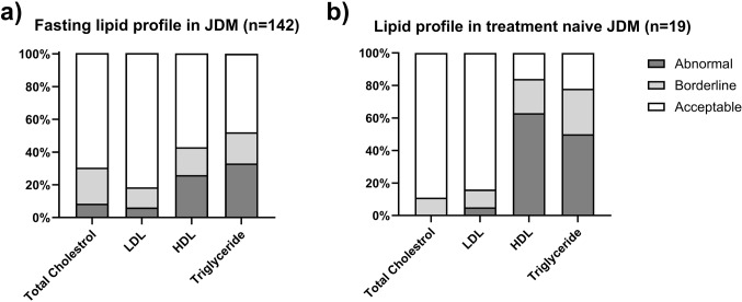
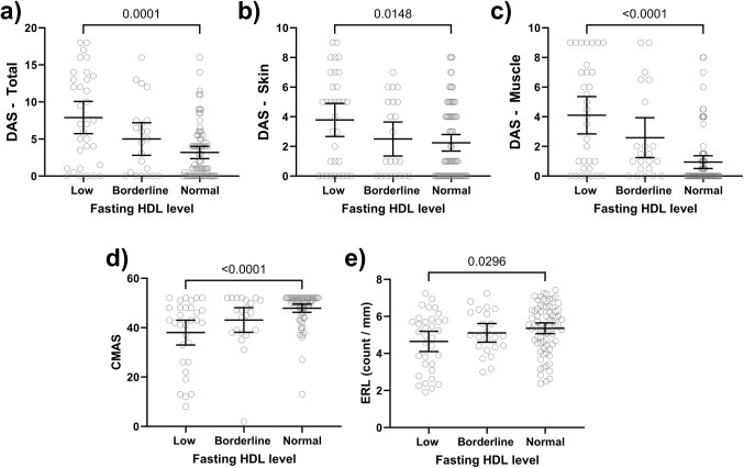
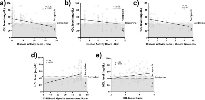
This study investigates the prevalence of dyslipidemia and its association with disease activity in children with Juvenile Dermatomyositis (JDM). A retrospective chart review of 142 JDM patients who had fasting lipid profiles was conducted. Clinical, and laboratory indicators of disease activity at the time of lipid assessment were obtained. JDM patients displayed a high prevalence (72%) of abnormal or borderline fasting lipid profiles, particularly involving HDL and triglycerides. Treatment-naïve patients exhibited the most significant dyslipidemia, with significantly lower median HDL levels compared to those on medication (30 vs. 49 mg/dL, p < 0.0001). HDL levels inversely correlated with various disease activity measures, including disease activity score (DAS) total (r= -0.38, p < 0.001), DAS muscle weakness (r= -0.5, p < 0.001), DAS skin (r= -0.25, p = 0.003), neopterin (r= -0.41, p < 0.001), ESR (r= -0.25, p = 0.006), and vWF Ag (r= -0.21, p = 0.02). In conclusion, JDM patients have a high prevalence of dyslipidemia, especially low HDL and elevated triglycerides. The severity of dyslipidemia (low HDL) correlates with disease activity, with treatment-naïve patients demonstrating the lowest HDL levels. These findings suggest the importance of annual lipid profile monitoring in JDM patients, potentially followed by early interventions such as dietary adjustments and exercise programs.

Keywords: Disease Activity scores; Dyslipidemia; HDL; Juvenile Dermatomyositis; Neopterin.


Conflict of interest statement

The authors declare no competing interests.

Figures

Figure 1
Figure 1
The prevalence of abnormal lipid profile in juvenile dermatomyositis (JDM). (A) 43% of JDM patients exhibited low or borderline-low HDL, while only 18% of them had elevated or borderline-high fasting LDL levels. Additionally, 52% of JDM patients displayed elevated or borderline-high fasting triglyceride levels. (B) Treatment-naïve JDM patients showed a higher prevalence of lipid profile abnormalities, with only 22% and 16% having normal triglyceride (TG) and HDL levels, respectively. However, total cholesterol and LDL cholesterol levels remained within the normal range for the majority of treatment-naïve individuals (70% and 82%, respectively).
Figure 2
Figure 2
Lipid profile in JDM based on the treatment status. (A) Treatment status (treatment-naïve, on medication, off medication) significantly impacted HDL levels with the treatment-naïve group displaying significantly lower HDL levels compared to the “on medication” group (median level 30 vs. 49, p < 0.0001). (B) There were no significant differences observed in low-density lipoprotein (LDL) among the different treatment groups. (C) TG levels did not show significant differences between the different treatment groups.
Figure 3
Figure 3
Disease activity indicators in JDM based on the HDL level. JDM patients with low HDL exhibited significantly higher median scores for DAS-T (A), DAS-S (B), and DAS-M (C) compared to the normal HDL group D) The low HDL group had a lower CMAS than the normal HDL group. E) Nailfold capillary assessment revealed a lower ERL in the low HDL.
Figure 4
Figure 4
Correlation analysis between HDL levels and JDM disease activity indicators. There was a negative correlation between HDL levels and disease activity scores, including DAS-T (A), DAS-S (B), and DAS-M (C). There was a positive correlation between HDL levels and CMAS (D) as well as ERL capillary count (E).

References

    1. Pachman, L. M., Nolan, B. E., DeRanieri, D. & Khojah, A. M. Juvenile Dermatomyositis: New clues to diagnosis and therapy. Curr. Treatm Opt. Rheumatol.7, 39–62. 10.1007/s40674-020-00168-5 (2021). - PMC - PubMed
    1. Mendez, E. P. et al. US incidence of juvenile dermatomyositis, 1995–1998: results from the National Institute of Arthritis and Musculoskeletal and skin diseases Registry. Arthritis Rheum.49, 300–305. 10.1002/art.11122 (2003). - PubMed
    1. Pachman, L. M. & Khojah, A. M. Advances in Juvenile Dermatomyositis: Myositis specific antibodies aid in understanding Disease Heterogeneity. J. Pediatr.195, 16–27. 10.1016/j.jpeds.2017.12.053 (2018). - PMC - PubMed
    1. Schwartz, T., Diederichsen, L. P., Lundberg, I. E., Sjaastad, I. & Sanner, H. Cardiac involvement in adult and juvenile idiopathic inflammatory myopathies. RMD Open.2, e000291. 10.1136/rmdopen-2016-000291 (2016). - PMC - PubMed
    1. Marie, I. Morbidity and mortality in adult polymyositis and dermatomyositis. Curr. Rheumatol. Rep.14, 275–285. 10.1007/s11926-012-0249-3 (2012). - PubMed

LinkOut - more resources