. 2024 Nov 4;31(1):99.
doi: 10.1186/s12929-024-01087-6.

Dental pulp mesenchymal stem cell (DPSCs)-derived soluble factors, produced under hypoxic conditions, support angiogenesis via endothelial cell activation and generation of M2-like macrophages

Affiliations

Dental pulp mesenchymal stem cell (DPSCs)-derived soluble factors, produced under hypoxic conditions, support angiogenesis via endothelial cell activation and generation of M2-like macrophages

Ludovica Barone et al. J Biomed Sci. .

Abstract

Background: Cell therapy has emerged as a revolutionary tool to repair damaged tissues by restoration of an adequate vasculature. Dental Pulp stem cells (DPSC), due to their easy biological access, ex vivo properties, and ability to support angiogenesis have been largely explored in regenerative medicine.

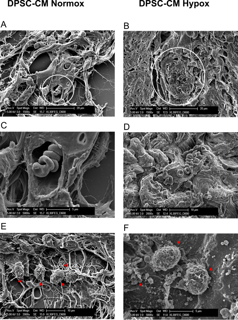
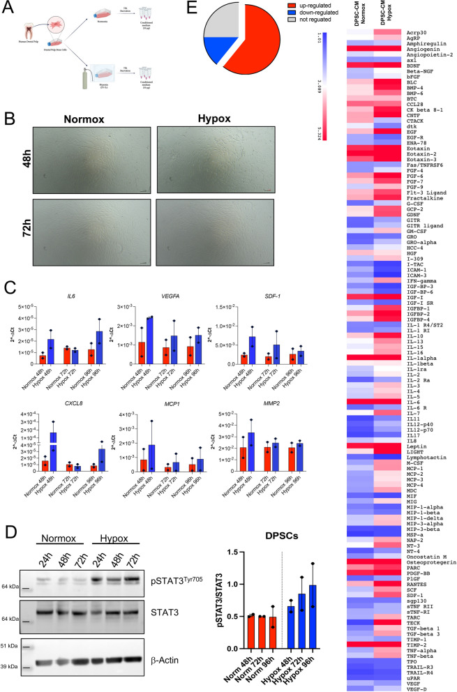
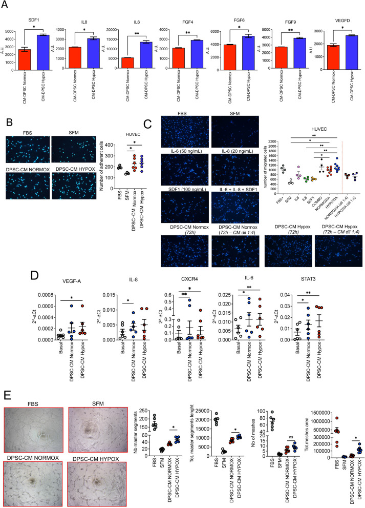
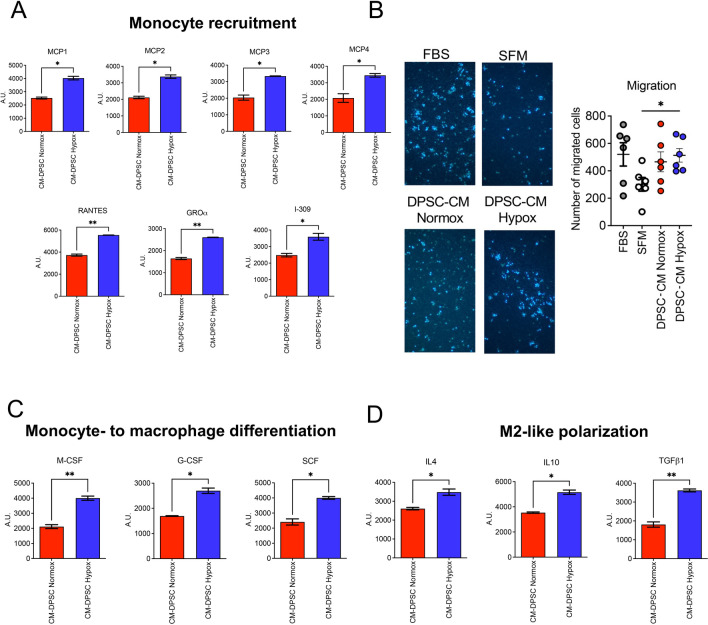
Methods: Here, we tested the capability of Dental Pulp Stem Cell-Conditioned medium (DPSC-CM), produced in normoxic (DPSC-CM Normox) or hypoxic (DPSC-CM Hypox) conditions, to support angiogenesis via their soluble factors. CMs were characterized by a secretome protein array, then used for in vivo and in vitro experiments. In in vivo experiments, DPSC-CMs were associated to an Ultimatrix sponge and injected in nude mice. After excision, Ultimatrix were assayed by immunohistochemistry, electron microscopy and flow cytometry, to evaluate the presence of endothelial, stromal, and immune cells. For in vitro procedures, DPSC-CMs were used on human umbilical-vein endothelial cells (HUVECs), to test their effects on cell adhesion, migration, tube formation, and on their capability to recruit human CD14+ monocytes.

Results: We found that DPSC-CM Hypox exert stronger pro-angiogenic activities, compared with DPSC-CM Normox, by increasing the frequency of CD31+ endothelial cells, the number of vessels and hemoglobin content in the Ultimatrix sponges. We observed that Utimatrix sponges associated with DPSC-CM Hypox or DPSC-CM Normox shared similar capability to recruit CD45- stromal cells, CD45+ leukocytes, F4/80+ macrophages, CD80+ M1-macrophages and CD206+ M2-macropages. We also observed that DPSC-CM Hypox and DPSC-CM Normox have similar capabilities to support HUVEC adhesion, migration, induction of a pro-angiogenic gene signature and the generation of capillary-like structures, together with the ability to recruit human CD14+ monocytes.

Conclusions: Our results provide evidence that DPSCs-CM, produced under hypoxic conditions, can be proposed as a tool able to support angiogenesis via macrophage polarization, suggesting its use to overcome the issues and restrictions associated with the use of staminal cells.

Keywords: Angiogenesis; Cell-free device; Dental pulp stem cells; Macrophage polarization; Mesenchymal stem cells; Secretome; Tissue engineering.


Conflict of interest statement

The authors declare that they have no competing interests.

Figures

Fig. 1
Fig. 1
Characterization of DPSCs. DPSCs at different culture passages (P2-P30) were characterized by (A, B) flow cytometry for CD90, CD105, CD73, CD45, EpCAM, CD31 surface markers; (C) real-time PCR for CD44, CD90, CD105, CD45, ALPL, DSPP, p16, p21 gene expression; (D) BrdU assay, for their proliferation rate
Fig. 2
Fig. 2
In vivo pro-angiogenic effects of DPSC-CMs produced in normoxia and hypoxia. The pro-angiogenic effects of DPSC-CMs were investigated by Ultimatrix sponge assay. A, B Representative excised Ultimatrix sponges associated with DPSC-CM Normox and DPSC-CM Hypox; C, D representative H&E images of sections from Ultimatrix sponges associated with DPSC-CM Normox and DPSC-CM Hypox showing vascular-like structures (circles) and mature blood vessels (arrows) respectively. Scale bar 20 μm; E hemoglobin content, detected by Drabkin’s assay, in sponges associated with DPSC-CM Normox and DPSC-CM Hypox; F vessel count in Ultimatrix sponges associated with DPSC-CM Normox and DPSC-CM Hypox; G flow cytometry analysis for the content of stromal CD45 cells, CD31+ endothelial cells, CD45+ total leukocytes, total F4/80+ macrophages, CD80+ M1 and CD206+ M2 macrophages in sponges associated with DPSC-CM Normox and DPSC-CM Hypox. Results are shown as mean ± SEM, t-student test, *p ≤ 0.05; **p ≤ 0.01. Experiments were performed using CMs from 3 donors
Fig. 3
Fig. 3
Ultrastructural analysis of DPSC-CM Ultimatrix sponges. Representative SEM images of sponges associated with DPSC-CM Normox (A, C, E) and DPSC-CM Hypox (B, D, F) are showed. Beside the collagen fibrils, is evident the presence of blood vessels, full of erythrocytes, within the scaffold (circle) (A, B). A magnification of the blood vessel in the picture A is shown in C. A capillary, full of erythrocytes, in longitudinal section, is represented in figure D. Macrophages (arrowheads) (E, F) and platelets (asterisk) (F) were also present. Scale bars are indicated in the pictures
Fig. 4
Fig. 4
Secretome analysis of DPSC-CM-produced in normoxya and hypoxia. We evaluated the secretome of DPSC-CM Normox and DPSC-CM Hypox by antibody-membrane arrays. A Schematic representation for the generation of the DPSC-CMs in normoxia and hypoxia; B Representative imagens showing the morphology of DPSCs maintained in normoxia nd hypoxia a 48 and 72 h; C qPCR showing the expression levels of hypoxia-inducible target genes (IL-6, VEGF-A,SDF1, CXCL8, MCP1, MMP2), on DPSCs cultured in normoxia or hypoxia, for 48–72–96 h; D western blot showing the activation of STAT3 pathway, as readout on an hypoxia-mediated signalling, in DPSCs cultured in normoxia or hypoxia, for 48–72–96 h; E analysis of the frequency of up, down, and not-regulated factors within the overall arrays and overall heatmap showing the modulation of factors present in C6 and C7 membrane-arrays
Fig. 5
Fig. 5
In vitro pro-angiogenic effects of DPSC-CMs produced in normoxia and hypoxia, compared to selected soluble factors. The pro-angiogenic effects of DPSC-CM Normox and DPSC-CM Hypox were tested in vitro on Human Umbilical-Vein Endothelial cells (HUVECs). A Graphs showing the most up-regulated pro-angiogenic factors in DPSC-CM Normox and DPSC-CM Hypox, as revealed by secretome analysis; B detection of HUVEC cell adhesion on fibronectin, upon stimulation with DPSC-CM Normox and DPSC-CM Hypox (magnification 10×); C detection of HUVEC cell migration on fibronectin towards DPSC-CM Normox and DPSC-CM Hypox, at 50 μg/mL of total protein or as 1:4 dilution, or IL-6 (50 ng/mL), IL-8 (20 ng/mL), SDF1 (100 ng/mL), or their combination (magnification 10×); D real-time PCR for pro-angiogenic factors VEGF-A, IL-8, CXCR4, IL-6, STAT3 expression of HUVECs stimulated with DPSC-CM Normox and DPSC-CM Hypox; E tube formation assay on HUVECs stimulated with DPSC-CM Normox and DPSC-CM Hypox (magnification 10×). Results are shown as mean ± SEM, t-student test or One-Way ANOVA, *p ≤ 0.05; **p ≤ 0.01. Basal: basal endothelial cell medium; FBS: foetal bovine serum (10%) supplemented medium; SFM: serum-free medium. Experiments were performed using CMs from 3 donors
Fig. 6
Fig. 6
Effects of DPSC-CMs produced in normoxia and hypoxia on monocytes. The effects of DPSC-CM Normox and DPSC-CM Hypox were tested on human CD14+ monocytes. Secretome analysis showing the most up-regulated soluble factors in DPSC-CM Normox and DPSC-CM Hypox related to (A) monocyte recruitment (MCP1-4, RANTES; GRO-α, I-309); (B) effects of DPSC-CM Normox and DPSC-CM Hypox on CD14+ monocyte migration (magnification 10×); secretome analysis showing the most up-regulated soluble factors in DPSC-CM Normox and DPSC-CM Hypox related to (C) monocyte to-macrophage differentiation (M-CSF, G-CSF, SCF) and (D) M2-like polarization (IL-4, IL-10, TGFβ1). Results are shown as mean ± SEM, t-Student test, *p ≤ 0.05; **p ≤ 0.01. FBS: foetal bovine serum (10%) supplemented medium; SFM: serum-free medium

Similar articles

Cited by

References

    1. Minton K. Connecting angiogenesis and autoimmunity. Nat Rev Immunol. 2019;19(10):596–7. - PubMed
    1. Bruno A, Pagani A, Pulze L, Albini A, Dallaglio K, Noonan DM, et al. Orchestration of angiogenesis by immune cells. Front Oncol. 2014;4:131. - PMC - PubMed
    1. Frantz S, Vincent KA, Feron O, Kelly RA. Innate immunity and angiogenesis. Circ Res. 2005;96(1):15–26. - PubMed
    1. Bhagwani A, Thompson AAR, Farkas L. When innate immunity meets angiogenesis-the role of toll-like receptors in endothelial cells and pulmonary hypertension. Front Med (Lausanne). 2020;7:352. - PMC - PubMed
    1. Varricchi G, Loffredo S, Galdiero MR, Marone G, Cristinziano L, Granata F, et al. Innate effector cells in angiogenesis and lymphangiogenesis. Curr Opin Immunol. 2018;53:152–60. - PubMed

Substances