

Left atrial function during exercise stress echocardiography as a sign of paroxysmal/persistent atrial fibrillation
- PMID: 39491022
- PMCID: PMC11533336
- DOI: 10.1186/s12947-024-00332-0
Left atrial function during exercise stress echocardiography as a sign of paroxysmal/persistent atrial fibrillation
Abstract
Objective: Atrial cardiomyopathy is closely associated with atrial fibrillation (AF), and some patients exhibit no dysfunction at rest but demonstrate evident changes in left atrial (LA) function and LA volume during exercise. This study aimed to identify distinguishing signs during exercise stress echocardiography (ESE) among patients in sinus rhythm (SR), with and without history of paroxysmal/persistent AF (PAF).
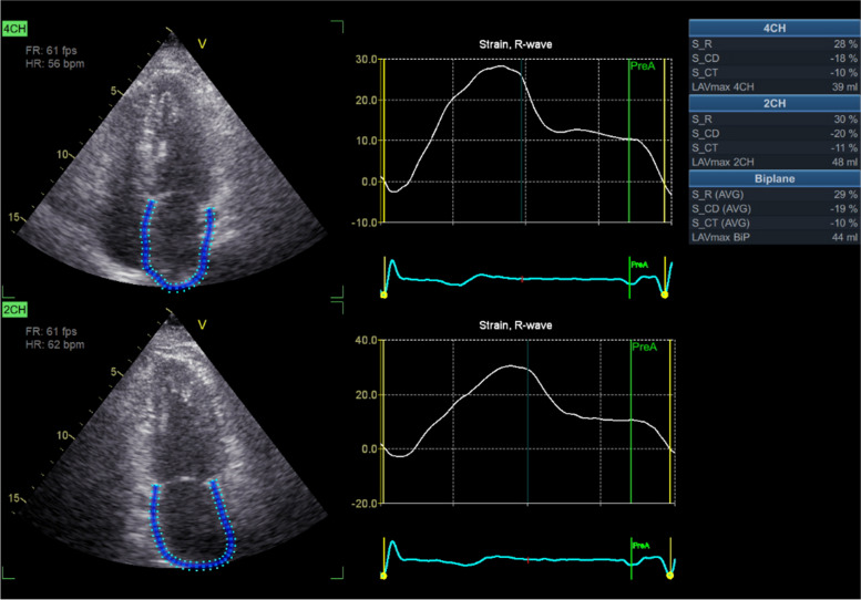
Methods: A prospective cohort of 1055 patients in SR was enrolled across 12 centers. The main study cohort was divided into two groups: the modeling group (n = 513) and the verification group (n = 542). All patients underwent ESE, which included B-lines, LA volume index (LAVi), and LA strain of the reservoir phase (LASr).
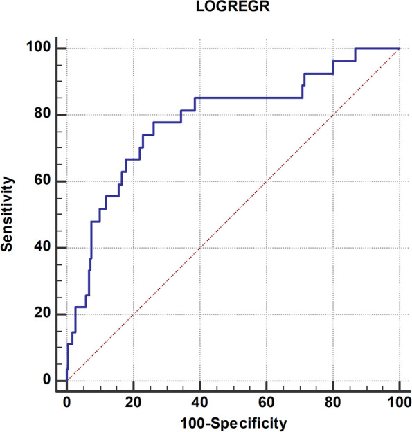
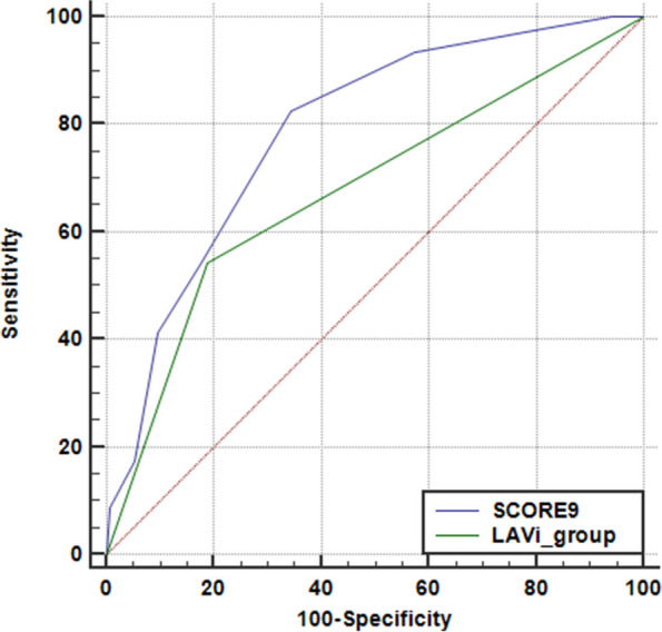
Results: Age, resting and stress LAVi and LASr, and B-lines were identified as a combination of detectors for PAF in both groups. In the entire cohort, aside from resting and stress LAVi and LASr, additional parameters differentiating PAF and non-PAF patients were the presence of systemic hypertension, exercise E/e' > 7, worse right ventricle (RV) contraction during exercise (∆ tricuspid annular plane systolic excursion < 5 mm), a lower left ventricular contractile reserve (< 1.6), and a reduced chronotropic reserve (heart rate reserve < 1.64). The composite score, summing all 9 items, yielded a score of > 4 as the best sensitivity (79%) and specificity (65%).
Conclusion: ESE can complement rest echocardiography in the identification of previous PAF in patients with SR through the evaluation of LA functional reservoir and volume reserve, LV chronotropic, diastolic, and systolic reserve, and RV contractile reserve.
Keywords: Atrial fibrillation; B-lines; Exercise stress echo; Left atrium; Reservoir strain.
© 2024. The Author(s).
Conflict of interest statement
The authors declare no competing interests.
Figures
References
-
- Hindricks G, Potpara T, Dagres N, Arbelo E, Bax JJ, Blomström-Lundqvist C, Boriani G, Castella M, Dan GA, Dilaveris PE, ESC Scientific Document Group, et al. 2020 ESC Guidelines for the diagnosis and management of atrial fibrillation developed in collaboration with the European Association for Cardio-Thoracic Surgery (EACTS): The Task Force for the diagnosis and management of atrial fibrillation of the European Society of Cardiology (ESC) Developed with the special contribution of the European Heart Rhythm Association (EHRA) of the ESC. Eur Heart J. 2021;42:373–498. - DOI - PubMed
-
- Morrone D, Arbucci R, Wierzbowska-Drabik K, Ciampi Q, Peteiro J, Agoston G, Varga A, Camarozano AC, Boshchenko A, Ryabova T, et al. Feasibility and functional correlates of left atrial volume changes during stress echocardiography in chronic coronary syndromes. Int J Cardiovasc Imaging. 2021;37:953–64. - DOI - PubMed
Publication types
MeSH terms
LinkOut - more resources
Full Text Sources
Medical
Research Materials
