. 2024 Dec;300(12):107954.
doi: 10.1016/j.jbc.2024.107954. Epub 2024 Nov 2.

The NEDD4-binding protein N4BP1 degrades mRNA substrates through the coding sequence independent of nonsense-mediated decay

Affiliations

The NEDD4-binding protein N4BP1 degrades mRNA substrates through the coding sequence independent of nonsense-mediated decay

Wen Zheng et al. J Biol Chem. 2024 Dec.

Abstract

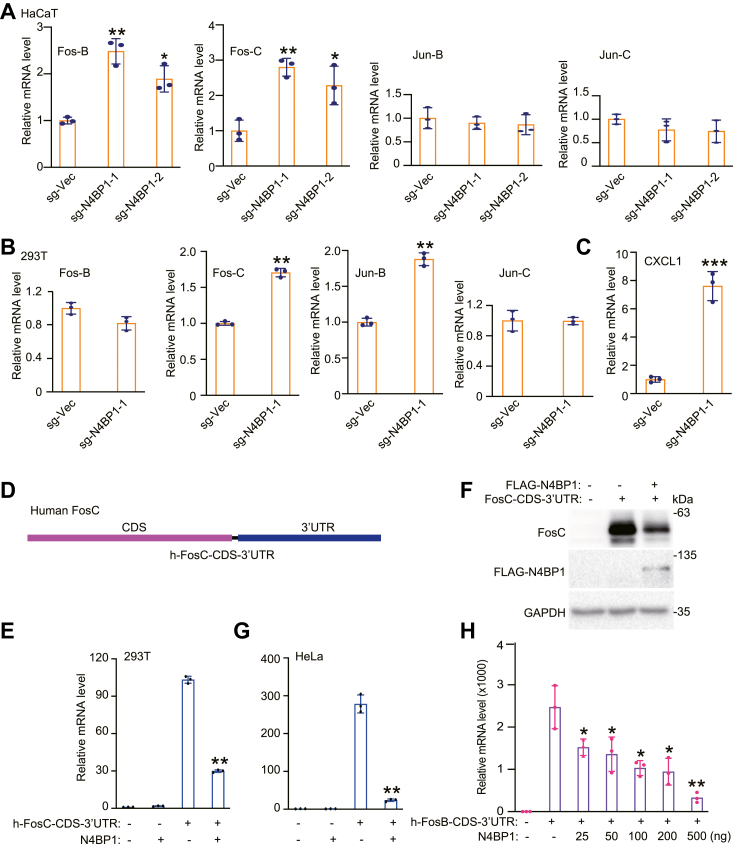
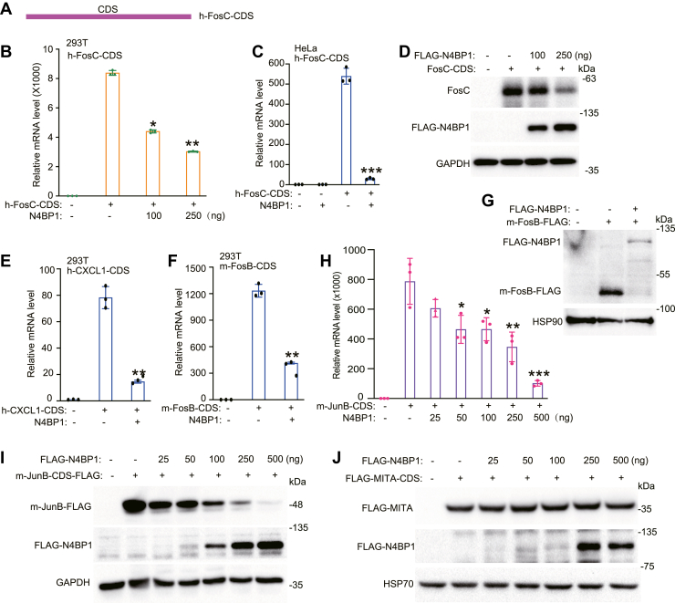
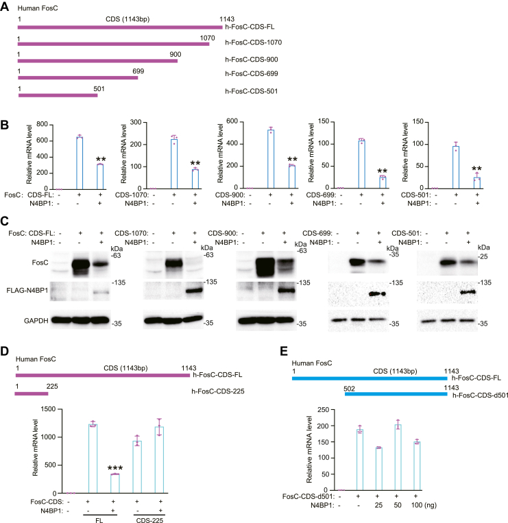
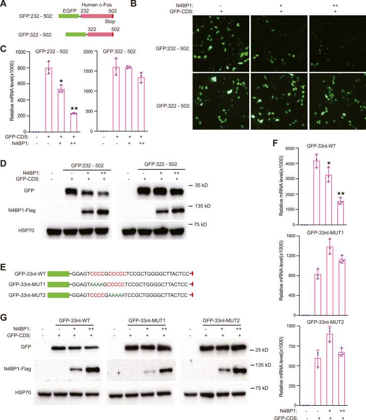
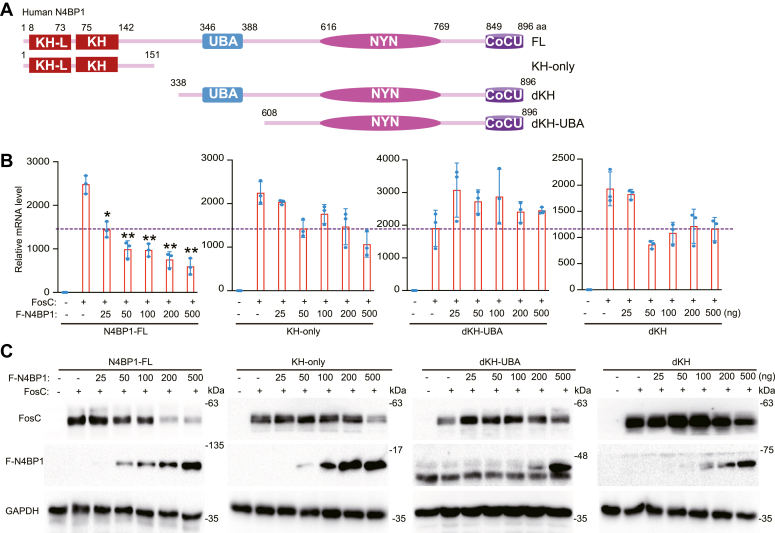
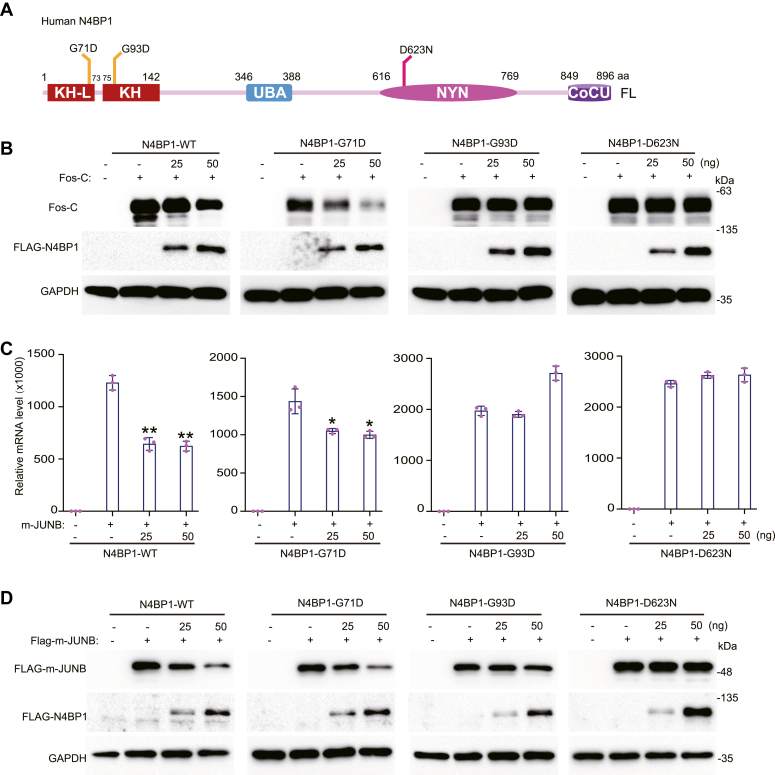
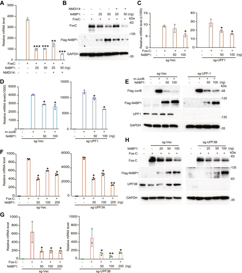
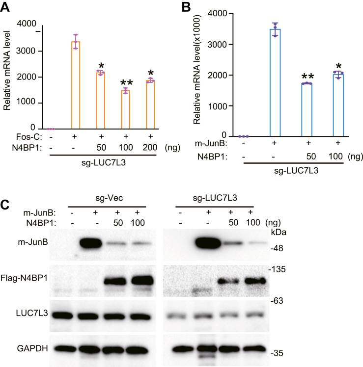
3'UTRs are recognized for their role in regulating mRNA turnover while the turnover of a specific group of mRNAs mediated by coding sequences (CDSs) remains poorly understood. N4BP1 is a critical inflammatory regulator in vivo with a molecular mechanism that is not yet clearly defined. Our study reveals that N4BP1 efficiently degrades its mRNA targets via CDS rather than the 3'-UTR. This CDS-dependent mRNA turnover mechanism appears to be a general feature of N4BP1, as evidenced by testing multiple mRNA substrates, such as Fos-C, Fos-B, Jun-B, and C-X-C motif chemokine ligand 1. Detailed mapping of the motif identified a crucial 33-nt (289-322) sequence near the 5'-end of Fos-C-CDS, where the presence of polyC is necessary for N4BP1-mediated degradation. Functional studies involving domain deletion and point mutations showed that both the K homology and N4BP1, YacP-like nuclease domains are essential for N4BP1 to restrict mRNA substrates. The function of N4BP1 in mRNA turnover is not dependent on nonsense-mediated decay as it efficiently restricts mRNA substrates even in cells deficient in UPF1, UPF3A, and UPF3B. Additionally, the function of N4BP1 is not reliant on LUC7L3 despite its known association with this protein. Our findings suggest that N4BP1 acts as an endoribonuclease to degrade mRNA substrates primarily through CDSs containing a C-rich motif.

Keywords: C-rich motif; Fos-C; KH domain; N4BP1; NYN domain; nonsense-mediated mRNA decay.


Conflict of interest statement

Conflict of interest The authors declare that they have no conflicts of interest with the contents of this article.

Figures

Figure 1
Figure 1
Identification N4BP1 mRNA targets.A, RT-PCR analysis of the mRNA levels of Fos-B, Fos-C, Jun-B, and Jun-C in WT and N4BP1−/− HaCaT cells. B, RT-PCR analysis of the mRNA levels of Fos-B, Fos-C, Jun-B, and Jun-C in WT and N4BP1−/− 293T cells. C, RT-PCR analysis of the mRNA level of CXCL1 in WT and N4BP1−/− 293T cells. D, schematic diagram of the mRNA structure of human Fos-C coding sequences and 3′-UTRs. E, RT-PCR analysis of the Fos-C level in 293T cells transfected with h-FosC-CDS-3′ UTR, f-prk-N4BP1 or their combination. F, Western blot analysis of Fos-C expression in 293T cells transfected with f-prk-N4BP1, h-FOSC-CDS-3′ UTR, or their combination. G, RT-PCR analysis of the Fos-C level in HeLa cells transfected with h-FosC-CDS-3′ UTR, f-prk-N4BP1, or their combination. H, RT-PCR analysis of Fos-B in 293T cells transfected with h-FosB-CDS-3′ UTR and different amount of f-prk-N4BP1. Data in (A and B) are representative of three independent experiments. Data in (E–H) are representative of four independent experiments. RT-PCR, n = 3, ∗p < 0.05; ∗∗p < 0.01; and ∗∗∗p < 0.001. CDS, coding sequence; CXCL1, C-X-C motif chemokine ligand 1; RT-PCR, reverse transcription-polymerase chain reaction.
Figure 2
Figure 2
N4BP1 restricts mRNA via coding sequences.A, schematic diagram of the structure of human Fos-C coding sequences (h-Fos-C-CDS). B, 293T cells were transfected with h-Fos-C-CDS and different amount of f-prk-N4BP1 for RT-PCR analysis. C, HeLa cells were transfected with h-Fos-C-CDS and f-prk-N4BP1 for RT-PCR analysis. D, 293T cells were transfected with h-Fos-C-CDS and different amount of f-prk-N4BP1 for WB analysis. E, 293T was transfected with h-CXCL1-CDS and f-prk-N4BP1, and the level of Fos-C was detected by RT-PCR. F, 293T cells transfected with m-Fos-B-CDS and f-prk-N4BP1 were used for RT-PCR analysis of m-Fos-B. G, 293T cells transfected with m-Fos-B-CDS and f-prk-N4BP1 were used for Western blot analysis of Flag-Fos-B and Flag-N4BP1. H, 293T cells were transfected with m-Jun-B-CDS and different amount of f-prk-N4BP1 for RT-PCR analysis of m-Jun-B. I, 293T cells were transfected with m-Jun-B-CDS and different amount of f-prk-N4BP1 for WB analysis of m-Jun-B. J, 293T cells were transfected with MITA-CDS and different amount of f-prk-N4BP1 for WB analysis of FLAG-MITA. Data in (B–I) are representative of three independent experiments. Data in (J) are representative of two independent experiments. RT-PCR, n = 3, ∗p < 0.05; ∗∗p < 0.01; and ∗∗∗p < 0.001. CDS, coding sequence; CXCL1, C-X-C motif chemokine ligand 1; RT-PCR, reverse transcription-polymerase chain reaction.
Figure 3
Figure 3
Mapping the mRNA motif in Fos-C-CDS.A, schematic diagram demonstrates the full length of human Fos-C-CDS and different mutants with deleted 3′-end. B, 293T was transfected with CDS-FL, CDS-1070, CDS-900, CDS-699, CDS-501 and f-PRK-N4BP1, and the level of Fos-C was detected by RT-PCR. C, detection of Fos-C by Western blot in 293T was transfected with CDS-FL, CDS-1070, CDS-900, CDS-699, CDS-501, and f-PRK-N4BP1. D, top: schematic indication of h-Fos-C-CDS-FL and h-FosC-CDS-225; below: 293T was transfected with 100 ng of CDS-FL, CDS-225, and 50 ng of f-prk-N4BP1, and the level of Fos-C was detected by RT-PCR. E, top: schematic demonstration of deletion of Fos-C-CDS at 5′-end. Bottom: 293T cells were transfected with 1 ug of FosC-CDS-d501 and 100 ng of f-prk-N4BP1 or 25 ng, 50 ng of f-prk-N4BP1 plus desired vector for a total of 100 ng of DNA. RT-PCR was used to detect Fos-C. Data in (B–E) are representative of three independent experiments. RT-PCR, n = 3, ∗p < 0.05; ∗∗p < 0.01; and ∗∗∗p < 0.001. CDS, coding sequence; RT-PCR, reverse transcription-polymerase chain reaction.
Figure 4
Figure 4
A polyC containing 33-nt region from Fos-C is sufficient to promote N4BP1-mediated targets degradation.A, schematic diagram showing the EGFP fusion plasmid of h-FosC-CDS (232–502) and h-FosC-CDS (322–502). B, the fluorescence intensity was observed by photographing under a fluorescence microscope with a 10× field of view. C, RT-PCR was used to detect the mRNA level of GFP with different fusion. The plasmids were transfected as indicated. D, Western blot analysis of GFP and N4BP1 expression in cells that transfected with plasmids encoding GFP with different fusion. E, schematic showing EGFP fusion core sequences and C-rich mutants. E, 293T cells were transfected with N4BP1 and GFP-33nt-WT or mutants, and the mRNA level of GFP were detected by RT-PCR. E, 293T cells were transfected with N4BP1 and GFP-33nt-WT or mutants, and the expressions of N4BP1 and GFP were detected by Western blot. Data in B–D, F, and G are representative of three independent experiments. RT-PCR, n = 3, ∗p < 0.05; ∗∗p < 0.01; and ∗∗∗p < 0.001. CDS, coding sequence; RT-PCR, reverse transcription-polymerase chain reaction.
Figure 5
Figure 5
N4BP1 degrades mRNA targets dependent on KH domain.A, schematic diagram demonstrates full-length N4BP1 or N4BP1 deletion mutants with FLAG tags at their N-terminal. B, 293T was transfected with different amount of N4BP1-FL, KH-only, dKH-UBA, dKH, along with Fos-C, and then the Fos-C was detected by RT-PCR. C, 293T was transfected with different amount of N4BP1-FL, KH-only, dKH-UBA, dKH, along with Fos-C, and then the Fos-C was detected by WB. Data in (B and C) are representative of three independent experiments. RT-PCR, n = 3, ∗p < 0.05; ∗∗p < 0.01; ∗∗∗p < 0.001. CDS, coding sequence; KH, K homology; RT-PCR, reverse transcription-polymerase chain reaction.
Figure 6
Figure 6
N4BP1 degrades mRNA targets dependent on KH and NYN domain.A, schematic diagram of point mutations in the KH and NYN domains of N4BP1. B, 293T was transfected with N4BP1-WT, N4BP1-G71D, N4BP1-G93D, N4BP1-D623N, and Fos-C, and then Fos-C and Flag-N4BP1 were analyzed by Western blot. C, 293T was transfected with N4BP1-WT, N4BP1-G71D, N4BP1-G93D, N4BP1-D623N and flag-m-JUNB, and then the mRNA level of m-JUNB was detected by RT-PCR. D, 293T was transfected with N4BP1-WT, N4BP1-G71D, N4BP1-G93D, N4BP1-D623N, and flag-m-JUNB, and then m-JUNB was detected by Western blot. Data in (B–D) are representative of three independent experiments. RT-PCR, n = 3, ∗p < 0.05; ∗∗p < 0.01; and ∗∗∗p < 0.001. KH, K homology; NYN, N4BP1, YacP-like nuclease; RT-PCR, reverse transcription-polymerase chain reaction.
Figure 7
Figure 7
N4BP1 restricts mRNA is independent on nonsense-mediated mRNA decay.A, 293T was transfected with f-prk-N4BP1 and Fos-C. After 8 h, the medium was replaced with a fresh medium along with or without 5 μM of NMDI14. Cells were harvested after 48 h. Fos-C was detected by RT-PCR. B, 293T was transfected with f-prk-N4BP1 and Fos-C. After 8 h, the medium was replaced with a fresh medium along with or without 5 mM of NMDI14. Cells were harvested after 48 h. Fos-C was detected by WB. C, f-prk-N4BP1 and Fos-C were transfected in sg-Vec cell line and sg-UPF1 cell line, respectively. The level of Fos-C was detected by RT-PCR. D, f-prk-N4BP1 and Jun-B were transfected in sg-Vec cell line and sg-UPF1 cell line, respectively. The level of Jun-B was detected by RT-PCR. E, f-prk-N4BP1 and Jun-B were transfected in sg-Vec cell line and sg-UPF1 cell line, respectively. The level of Jun-B was detected by WB. F, f-prk-N4BP1 and Fos-C were transfected in sg-Vec cell line and sg-UPF3A cell line, respectively. The level of Fos-C was detected by RT-PCR. G, f-prk-N4BP1 and Fos-C were transfected in sg-Vec cell line and sg-UPF3B cell line, respectively. The level of Fos-C was detected by RT-PCR. H, f-prk-N4BP1 and Fos-C were transfected in sg-Vec cell line and sg-UPF3B cell line, respectively. The level of Fos-C was detected by WB. Data in (A and B) are representative of two independent experiments. Data in (C–H) are representative of three independent experiments. RT-PCR, n = 3, ∗p < 0.05; ∗∗p < 0.01; and ∗∗∗p < 0.001. RT-PCR, reverse transcription-polymerase chain reaction; UPF1, upstream frameshift 1.
Figure 8
Figure 8
N4BP1 restricts mRNA is independent of LUC7L3.A, sg-LUC7L3 cell line was transfected with f-prk-N4BP1 and Fos-C, and Fos-C was detected by RT-PCR. B, sg-LUC7L3 cell line was transfected with f-prk-N4BP1 and Flag-m-Jun-B, and m-JUNB was detected by RT-PCR. C, control and sg-LUC7L3 cells were transfected with f-prk-N4BP1 and Jun-B, and the level of Jun-B was detected by WB. The control sample of sg-Vec is the same with it used in Figure 7E. The FLAG-N4BP1 panel of sg-Vec in Figure 7E was reused at here. Data in (A–C) are representative of three independent experiments. RT-PCR, n = 3, ∗p < 0.05; ∗∗p < 0.01; and ∗∗∗p < 0.001. RT-PCR, reverse transcription-polymerase chain reaction.

Similar articles

Cited by

References

    1. van Hoof A., Wagner E.J. A brief survey of mRNA surveillance. Trends Biochem. Sci. 2011;36:585–592. - PMC - PubMed
    1. Hilleren P., Parker R. Mechanisms of mRNA surveillance in eukaryotes. Annu. Rev. Genet. 1999;33:229–260. - PubMed
    1. Munchel S.E., Shultzaberger R.K., Takizawa N., Weis K. Dynamic profiling of mRNA turnover reveals gene-specific and system-wide regulation of mRNA decay. Mol. Biol. Cell. 2011;22:2787–2795. - PMC - PubMed
    1. Matoulkova E., Michalova E., Vojtesek B., Hrstka R. The role of the 3' untranslated region in post-transcriptional regulation of protein expression in mammalian cells. RNA Biol. 2012;9:563–576. - PubMed
    1. Hong D., Jeong S. 3'UTR diversity: expanding repertoire of RNA alterations in human mRNAs. Mol. Cells. 2023;46:48–56. - PMC - PubMed

Publication types

LinkOut - more resources