. 2024 Dec 1;17(12):dmm050716.
doi: 10.1242/dmm.050716. Epub 2024 Dec 13.

The role of mesenchymal cells in cholangiocarcinoma

Affiliations

The role of mesenchymal cells in cholangiocarcinoma

Mireia Sueca-Comes et al. Dis Model Mech. .

Abstract

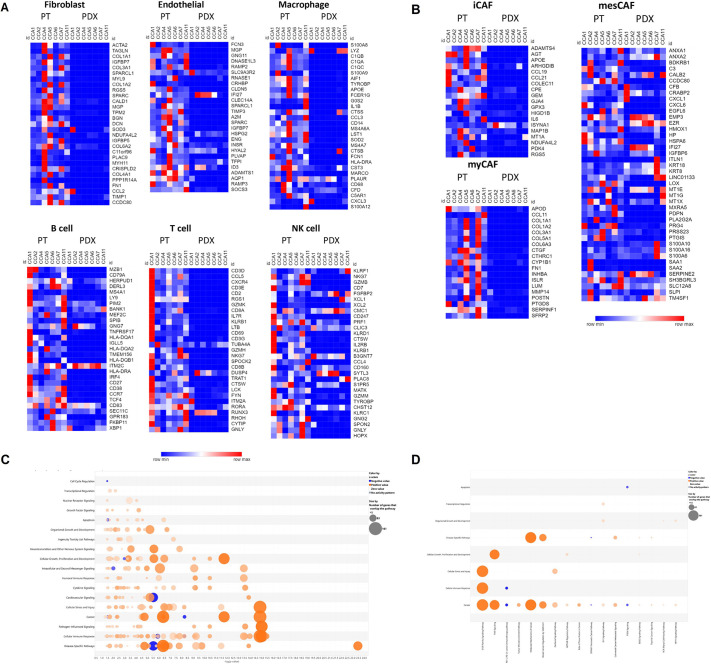
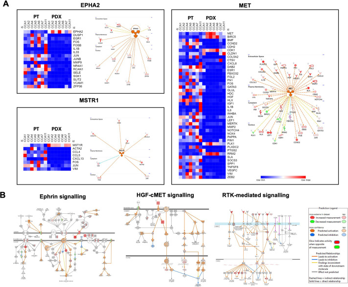
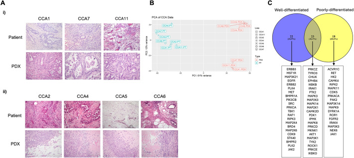
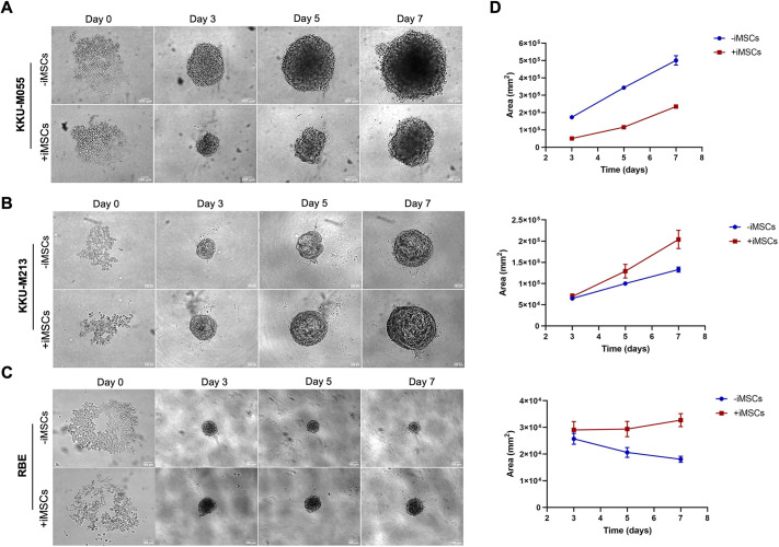
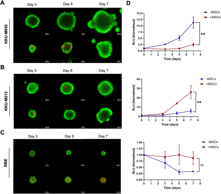
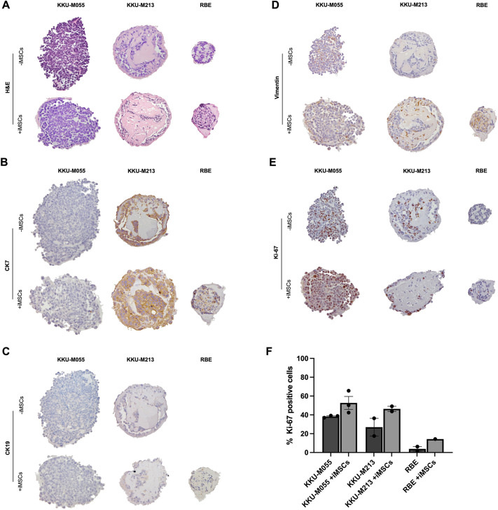
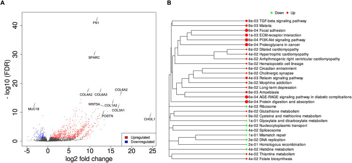
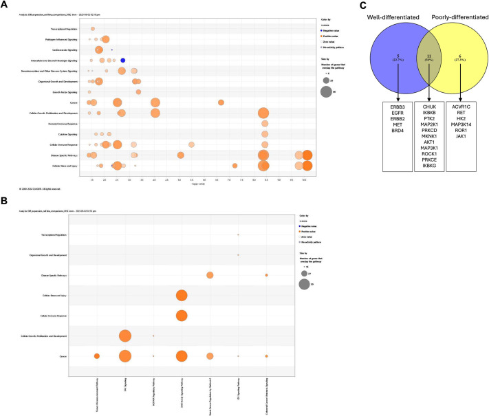
The tumour microenvironment (TME) significantly influences tumour formation and progression through dynamic interactions. Cholangiocarcinoma (CCA), a highly desmoplastic tumour, lacks early diagnostic biomarkers and has limited effective treatments owing to incomplete understanding of its molecular pathogenesis. Investigating the role of the TME in CCA progression could lead to better therapies. RNA sequencing was performed on seven CCA patient-derived xenografts (PDXs) and their corresponding patient samples. Differential expression analysis was conducted, and Qiagen Ingenuity Pathway Analysis was used to predict dysregulated pathways and upstream regulators. PDX- and cell line-derived spheroids, with and without immortalised mesenchymal stem cells, were grown and analysed for morphology, growth and viability. Histological analysis confirmed biliary phenotypes. RNA sequencing indicated upregulation of extracellular matrix-receptor interaction and PI3K-AKT pathways in the presence of mesenchymal cells, with several genes linked to poor survival. Mesenchymal cells restored the activity of inhibited cancer-associated kinases. Thus, adding mesenchymal cells to CCA spheroid models restored key paracrine signalling pathways lost in PDXs, enhancing tumour growth and viability. These findings highlight the importance of including stromal components in cancer models to improve pre-clinical studies.

Keywords: Cholangiocarcinoma; Mesenchymal stem cells; PDX models; Signalling pathways; Tumour microenvironment.


Conflict of interest statement

Competing interests The authors declare no competing or financial interests.

Figures

Fig. 1.
Fig. 1.
Loss of human stromal cells and dysregulation of cancer-associated pathways in CCA PDXs. (A,B) Heatmaps of genes identified as being associated with different populations of stromal cells (A) or cancer-associated fibroblasts (B) through single-cell analysis (Song et al., 2022; Affo et al., 2021). Blue and red indicate downregulated and upregulated genes, respectively. (C,D) Bubble charts showing canonical pathways associated with signalling (C) or with cancer (D) that were significantly dysregulated in patient tissues compared to PDXs. Orange and blue bubbles indicate activated and inhibited pathways, respectively; the size of the bubble indicates the number of genes in each gene set. CCA, cholangiocarcinoma; iCAF, immune-related cancer-associated fibroblast (CAF); myCAF, myofibroblast-like CAF; mesCAF, mesenchymal CAF; NK, natural killer; PDX, patient-derived xenograft; PT, patient tissue.
Fig. 2.
Fig. 2.
Dysregulation of paracrine signalling pathways in cancer cells within CCA PDXs. (A) Heatmaps of genes involved in the pathways downstream of the top 3 inhibited cancer-associated kinases (ICAKs; kinases predicted to be inhibited in the PDXs in spite of maintained expression) – MET, EPHA2 and MSTR1 – and diagrams of the respective networks. Blue and red indicate downregulated and upregulated genes, respectively. (B) Diagrams of three pathways involving these and other kinases. Pathways are provided by Qiagen Ingenuity Pathway Analysis and were overlaid with information to demonstrate molecules that were overexpressed or underexpressed and/or predicted to be activated/inhibited in the patient tissues compared to the PDXs.
Fig. 3.
Fig. 3.
Classification and dysregulation of upstream regulators in CCA PDXs. Histology and Upstream Regulator Analysis for two PDX subtypes. (A) Haematoxylin and Eosin (H&E) staining of patient and PDX tissues for two subtypes of PDX: well-differentiated (i) and poorly differentiated (ii). Objective, 20×. Scale bars: 200 μm. (B) Principal component analysis plot based on gene expression in patient and PDX samples, indicating two clusters of PDXs. PC, principal component. (C) Venn diagram to illustrate ICAKs found to be common or unique to the two PDX subtypes.
Fig. 4.
Fig. 4.
Effects of iMSCs on CCA cells in spheroid co-culture. (A-C) CCA cells [KKU-M055 (A), KKU-M213 (B) and RBE (C)] were cultured as spheroids alone (mono-culture) or iMSCs at a 1:2 ratio (cancer cells:iMSCs) (co-culture) in an ultra-low attachment (ULA) 96-well round-bottom plate at a final cell density of 1000 cells per well with 1001 μg/ml basement membrane extract (BME). The effect of co­ culture with iMSCs was assessed using brightfield microscopy at day 0, 3, 5 and 7 at 10× magnification. Representative spheroids are shown. Scale bars: 100 μm. The spheroids were analysed after initiation to determine area based on image analysis of brightfield micrographs captured at each timepoint from three independent replicates (mean±s.e.m.). iMSC, immortalised mesenchymal stem cell.
Fig. 5.
Fig. 5.
Cell viability of CCA spheroids as mono-cultures and iMSC co-cultures. (A-C) CCA cells [KKU-M055 (A), KKU-M213 (B) and RBE (C)] were cultured as spheroids alone (mono-culture) or iMSCs at a 1:2 ratio (cancer cells:iMSCs) (co-culture) in a ULA 96-well round-bottom plate at a final cell density of 1000 cells per well with 100 μg/ml BME. Live/dead cell staining was carried out using calcein acetoxymethyl (green, live cells) and ethidium homodimer (red, dead cells) at days 3, 5 and 7. Representative spheroids are shown. Scale bars: 100 μm. Progressive growth of the spheroid was monitored by adding D-luciferin at day 0, 3, 5 and 7. Values were normalised to day 0 (mean±s.e.m.). Co-cultures were compared to the mono-culture via paired two-tailed Student's t-test (n=2). ns, not significant (P>0.05); **P<0.01. RLU, relative luminescence units.
Fig. 6.
Fig. 6.
Morphology and protein expression of CCA spheroids. (A-E) CCA cells (KKU-M055, KKU-M213 and RBE) were cultured as spheroids alone (mono-culture) or iMSCs at a 1:2 (cancer cells:iMSCs) ratio (co-culture) in a ULA 96-well round-bottom plate at a final cell density of 1000 cells per well with 100 µg/ml BME. The spheroids were fixed at day 5 of culture, paraffin embedded, sectioned and stained with Haematoxylin and Eosin (H&E) (A), or stained for CK7 (B), CK19 (C), vimentin (D) and Ki-67 (E). (F) Ki-67 expression was quantified by manually counting the positive cells and cancer cells and calculating the ratio of positively stained cancer cells over the total of cancer cells. At least six spheroids were embedded in the array from each condition. Representative spheroids are shown at 20× magnification.
Fig. 7.
Fig. 7.
Differential expression analysis using DESeq2 for the KKU-M213-derived spheroids. (A) The differentially expressed genes between mono-culture and iMSCs co-culture were separated based on their false discovery rate (FDR) and fold change in a volcano plot. The upregulated and downregulated genes are represented in red and blue, respectively. (B) A gene set enrichment analysis (GSEA) was performed, and Kyoto Encyclopedia of Genes and Genomes (KEGG)-enrichment plots of representative gene sets from activated pathway are shown. The names of KEGG terms are listed, and the downregulated and upregulated pathways are represented in green and red, respectively. The length of the horizontal graph represents the gene ratio. The area of circle in the graph represents the fold-change value. These graphs were generated using integrated differential expression and pathway analysis (iDEP).
Fig. 8.
Fig. 8.
Activation of ICAKs in MSC co-cultures and restoration of POX-type specific signalling pathways. (A,B) Bubble charts showing canonical pathways associated with signalling (A) or with cancer (B) that were significantly activated in co-culture compared with mono-culture spheroids. Orange and blue bubbles indicate activated and inhibited pathways, respectively; the size of the bubble indicates the number of genes in each gene set. (C) Venn diagram to indicate inhibited cancer-associated kinases (ICAKs) common to or unique to well-differentiated or poorly differentiated PDXs, which were predicted to be activated in the co-culture compared to the mono-culture spheroids.

References

    1. Affo, S., Nair, A., Brundu, F., Ravichandra, A., Bhattacharjee, S., Matsuda, M., Chin, L., Filliol, A., Wen, W., Song, X.et al. (2021). Promotion of cholangiocarcinoma growth by diverse cancer-associated fibroblast subpopulations. Cancer Cell 39, 866-882.e11. 10.1016/j.ccell.2021.03.012 - DOI - PMC - PubMed
    1. Ai, Y., Wu, S., Zou, C. and Wei, H. (2020). LINC00941 promotes oral squamous cell carcinoma progression via activating CAPRIN2 and canonical WNT/β-catenin signaling pathway. J. Cell. Mol. Med. 24, 10512-10524. 10.1111/jcmm.15667 - DOI - PMC - PubMed
    1. Allen, I. C., Eden, K., Heid, B. and Holl, E. K. (2017). Map3K14 signaling attenuates the development of colorectal cancer through activation of the non-canonical NF-κB signaling cascade. J. Immunol. 198, 197.6-197.6. 10.4049/jimmunol.198.Supp.197.6 - DOI
    1. Antoni, D., Burckel, H., Josset, E. and Noel, G. (2015). Three-dimensional cell culture: a breakthrough in vivo. Int. J. Mol. Sci. 16, 5517-5527. 10.3390/ijms16035517 - DOI - PMC - PubMed
    1. Banales, J. M., Marin, J. J. G., Lamarca, A., Rodrigues, M., Khan, S. A., Roberts, L. R., Cardinale, V., Carpino, G., Andersen, J. B., Braconi, C.et al. (2020). Cholangiocarcinoma 2020: the next horizon in mechanisms and management. Nat. Rev. Gastroenterol. Hepatol. 17, 557-588. 10.1038/s41575-020-0310-z - DOI - PMC - PubMed

MeSH terms

Substances