Review
. 2024 Oct 31;5(11):e784.
doi: 10.1002/mco2.784. eCollection 2024 Nov.

Neuroscience in peripheral cancers: tumors hijacking nerves and neuroimmune crosstalk

Affiliations
Review

Neuroscience in peripheral cancers: tumors hijacking nerves and neuroimmune crosstalk

Hua-Yang Fan et al. MedComm (2020). .

Abstract

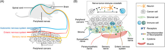
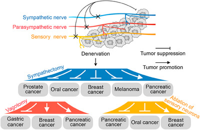
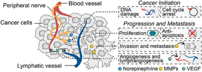
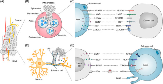
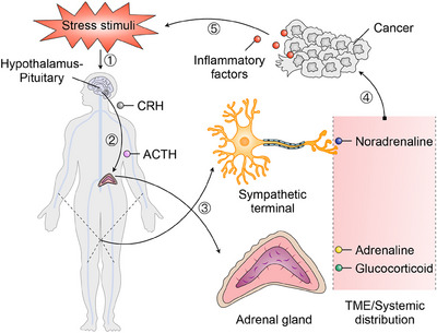
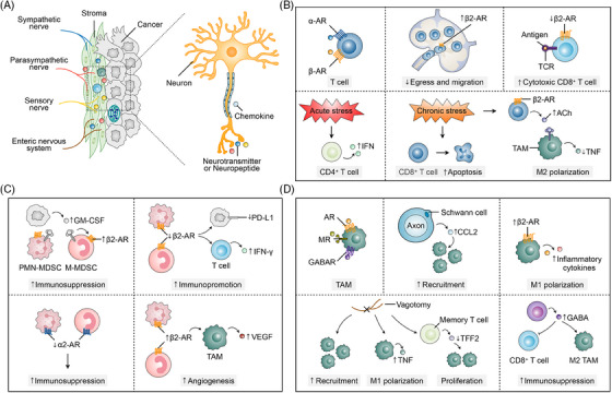
Cancer neuroscience is an emerging field that investigates the intricate relationship between the nervous system and cancer, gaining increasing recognition for its importance. The central nervous system governs the development of the nervous system and directly affects brain tumors, and the peripheral nervous system (PNS) shapes the tumor microenvironment (TME) of peripheral tumors. Both systems are crucial in cancer initiation and progression, with recent studies revealing a more intricate role of the PNS within the TME. Tumors not only invade nerves but also persuade them through remodeling to further promote malignancy, creating a bidirectional interaction between nerves and cancers. Notably, immune cells also contribute to this communication, forming a triangular relationship that influences protumor inflammation and the effectiveness of immunotherapy. This review delves into the intricate mechanisms connecting the PNS and tumors, focusing on how various immune cell types influence nerve‒tumor interactions, emphasizing the clinical relevance of nerve‒tumor and nerve‒immune dynamics. By deepening our understanding of the interplay between nerves, cancer, and immune cells, this review has the potential to reshape tumor biology insights, inspire innovative therapies, and improve clinical outcomes for cancer patients.

Keywords: cancer neuroscience; cancer therapy; neuroimmune crosstalk; peripheral nervous system; tumor microenvironment; tumor‒nerve interactions.


Conflict of interest statement

The authors declare they have no conflicts of interest.

Figures

FIGURE 1
FIGURE 1
Schematic diagram of peripheral nerves intertwined with peripheral cancers. (A) The sympathetic, parasympathetic, and sensory nerves play a regulatory role in peripheral cancers. (B) This diagram shows how nerves, tumor cells, immune cells, and other components interact within the tumor microenvironment (TME) of peripheral cancers. Various types of nerves—sympathetic, parasympathetic, sensory, motor, and enteric—enter the tissue around tumor cells. The diagram highlights how these nerves communicate with cancer cells by releasing neurotransmitters and neuropeptides, which in turn influence tumor growth and the immune response.
FIGURE 2
FIGURE 2
The impact of denervation on peripheral cancers. This diagram illustrates how denervation of different nerve types—sympathetic, parasympathetic, and sensory—affects tumor growth in different peripheral cancers, highlighting the diverse effects based on nerve type and cancer type.
FIGURE 3
FIGURE 3
Neural regulation of tumor initiation and progression. Neural signals regulate tumorigenesis, including their effects on cellular DNA damage and the cell cycle, as well as tumor progression, including cell proliferation, anti‐apoptosis, invasion, metastasis, angiogenesis, and lymphangiogenesis.
FIGURE 4
FIGURE 4
Perineural infiltration (PNI) and molecular interactions between Schwann cells and cancer cells. (A) Diagram shows how nerves interact with peripheral cancers. (B) A cross‐section of a nerve shows its protective layers—epineurium, perineurium, and endoneurium—surrounding the axons within a fascicle. The diagram illustrates how cancer cells infiltrate these layers during PNI. (C) Molecular signaling between Schwann cells and cancer cells. This section highlights the key molecular interactions driving communication between Schwann cells and cancer cells. Molecules such as neural cell adhesion molecule 1 (NCAM1), myelin‐associated glycoprotein (MAG), L1 cell adhesion molecule (L1CAM), glial cell line‐derived neurotrophic factor (GDNF), nerve growth factor (NGF), and fractalkine (CX3CL1) on Schwann cells interact with cancer cell receptors or ligands, including E‐cadherin (E‐Cad), Mucin 1 (MUC1), L1CAM, rearranged during transfection (RET), TRKA, and C‒X3‒C motif chemokine receptor 1 (CX3CR1). (D) The figure shows how cancer cells interact with Schwann cells and axons, highlighting the formation of the tumor‐activated Schwann cell tracks (TAST) by Schwann cells. (E) The figure illustrates the molecular crosstalk between cancer cells, Schwann cells, and axons. It highlights key signaling pathways through which cancer cells communicate with neurons. Cancer cells secrete growth factors such as GDNF, NGF, and brain‐derived neurotrophic factor (BDNF), which bind to their respective receptors (GFRα, TRKA, and TRKB) on Schwann cells and axons, promoting neuronal growth and cancer invasion. Additionally, cancer cells release extracellular vehicles (EVs) containing microRNAs (miRNAs), which contribute to neural reprogramming. The figure also showcases the TAST formed by Schwann cells, aiding in cancer progression.
FIGURE 5
FIGURE 5
The stress response and its influence on the tumor microenvironment (TME). This schematic illustrates the biological pathway by which stress stimuli influence tumor progression via the hypothalamic‒pituitary‒adrenal (HPA) axis and the sympathetic nervous system. Upon exposure to stress, the hypothalamus releases corticotropin‐releasing hormone (CRH), which stimulates the anterior pituitary gland to secrete adrenocorticotropic hormone (ACTH) into the bloodstream. ACTH then prompts the adrenal glands to release adrenaline and glucocorticoids, which are distributed systemically, including to the TME. Simultaneously, the sympathetic nervous system is activated, releasing noradrenaline from sympathetic nerve terminals that innervate the tumor environment. These stress hormones, together with inflammatory factors, promote cancer cell survival, proliferation, and tumor progression by modulating the inflammatory conditions within the TME.
FIGURE 6
FIGURE 6
Neuro‐immune interactions in the tumor microenvironment (TME). (A) The diagram on the left shows sympathetic, parasympathetic, sensory, and enteric nerves infiltrating the tumor stroma. These neurons release neurotransmitters and neuropeptides, which interact with cancer cells and immune cells via chemokine signaling, influencing both tumor progression and immune responses. (B) T cells express both α‐ and β‐adrenergic receptors (β‐ARs), and their activation triggers distinct immune responses. Increased β2‐AR signaling impairs T‐cell egress and migration from lymphoid organs, limiting their ability to reach tumor sites. Conversely, the downregulation of β2‐AR on CD8+ T cells enhances their cytotoxic functions and improves antigen recognition through interactions with T‐cell receptor (TCR). Acute stress can boost immune responses by promoting interferon (IFN) production in CD4+ T cells, while chronic stress has the opposite effect, inducing apoptosis in CD8+ T cells and weakening immune surveillance, thereby promoting tumor progression. Moreover, chronic stress and β2‐AR activation increase acetylcholine (ACh) production, which drives M2 polarization of tumor‐associated macrophages (TAMs) and suppresses tumor necrosis factor (TNF) production, creating an immunosuppressive tumor microenvironment. (C) β2‐AR signaling in myeloid‐derived suppressor cells (MDSCs) enhances their immunosuppressive activity by increasing granulocyte‒macrophage colony‐stimulating factor (GM‐CSF) production. Conversely, reduced β2‐AR signaling lowers programmed death‐ligand 1 (PD‐L1) expression and boosts IFN‐γ production in T cells, leading to enhanced immune activation. Additionally, α2‐AR signaling in MDSCs further strengthens their immunosuppressive effects, promoting tumor immune evasion. In TAMs, β2‐AR activation supports angiogenesis by increasing vascular endothelial growth factor (VEGF) secretion, which facilitates tumor growth. (D) AR, MR, and GABAR on TAMs regulate their activation and function in the tumor microenvironment. Schwann cells, in conjunction with axons, release CCL2 to recruit TAMs to the tumor site, increasing immune cell infiltration. β2‐AR signaling in TAMs elevates the production of inflammatory cytokines, promoting M1 polarization and a pro‐inflammatory response. Vagotomy enhances TAM recruitment and M1 polarization by boosting TNF production, thereby supporting an anti‐tumor immune response. It also reduces TFF2 expression in memory T cells, which boosts their proliferation. Conversely, GABA production by CD8+ T cells promotes TAM polarization towards the M2 phenotype, leading to immunosuppression in the TME.

Similar articles

Cited by

References

    1. Mancusi R, Monje M. The neuroscience of cancer. Nature. 2023;618(7965):467‐479. - PMC - PubMed
    1. Hanahan D, Monje M. Cancer hallmarks intersect with neuroscience in the tumor microenvironment. Cancer Cell. 2023;41(3):573‐580. - PMC - PubMed
    1. Winkler F, Venkatesh HS, Amit M, et al. Cancer neuroscience: state of the field, emerging directions. Cell. 2023;186(8):1689‐1707. - PMC - PubMed
    1. Wu J, Heidelberg RE, Gajjar A. Adolescents and young adults with cancer: CNS tumors. J Clin Oncol. 2024;42(6):686‐695. - PMC - PubMed
    1. Magnon C, Hondermarck H. The neural addiction of cancer. Nat Rev Cancer. 2023;23(5):317‐334. - PubMed

LinkOut - more resources