

A Rare Presentation of Myoepithelioma of the Parotid Gland Manifesting as an Infra-Auricular Swelling
- PMID: 39493007
- PMCID: PMC11530963
- DOI: 10.7759/cureus.70746
A Rare Presentation of Myoepithelioma of the Parotid Gland Manifesting as an Infra-Auricular Swelling
Abstract
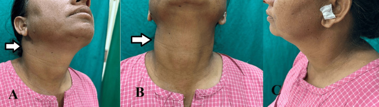
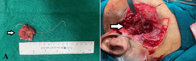
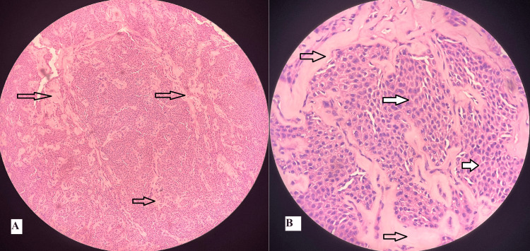
This report describes an uncommon tumor of the salivary glands, myoepithelioma, that primarily affects major and minor glands, with a notable predilection for the parotid gland. Typically benign, this tumor arises from aberrant myoepithelial cells situated between the basement membrane and acinar cells. Myoepitheliomas are considered a subset of pleomorphic adenomas, distinguished by excessive myoepithelial cell growth. Despite their initial discovery, the precise histopathological and immunohistochemical characteristics of these tumors remain elusive, posing a diagnostic challenge because of their complex nature. We discuss a case of a 42-year-old female who had a 2 x 2 cm lump in the right infra-auricular area. The lump was examined with ultrasonography (USG) and later surgically removed. The initial frozen section analysis indicated an oncocytic lesion, but further histopathological and immunohistochemical evaluations confirmed that it was a myoepithelioma of the parotid gland.
Keywords: benign parotid tumors; myoepithelioma; parotid myoepithelioma; rare case report; unusual presentation.
Copyright © 2024, Kalra et al.
Conflict of interest statement
Human subjects: Consent was obtained or waived by all participants in this study. Conflicts of interest: In compliance with the ICMJE uniform disclosure form, all authors declare the following: Payment/services info: All authors have declared that no financial support was received from any organization for the submitted work. Financial relationships: All authors have declared that they have no financial relationships at present or within the previous three years with any organizations that might have an interest in the submitted work. Other relationships: All authors have declared that there are no other relationships or activities that could appear to have influenced the submitted work.
Figures
Similar articles
-
Myoepithelioma of the Parotid Gland: A Case Report and Literature Review.Indian J Otolaryngol Head Neck Surg. 2022 Dec;74(Suppl 3):6087-6090. doi: 10.1007/s12070-021-02763-x. Epub 2021 Jul 9. Indian J Otolaryngol Head Neck Surg. 2022. PMID: 36742563 Free PMC article.
-
Myoepithelioma of the Parotid Gland: A Case Report with Review of the Literature and Classic Histopathology.Case Rep Otolaryngol. 2017;2017:6036179. doi: 10.1155/2017/6036179. Epub 2017 Aug 16. Case Rep Otolaryngol. 2017. PMID: 28900549 Free PMC article.
-
Myoepithelioma of the Palatal Minor Salivary Gland: A Case Report.Cureus. 2024 Mar 17;16(3):e56305. doi: 10.7759/cureus.56305. eCollection 2024 Mar. Cureus. 2024. PMID: 38629005 Free PMC article.
-
Salivary gland myoepithelial carcinoma.Clin Transl Oncol. 2015 Nov;17(11):847-55. doi: 10.1007/s12094-015-1329-4. Epub 2015 Jul 2. Clin Transl Oncol. 2015. PMID: 26133522 Review.
-
Mucinous myoepithelioma, a recently described new myoepithelioma variant.Head Neck Pathol. 2013 Jul;7 Suppl 1(Suppl 1):S85-9. doi: 10.1007/s12105-013-0464-x. Epub 2013 Jul 3. Head Neck Pathol. 2013. PMID: 23821216 Free PMC article. Review.
References
-
- Myoepithelioma of parotid: a case report and review of literature. Kapoor A, Rajput PS, Bagri PK, Beniwal S, Kumar V, Kumar HS. J Oral Res Rev. 2014;6:53–56.
-
- Myoepithelioma of the parotid gland: a rare case. Yenidünya S, Haltaş H, Bayrak R, Aktaş D. https://dergipark.org.tr/en/download/article-file/2016952 Ann Health Sci Res. 2012;1:49–51.
-
- Salivary gland myoepithelioma with focal capsular invasion. Gun BD, Ozdamar SO, Bahadir B, Uzun L. Ear Nose Throat J. 2009;88:1005–1009. - PubMed
-
- Myoepithelioma of the parotid gland: case report and review of literature. Politi M, Toro C, Zerman N, Mariuzzi L, Robiony M. Oral Oncol Extra. 2005;1:104–108.
Publication types
LinkOut - more resources
Full Text Sources