

The Radiographic Characteristics of Mandibular Wisdom Teeth That Can Cause Severe Deep Neck Infection
- PMID: 39493048
- PMCID: PMC11531351
- DOI: 10.7759/cureus.70791
The Radiographic Characteristics of Mandibular Wisdom Teeth That Can Cause Severe Deep Neck Infection
Abstract
Purpose: Mandibular wisdom teeth can occasionally cause infections, which can progress to severe deep neck infections (DNIs) including deep neck abscesses or necrotizing soft tissue infections, which are fatal. This study aimed to identify the radiographic characteristics of mandibular wisdom teeth that developed severe DNIs.
Methods: This study included patients who were admitted for the treatment of severe mandibular wisdom tooth infection between July 2012 and June 2024 at a single center. Patient characteristics, clinical data, and radiographic findings were analyzed and compared between the severe DNI group and mild DNI group including patients with cellulitis or superficial abscess. P < 0.05 was considered significant.
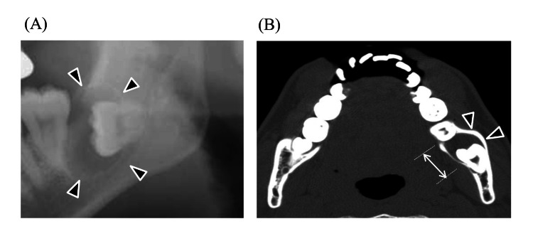
Results: Nineteen of 42 patients (45.2%) were included in the severe DNI group. The multivariate analysis showed that the highest odds ratio (OR) was for the presence of a radicular cyst (OR=17.7), followed by the presence of a dentigerous cyst (OR =14.5). The most common mandibular wisdom tooth with a dentigerous cyst in patients with severe DNIs was inverted according to Winter's classification and type IIIC in the Pell and Gregory classification.
Conclusion: Radiographic characteristics associated with severe DNIs included the presence of radicular and dentigerous cysts in the mandibular wisdom teeth. Especially in dentigerous cysts, deeply impacted teeth should be taken attention.
Keywords: deep neck abscess; deep neck infections; dentigerous cyst; mandibular wisdom tooth; necrotizing soft tissue infection; radicular cyst.
Copyright © 2024, Iwata et al.
Conflict of interest statement
Human subjects: Consent was obtained or waived by all participants in this study. Kakogawa Central City Hospital issued approval 2019-85. This study was conducted in accordance with the 1964 Declaration of Helsinki. The ethics committee approved the study and gave us administrative permission to access the data used in this study. Since this was a retrospective study, the research plan was published on the homepage of the participating hospitals according to the instructions of the IRB, in accordance with the guaranteed opt-out opportunity. Animal subjects: All authors have confirmed that this study did not involve animal subjects or tissue. Conflicts of interest: In compliance with the ICMJE uniform disclosure form, all authors declare the following: Payment/services info: All authors have declared that no financial support was received from any organization for the submitted work. Financial relationships: All authors have declared that they have no financial relationships at present or within the previous three years with any organizations that might have an interest in the submitted work. Other relationships: All authors have declared that there are no other relationships or activities that could appear to have influenced the submitted work.
Figures


References
-
- The assessment and management of deep neck space infections in adults: a systematic review and qualitative evidence synthesis. Sheikh Z, Yu B, Heywood E, Quraishi N, Quraishi S. Clin Otolaryngol. 2023;48:540–562. - PubMed
-
- Risk factors for transcervical incision and drainage of pediatric deep neck infections. Hah YM, Jung AR, Lee YC, Eun YG. J Pediatr Surg. 2018;53:666–670. - PubMed
-
- Imaging assessment of deep neck spaces infections: an anatomical approach. Caprioli S, Tagliafico A, Fiannacca M, et al. Radiol Med. 2023;128:81–92. - PubMed
-
- Submandibular triangle - in normal conditions and in acute purulent inflammatory diseases. Yotsova R. Varna Med Forum. 2023;12:180–185.
LinkOut - more resources
Full Text Sources