. 2024 Oct 18:14:1474007.
doi: 10.3389/fonc.2024.1474007. eCollection 2024.

Vδ2 T-cell engagers bivalent for Vδ2-TCR binding provide anti-tumor immunity and support robust Vγ9Vδ2 T-cell expansion

Affiliations

Vδ2 T-cell engagers bivalent for Vδ2-TCR binding provide anti-tumor immunity and support robust Vγ9Vδ2 T-cell expansion

Lisa A King et al. Front Oncol. .

Abstract

Background: Vγ9Vδ2 T-cells are antitumor immune effector cells that can detect metabolic dysregulation in cancer cells through phosphoantigen-induced conformational changes in the butyrophilin (BTN) 2A1/3A1 complex. In order to clinically exploit the anticancer properties of Vγ9Vδ2 T-cells, various approaches have been studied including phosphoantigen stimulation, agonistic BTN3A-specific antibodies, adoptive transfer of expanded Vγ9Vδ2 T-cells, and more recently bispecific antibodies. While Vγ9Vδ2 T-cells constitute a sizeable population, typically making up ~1-10% of the total T cell population, lower numbers have been observed with increasing age and in the context of disease.

Methods: We evaluated whether bivalent single domain antibodies (VHHs) that link Vδ2-TCR specific VHHs with different affinities could support Vγ9Vδ2 T-cell expansion and could be incorporated in a bispecific engager format when additionally linked to a tumor antigen specific VHH.

Results: Bivalent VHHs that link a high and low affinity Vδ2-TCR specific VHH can support Vγ9Vδ2 T-cell expansion. The majority of Vγ9Vδ2 T-cells that expanded following exposure to these bivalent VHHs had an effector or central memory phenotype and expressed relatively low levels of PD-1. Bispecific engagers that incorporated the bivalent Vδ2-TCR specific VHH as well as a tumor antigen specific VHH triggered antitumor effector functions and supported expansion of Vγ9Vδ2 T-cells in vitro and in an in vivo model in NOG-hIL-15 mice.

Conclusion: By enhancing the number of Vγ9Vδ2 T-cells available to exert antitumor effector functions, these novel Vδ2-bivalent bispecific T cell engagers may promote the overall efficacy of bispecific Vγ9Vδ2 T-cell engagement, particularly in patients with relatively low levels of Vγ9Vδ2 T-cells.

Keywords: Vγ9Vδ2 T-cells; bispecific T-cell engager; cancer; expansion; immunotherapy; single domain antibody.


Conflict of interest statement

NV. TG and HV own LAVA Therapeutics NV shares. DH, PY, VI-G, PH and HV are/were employed by LAVA Therapeutics NV. TG is scientific advisor to LAVA Therapeutics NV. The authors declare that this study received funding from Lava Therapeutics NV. LK, MJ and MV were funded by LAVA Therapeutics NV.

Figures

Figure 1
Figure 1
Bivalent Vδ2-TCR-specific VHHs support enrichment and expansion of Vγ9Vδ2 T-cells. (A) Binding of bivalent Vδ2-VHHs to Vγ9Vδ2 T-cells assessed using flow cytometry (n=3) or ELISA (n=2). (B) Overview of experimental design to assess expansion. (C) Representative gating strategy to asses enriched and expanded Vγ9Vδ2 T-cells within PBMC. (D) Enrichment (left panel) and fold expansion (right panel) of Vγ9Vδ2 T-cells during an 8 day culture of healthy donor PBMC in the presence or absence of 1 nM bivalent Vδ2-VHHs or 10 µM pamidronate (n=3-6). (E) Binding of the 6H4-5C7 bivalent VHH with either a 5, 10 or 20 AA linker to Vγ9Vδ2 T-cells (n=3). (F) Enrichment (left panel) and fold expansion (right panel) of Vγ9Vδ2 T-cells during an 8 day culture of healthy donor PBMC in the presence or absence of 0.001, 0.1, 1 or 100 nM of the 6H4-5C7 bivalent VHH with either 5, 10 or 20 AA linker or 10 µM pamidronate. Data in (A) (left panel) and (D–F) assessed using flow cytometry. Data in A (right panel) assessed using ELISA. Data represent mean and SEM (A, E) or individual data-points are indicated using open circles and box and whisker plots indicate the median, 25th to 75th percentiles and minimum to maximum (D, F). Two-way ANOVA with Tukey’s multiple comparisons test (F) were used for statistical analysis; P=< 0.05: *.
Figure 2
Figure 2
TAA-Vδ2hi-lo bsVHH and TAA-Vδ2hi-lo bsVHH-Fc support Vγ9Vδ2 T-cell expansion and trigger Vγ9Vδ2 T-cell degranulation and tumor cell lysis. (A) Illustration of the TAA-Vδ2hi-lo bsVHH with and without Fc domain. (B) Binding of the TAA-Vδ2hi-lo bsVHH with and without Fc to Vγ9Vδ2 T-cells. Data represent mean and SEM (n=3). (C) Enrichment (left panel) and fold expansion (right panel) of Vγ9Vδ2 T-cells during an 8 day culture of healthy donor PBMC in the presence or absence of 1 nM TAA-Vδ2hi-lo bsVHH, 100nM TAA-Vδ2hi-lo bsVHH-Fc or 10 µM pamidronate. Individual data-points are indicated using open circles and box and whisker plots indicate the median, 25th to 75th percentiles and minimum to maximum (n=15-30). (D) Binding of the TAA-Vδ2hi-lo bsVHH and TAA-Vδ2hi-lo bsVHH-Fc to SW480 (EGFR+), 22Rv1 (PSMA+) or MM.1s.CD1d (CD1d+) tumor cells. Data represent mean and SEM (n=3). (E) Vγ9Vδ2 T-cell CD107a expression after 24 hr co-cultures of Vγ9Vδ2 T-cells and SW480, 22Rv1 or MM.1s.CD1d tumor cells (1:1 E:T ratio) ± concentration range of the TAA-Vδ2hi-lo bsVHH and TAA-Vδ2hi-lo bsVHH-Fc. Data represent mean and SEM (n=3). (F) Lysis of SW480, 22Rv1 or MM.1s.CD1d tumor cells after 24 hr incubation with Vγ9Vδ2 T-cells (1:1 E:T ratio) ± concentration range of the TAA-Vδ2hi-lo bsVHH and TAA-Vδ2hi-lo bsVHH-Fc. Data represent mean and SEM (n=3-6). Data generated using flow cytometry. One-way ANOVA with Tukey’s multiple comparisons test was used for statistical analysis and asterisks are shown compared to IL-2 control; P=< 0.01: **, P=< 0.001: ***, P=< 0.0001: ****.
Figure 3
Figure 3
Vγ9Vδ2 T-cells expanded using TAA-Vδ2hi-lo bsVHH and TAA-Vδ2hi-lo bsVHH-Fc are activated and induce tumor lysis when exposed to TAA-expressing tumor cells. (A) Schematic overview of method used for PBMC culture, subsequent assessment of Vγ9Vδ2 T-cell expansion, as well as co-culture of enriched Vγ9Vδ2 T-cells and tumor cells. (B–E) Vγ9Vδ2 T-cell CD25 expression (A; n=4-12), CD107a expression (B; n=4-12), specific tumor cell lysis (C; n=4-12) and levels of IL-2, TNF and IFN-γ (pg/ml, D; n=4-5) in 24hr co-cultures of SW480 (EGFR+), 22Rv1 (PSMA+) or MM.1s.CD1d (CD1d+) tumor cells with Vγ9Vδ2 T-cells enriched (purity > 60%) from 8 day cultures of healthy donor PBMC in the presence or absence of 1 nM TAA-Vδ2hi-lo bsVHH, 100 nM TAA-Vδ2hi-lo bsVHH-Fc or 10 µM pamidronate (1:1 E:T ratio). Data generated through flow cytometry (B–D) or CBA (E). Individual data-points are indicated using open circles and box and whisker plots indicate the median, 25th to 75th percentiles and minimum to maximum. One-way ANOVA with Tukey’s multiple comparisons test was used for statistical analysis; P=< 0.05: *, P=< 0.01: **, P=< 0.001: ***, P=< 0.0001: ****.
Figure 4
Figure 4
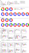
Bivalent Vδ2hi-lo VHH, TAA-Vδ2hi-lo bsVHH and TAA-Vδ2hi-lo bsVHH-Fc support Vγ9Vδ2 T-cell expansion in cancer patient PBMC and expanded Vγ9Vδ2 T-cells display a memory dominated phenotype. (A) Enrichment (left panel) and fold expansion (right panel) of Vγ9Vδ2 T-cells during an 8 day culture of cancer patient PBMC in the presence or absence of 1nM bivalent Vδ2hi-lo VHH, 1 nM TAA-Vδ2hi-lo bsVHH, 100nM TAA-Vδ2hi-lo bsVHH-Fc or 10 µM pamidronate (n=5-10). (B, C) Proportion (%) of central memory (CD27+ CD45RA-), effector memory (CD27- CD45RA-), terminally differentiated (CD27- CD45RA+) or naïve (CD27+ CD45RA+) cells within total Vγ9Vδ2 T-cell population before (baseline) and after an 8 day culture in the presence or absence of 1 nM TAA-Vδ2hi-lo bsVHH, 100nM TAA-Vδ2hi-lo bsVHH-Fc or 10 µM pamidronate (n=5-16) using healthy donor PBMC (B) or cancer patient PBMC (C). (D, E) Expression of CD25, HLA-DR, DNAM-1, NKG2D, NKG2A, PD-1, CTLA-4 and TIGIT on Vγ9Vδ2 T-cells expanded for 8 days in the presence or absence of 1 nM TAA-Vδ2hi-lo bsVHH, 100nM TAA-Vδ2hi-lo bsVHH-Fc or 10 µM pamidronate (data reflect % positive of total Vγ9Vδ2 T-cell fraction; n=5-17) using healthy donor PBMC (D) or cancer patient PBMC (E). Data generated using flow cytometry. Individual data-points are indicated using open circles and box and whisker plots indicate the median, 25th to 75th percentiles and minimum to maximum (A, D, E). One-way ANOVA with Tukey’s multiple comparisons test was used for statistical analysis and asterisks are shown compared to IL-2 control; P=< 0.05: *, P=< 0.01: **, P=< 0.001: ***, P=< 0.0001: ****.
Figure 5
Figure 5
CD1d-Vδ2hi-lo bsVHH induces Vγ9Vδ2 T-cell expansion in NOG-hIL-15 mice. NOG-hIL-15 mice were sub-lethally irradiated and i.v. inoculated with human PBMC on day 0 and treated i.p. with PBS (control group) or 0.5 mg/kg CD1d-Vδ2hi-lo bsVHH on day 0 and 4 (n=4 mice per group). (A) Timeline of the in vivo study. (B) Enrichment (left panel) and fold expansion (right panel) of human PBMC-derived Vγ9Vδ2 T-cells in vivo 8 days after PBMC inoculation and two doses of i.p. injection with PBS or CD1d-Vδ2hi-lo bsVHH. (C) Enrichment of human PBMC-derived Vγ9Vδ2 T-cells in spleen, liver and lungs 8 days after PBMC inoculation and two doses of i.p. injection with PBS or CD1d-Vδ2hi-lo bsVHH. (D) Phenotype of expanded human PBMC-derived Vγ9Vδ2 T-cells in vivo from blood, spleen, liver and lungs 8 days after PBMC inoculation and two doses of i.p. injection with PBS or CD1d-Vδ2hi-lo bsVHH. Data generated using flow cytometry. Individual data-points are indicated using open circles and box and whisker plots indicate the median, 25th to 75th percentiles and minimum to maximum. Unpaired t tests were used for statistical analysis; P=< 0.01: **, P=< 0.001: ***.

Similar articles

References

    1. Melandri D, Zlatareva I, Chaleil RAG, Dart RJ, Chancellor A, Nussbaumer O, et al. . The gammadeltaTCR combines innate immunity with adaptive immunity by utilizing spatially distinct regions for agonist selection and antigen responsiveness. Nat Immunol. (2018) 19:1352–65. doi: 10.1038/s41590-018-0253-5 - DOI - PMC - PubMed
    1. Harly C, Guillaume Y, Nedellec S, Peigne CM, Monkkonen H, Monkkonen J, et al. . Key implication of CD277/butyrophilin-3 (BTN3A) in cellular stress sensing by a major human gammadelta T-cell subset. Blood. (2012) 120:2269–79. doi: 10.1182/blood-2012-05-430470 - DOI - PMC - PubMed
    1. Rigau M, Ostrouska S, Fulford TS, Johnson DN, Woods K, Ruan Z, et al. . Butyrophilin 2A1 is essential for phosphoantigen reactivity by gammadelta T cells. Science. (2020) 367. doi: 10.1126/science.aay5516 - DOI - PubMed
    1. Sebestyen Z, Prinz I, Déchanet-Merville J, Silva-Santos B, Kuball J. Translating gammadelta (γδ) T cells and their receptors into cancer cell therapies. Nat Rev Drug Discovery. (2020) 19:169–84. doi: 10.1038/s41573-019-0038-z - DOI - PubMed
    1. Holmen Olofsson G, Idorn M, Carnaz Simoes AM, Aehnlich P, Skadborg SK, Noessner E, et al. . Vgamma9Vdelta2 T cells concurrently kill cancer cells and cross-present tumor antigens. Front Immunol. (2021) 12:645131. doi: 10.3389/fimmu.2021.645131 - DOI - PMC - PubMed

LinkOut - more resources