. 2024 Oct 29:17:7893-7912.
doi: 10.2147/JIR.S483652. eCollection 2024.

The Mechanism by Which Cyperus rotundus Ameliorates Osteoarthritis: A Work Based on Network Pharmacology

Affiliations

The Mechanism by Which Cyperus rotundus Ameliorates Osteoarthritis: A Work Based on Network Pharmacology

Min-Dong Du et al. J Inflamm Res. .

Abstract

Background: Cyperus rotundus (CR) is widely used in traditional Chinese medicine to prevent and treat a variety of diseases. However, its functions and mechanism of action in osteoarthritis (OA) has not been elucidated. Here, a comprehensive strategy combining network pharmacology, molecular docking, molecular dynamics simulation and in vitro experiments was used to address this issue.

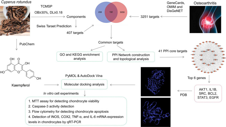
Methods: The bioactive ingredients of CR were screened in TCMSP database, and the potential targets of these ingredients were obtained through Swiss Target Prediction database. Genes in OA pathogenesis were collected through GeneCards, OMIM and DisGeNET databases. Gene Ontology (GO) analysis and Kyoto Encyclopedia of Genes and Genomes (KEGG) enrichment analysis were performed using DAVID database. STRING database and Cytoscape 3.10 software were used to construct "component-target-pathway" network, and predict the core targets affected by CR. The binding affinity between bioactive components and the core targets was evaluated by molecular docking and molecular dynamics simulation. The therapeutic activity of kaempferol on chondrocytes in inflammatory conditions was verified by in vitro experiments.

Results: Fifteen CR bioactive ingredients were obtained, targeting 192 OA-related genes. A series of biological processes, cell components, molecular functions and pathways were predicted to be modulated by CR components. The core targets of CR in OA treatment were AKT serine/threonine kinase 1 (AKT1), interleukin 1 beta (IL1B), SRC proto-oncogene, non-receptor tyrosine kinase (SRC), BCL2 apoptosis regulator (BCL2), signal transducer and activator of transcription 3 (STAT3), epidermal growth factor receptor (EGFR), hypoxia-inducible factor 1 subunit alpha (HIF1A), matrix metallopeptidase 9 (MMP9), estrogen receptor 1 (ESR1) and PPARG orthologs from vertebrates (PPARG), and the main bioactive ingredients of CR showed good binding affinity with these targets. In addition, kaempferol, one of the CR bioactive components, weakens the effects of IL-1β on the viability, apoptosis and inflammation of chondrocytes.

Conclusion: Theoretically, CR has great potential to ameliorate the symptoms and progression of OA, via multiple components, multiple targets, and multiple downstream pathways.

Keywords: Cyperus rotundus; molecular docking; network pharmacology; osteoarthritis.


Conflict of interest statement

The authors declare that they have no competing interests.

Figures

None
Graphical abstract
Figure 1
Figure 1
Chemical structure of 15 CR bioactive ingredients.
Figure 2
Figure 2
Screening of the target of CR in OA treatment. (A) Venn diagram of CR’s active ingredients and genes in OA pathogenesis. (B) The “bioactive ingredient-target” was constructed using Cytoscape 3.10 software. Green diamond-shaped nodes represent CR, pink purple triangular nodes represent OA, yellow circular nodes represent active ingredients, and V-shaped nodes represent targets. (C) Top 10 CR bioactive ingredients were sorted by degree value.
Figure 3
Figure 3
GO and KEGG enrichment analysis of the target of OA treated by CR. (A) GO analysis was performed to predict the biological processes / cellular components / molecular functions affected by CR. Red represents biological processes, blue represents cellular components, and dark green represents molecular functions. The horizontal axis shows the P value and the number of enriched genes respectively, and the vertical axis is the name of the term. (B) KEGG analysis was performed to predict the pathways affected by CR. The horizontal axis is the number of genes, and the vertical axis is the name of the term. (C) Classification summary diagram of the top 30 KEGG pathways. (D) The potential targets of CR in PI3K/AKT pathway was shown with KEGG Pathview. The targets of CR treatment for OA are colored with red.
Figure 4
Figure 4
“Component-target-pathway” network diagram established by Cytoscape 3.10 software.
Figure 5
Figure 5
PPI network of the targets of CR in OA treatment. (A) The PPI network of 192 targets according to the STRING database. (B) PPI network was analyzed using Cytoscape 3.10 software, to show the crucial targets affected by CR. The higher the degree value, the darker the color.
Figure 6
Figure 6
Screening of core targets in PPI network. (AC) Screening flowchart of Centiscape 2.2 plug-in. (D) Core targets of PPI network nodes. The larger the degree value, the larger the font.
Figure 7
Figure 7
Molecular docking of key components of CR and core targets of OA. Light blue indicates amino acid residues around the binding bag, and dark blue indicates the bioactive ingredient.
Figure 8
Figure 8
Molecular dynamics simulation showed stable binding relationship between kaempferol and AKT1. (A) Evolution of RMSDs during 100 ns molecular dynamics simulations of kaempferol (ligand) and AKT1 (pocket). (B) RMSFs of the backbones in kaempferol/AKT1 complex. (C) The results of MM/PBSA free energy calculation (kcal/mol). (D) Energy decomposition was performed to show the contributions of the amino acid residues to form the complex. (E) The conformation images of the complex during molecular dynamics simulation.
Figure 9
Figure 9
Kaempferol reversed the effects of IL-1β treatment on chondrocytes. (A) The viability of chondrocytes treated with or without 10 ng/mL IL-1β was measured by MTT assay. (B) MTT assay was used to detect the viability of mouse chondrocytes treated with kaempferol at different concentrations (0, 10, 25, 50, 80 and 100 μM) for 24 h.) (C) MTT assay was used to evaluate the effects of kaolin treatment at different concentrations (50 and 100 μM) on IL-1β-induced chondrocyte viability. (D) The activity of caspase-3 was detected by colorimetry. (E and F) Flow cytometry was used to evaluate the effect of kaempferol treatment at different concentrations (50 and 100 μM) on IL-1β-induced apoptosis of chondrocytes. (GJ) The mRNA expression levels of INOS, COX2, TNF-α and IL-6 in IL-1β-induced chondrocytes were detected by qRT-PCR at different concentrations (50 and 100 μM) kaolin.*P<0.05, **P<0.01, ***P<0.001.

References

    1. Ma L, Zhao X, Liu Y, Wu J, Yang X, Jin Q. Dihydroartemisinin attenuates osteoarthritis by inhibiting abnormal bone remodeling and angiogenesis in subchondral bone. Int J Mol Med. 2021;47(3):04855. doi:10.3892/ijmm.2021.4855 - DOI - PMC - PubMed
    1. Sun T, Wang F, Hu G, Li Z. Salvianolic acid B activates chondrocytes autophagy and reduces chondrocyte apoptosis in obese mice via the KCNQ1OT1/miR-128-3p/SIRT1 signaling pathways. Nutr Metab. 2022;19(1):53. doi:10.1186/s12986-022-00686-0 - DOI - PMC - PubMed
    1. Wang XY, Bao CC, An R, et al. Evaluation of the effect of physical therapy on pain and dysfunction of knee osteoarthritis based on fNIRS: a randomized controlled trial protocol. BMC Musculoskelet Disord. 2023;24(1):152. doi:10.1186/s12891-022-06074-2 - DOI - PMC - PubMed
    1. Chen Z, Ge Y, Zhou L, et al. Pain relief and cartilage repair by Nanofat against osteoarthritis: preclinical and clinical evidence. Stem Cell Res Ther. 2021;12(1):477. doi:10.1186/s13287-021-02538-9 - DOI - PMC - PubMed
    1. Cross M, Smith E, Hoy D, et al. The global burden of hip and knee osteoarthritis: estimates from the global burden of disease 2010 study. Ann Rheum Dis. 2014;73(7):1323–1330. doi:10.1136/annrheumdis-2013-204763 - DOI - PubMed