. 2024 Oct 21;20(14):5764-5778.
doi: 10.7150/ijbs.101127. eCollection 2024.

AIMP1-Derived Peptide Secreted from Hair Follicle Stem Cells Promotes Hair Growth by Activating Dermal Papilla Cells

Affiliations

AIMP1-Derived Peptide Secreted from Hair Follicle Stem Cells Promotes Hair Growth by Activating Dermal Papilla Cells

YounHa Kim et al. Int J Biol Sci. .

Abstract

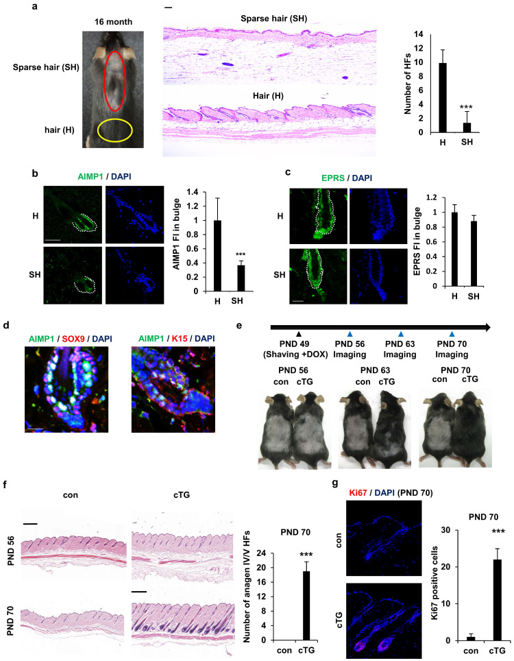
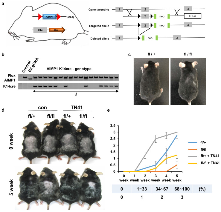
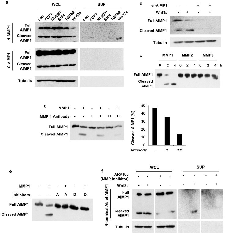
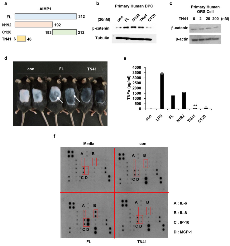
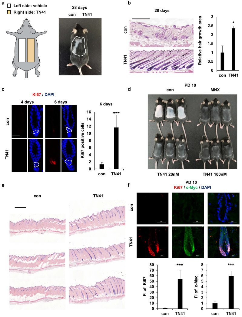
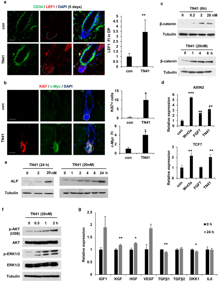
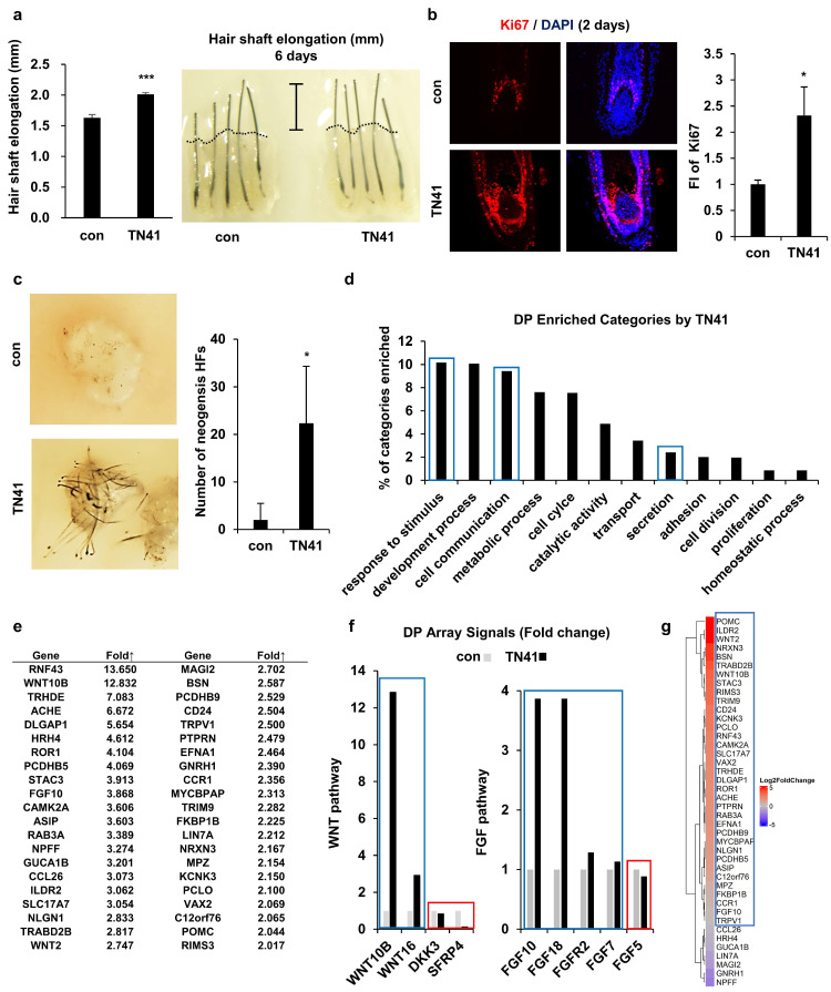
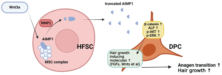
Hair follicle stem cells (HFSCs) and dermal papilla cells (DPCs) are crucial in the biogenesis and maintenance of hair follicles (HFs). This study demonstrated that a fragment derived from aminoacyl-tRNA synthetase-interacting multifunctional protein1 (AIMP1) secreted from HFSCs activated DPCs and maintained HF homeostasis. A histological analysis revealed that AIMP1 levels in HF decreased with hair loss. Hair regrowth in AIMP1-induced mice was faster than in non-induced mice. Deletion mapping revealed 41 amino acids (TN41, aa 6-46) as the active region of AIMP1. The N-terminal peptide fragment of AIMP1 generated by MMP1 was secreted from Wnt-treated HFSCs to activate DPCs. TN41 activated Akt and ERK, increased β-catenin, and enhanced DPC activation. TN41 promoted hair shaft elongation in cultured human HFs and improved the hair-inducing activity of cultured DPC spheroids. Our findings suggest that the AIMP1 fragment secreted from HFSCs stimulates active hair regrowth through activating DPCs.

Keywords: AIMP1; dermal papilla cell; hair follicle stem cell; hair growth.


Conflict of interest statement

Competing Interests: The authors have declared that no competing interest exists.

Figures

Figure 1
Figure 1
AIMP1 for hair follicle maintenance. (a) Representative and histological pictures of C57BL/6 (n = 4). HFs counts from each region. (b) IF images of AIMP1 in the hair follicle. Relative fluorescence intensity (FI) was measured using Image J. (c) IF images of EPRS in the hair follicle from H and SH regions. Bulge was surrounded by dotted line. Relative FI of EPRS in the bulge was measured using Image J software. (d) IF images of AIMP1, SOX9, and K15 in the bulge region from 2-month-old C57BL/6. (e) The design for analyzing hair growth and representative pictures of mice taken at indicated days (n = 2). (f) H&E image from each mouse. HFs counts from each mouse. (g) IF staining of Ki67 and counts of Ki67 positive cells from each mouse. The bars: 20 μm (c, d), 50 μm (a, b and g) and 500 μm (c, f). Error bars indicate mean +/- SD. ***: P < 0.001.
Figure 2
Figure 2
A. Schematic representation of skin-specific AIMP1 conditional KO mice, AIMP1-KRT14-cre (fl/fl). (a) Diagram of the mouse AIMP1 locus, targeted allele, and deleted allele. Exon 2 was flanked by the loxP sequence via homologous recombination with the targeting constructs. Flanked exon2 was deleted in the presence of the Cre enzyme. The absence of exon2 disrupts the 1st ATG of the AIMP1 coding sequence. (b) Validation of AIMP1 deletion using genotyping. (c) Representative KRT14-cre; AIMP1 fl/+ (fl/+) and KRT14-cre; AIMP1 fl/fl (fl/fl) mice at 6 months. (d) Representative images of fl/+ and fl/fl mice at 0 and 5 weeks after shaving. Mice were clipped on PND 49 and treated with 100 nM of TN41 or the vehicle (con) once daily (≥2 mice per group). (e) Hair growth was measured at 0, 1, 2, 3, 4, and 5 weeks and analyzed using ImageJ software (≥4 mice per group). TN41 (100 nM). Hair growth scoring; 0%: 0, 1-33%: 1, 34-67%: 2, 68-100%. Error bars indicate mean +/- SD. ***: P < 0.001.
Figure 3
Figure 3
Secretion of AIMP1 N-terminal fragment from Wnt-activated HFSCs. (a) AIMP1 secretion by incubating HFSCs under different conditions. The proteins in the whole cell lysates (WCL) and supernatant (SUP) were subjected to WB analysis with the antibodies specific to the N-terminal (N-AIMP1) and C-terminal regions of AIMP1 (C-AIMP1). Wnt3a: 200 ng/ml, FGF7: 100 ng/ml, Noggin: 200 ng/ml, TGFβ2: 10 ng/ml, SHH: 200 ng/ml. (b) HFSCs were transfected with AIMP1 siRNA (10 pmol) and treated with Wnt3a (200 ng/ml). WB was performed with the N-AIMP1 antibody. (c) AIMP1(400 μg) was incubated with each of MMP1 (72.9 ng), MMP2 (100 ng), and MMP9 (106 ng) at 37 ℃ for 4 h and WB analysis with the N-AIMP1 antibody. (d) AIMP1 was incubated with recombinant MMP1 and different amounts of anti-MMP1 antibody. AIMP1 protein was detected by N-AIMP1 Antibody. (e) AIMP1 was incubated with MMP1 in the absence or presence of ARP100 (A, 1.5 μg, MMP1 and MMP2 inhibitor) and doxycycline hyclate (D, 1 μg, MMP1, MMP9 and MMP12 inhibitor). (f) Secretion of AIMP1 fragment was confirmed by WB in the absence or presence of ARP100.
Figure 4
Figure 4
Determination of the peptide region of AIMP1 responsible for the hair growing activity without inflammation. (a) Diagram of fragments of AIMP1. FL: Full length, N192: 1-192aa, C120: 193-312aa, TN41: 6-46aa. (b-c) β-catenin induction was tested by human DPC and ORS cells with AIMP1 full length and fragments. Human primary DPCs were treated with FL, N192, TN41, or C120 (20 nM each). Human primary ORS cell was treated with each concentration of TN41. WB were performed with each antibody. (d) Mouse back skins were depilated at PND 49, and images were taken PD10. White arrows indicate the skin regions showing inflammatory response. (e) Effects of AIMP1 fragments on TNF-α secretion from RAW 264.7 cells. (f) Effects of FL and TN41 on cytokines secretion from DPC. Media: culture medium, con: cultured medium containing vehicle. The cytokines secreted by FL but not by TN41 are in red boxes.
Figure 5
Figure 5
Effect of an AIMP1-derived peptide on hair growth. (a) Mouse back skins were clipped at PND 49. The left and right halves of the back skin were treated with vehicle, TN41 (100 nM), respectively. (b) Representative H&E image at 28 days after clipping (n = 3). HFs area was measured. (c) IF images of Ki67 and counts of Ki67- positive cells in hair follicles. (d) Mice were depilated on PND 49 and treated with the vehicle, 3% MNX and TN41. Pictures were taken on PD10 (n = 3). (e) H&E image of control and the TN41 (100 nM) treated dorsal skin. (f) Representative IF staining images for c-Myc and Ki67 in hair follicles and measured relative FI. The bars: 200 μm (b), 20 μm (c, d and f), 500 μm (e). Error bars: mean ± SD. ***: P < 0.001.
Figure 6
Figure 6
TN41 induced Activation of the MAPK pathway in DPCs. (a) IF images of CD34 (HFSC marker) and LEF1 in hair follicles from TN41-treated and vehicle-treated mice. Relative LEF1 FI was measured in the DPC. (b) IF images of Ki67 and c-Myc in the hair follicle. Ki67-positive cell was counted and the relative FI of c-Myc was measured in the DPC. (c and e, f) DPCs were treated with time and dose-dependent TN41, and WB were performed via each antibody. (d) DPCs were treated with 5.1 nM of Wnt3a, 8.9 nM of FGF7, and 20 nM of TN41 for 14 h, and relative gene expression levels were measured by quantitative PCR. (g) Regulatory effect of TN41 on the expression of various growth factors in DPCs. The bars: 20 μm (a and b). Error bars: mean ±SD. ***: P < 0.001, **: P < 0.01, *: P < 0.05.
Figure 7
Figure 7
Effects of TN41 on human DPCs. (a) Effects of TN41 in human hair shaft elongation. Human HFs were cultured for 6 days with TN41, or the vehicle and hair length were measured (3 donors). (b) IF image of Ki67 in human hair follicles and relative FI was measured by Image J. (c) 3D-cultured DPCs with or without TN41 were injected into the hypodermis of nude mice. Images were obtained after 2 weeks (injection site = 3) and measured neogenesis HF numbers. (d) Gene categories enriched in TN41-treated DPCs measured by RNA sequence. Extracellular signaling molecules were among the most enriched (blue box). (e) List of upregulated DPC genes in the extracellular signaling category. (f) TN41 treatment induced Wnt and FGF pathway genes in DPC. The microarray signal values of Wnt and FGF pathway members displayed temporal differences. (g) RNA-seq heatmap depiction of 42 DPC genes upregulated by TN41 in DPCs co-cultured with HFSCs and treated with vesicles and Wnt3a (shown in Fig. 7e). The fold changes (FC) were shown by color gradient. Blue box: positive genes, red box: negative genes. Error bars: mean ± SD. ***: P < 0.001, *: P < 0.05.
Figure 8
Figure 8
The schematic diagram illustrating how truncated AIMP1 stimulates DPCs to promote hair growth. Wnt3a stimulation induces AIMP1 cleavage via MMP1 activation in hair follicle stem cells (HFSCs), leading to the release of a truncated form of AIMP1. The secreted AIMP1 enhances hair growth by upregulating hair growth-promoting molecules in dermal papilla cells (DPCs), subsequently activating β-catenin, ALP, p-AKT, and p-ERK signaling pathways, thereby facilitating anagen transition and promoting hair growth.

References

    1. Oh JW, Kloepper J, Langan EA, Kim Y, Yeo J, Kim MJ. et al. A Guide to Studying Human Hair Follicle Cycling In Vivo. J Invest Dermatol. 2016;136:34–44. - PMC - PubMed
    1. Obara K, Tohgi N, Mii S, Hamada Y, Arakawa N, Aki R. et al. Hair-follicle-associated pluripotent stem cells derived from cryopreserved intact human hair follicles sustain multilineage differentiation potential. Sci Rep. 2019;9:9326. - PMC - PubMed
    1. Hsu YC, Li L, Fuchs E. Emerging interactions between skin stem cells and their niches. Nat Med. 2014;20:847–56. - PMC - PubMed
    1. Giangreco A, Qin M, Pintar JE, Watt FM. Epidermal stem cells are retained in vivo throughout skin aging. Aging Cell. 2008;7:250–9. - PMC - PubMed
    1. Ji J, Ho BS, Qian G, Xie XM, Bigliardi PL, Bigliardi-Qi M. Aging in hair follicle stem cells and niche microenvironment. J Dermatol. 2017;44:1097–104. - PubMed