. 2024 Nov 5;13(21):e035193.
doi: 10.1161/JAHA.124.035193. Epub 2024 Nov 4.

Sestrin2 Attenuates Myocardial Endoplasmic Reticulum Stress and Cardiac Dysfunction During Ischemia/Reperfusion Injury

Affiliations

Sestrin2 Attenuates Myocardial Endoplasmic Reticulum Stress and Cardiac Dysfunction During Ischemia/Reperfusion Injury

Xuan Li et al. J Am Heart Assoc. .

Abstract

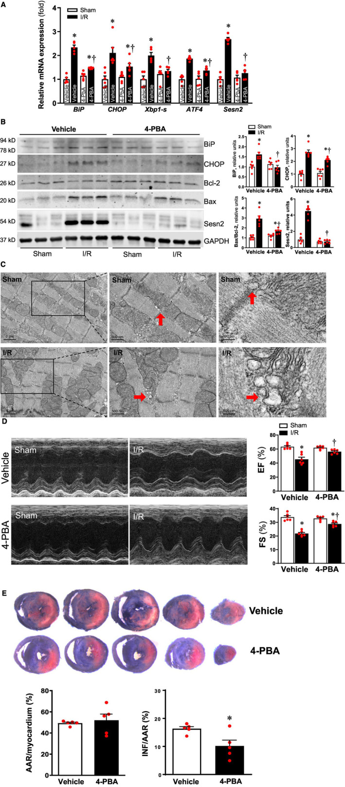
Background: Sesn2 (Sestrin2) is a stress-induced protein that provides protective effects during myocardial ischemia and reperfusion (I/R) injury, while endoplasmic reticulum (ER) stress may be a pivotal mediator of I/R injury. The goal of this study was to determine whether Sesn2-mTOR (mammalian target of rapamycin) signaling regulates ER stress during myocardial I/R.

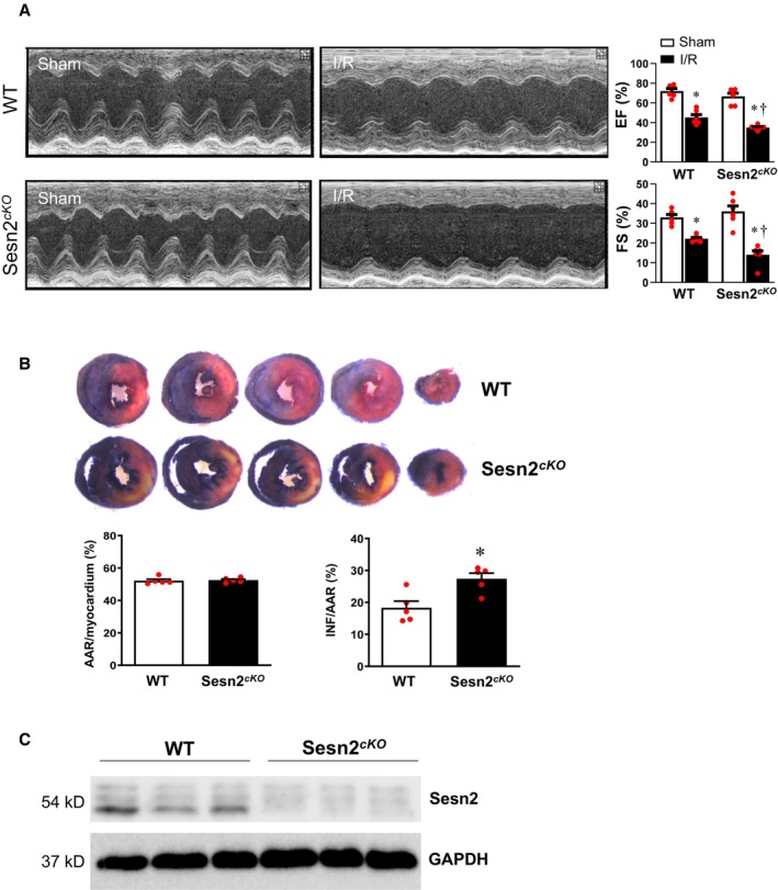
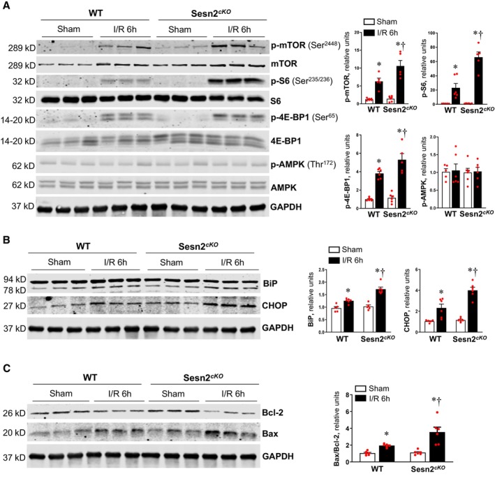
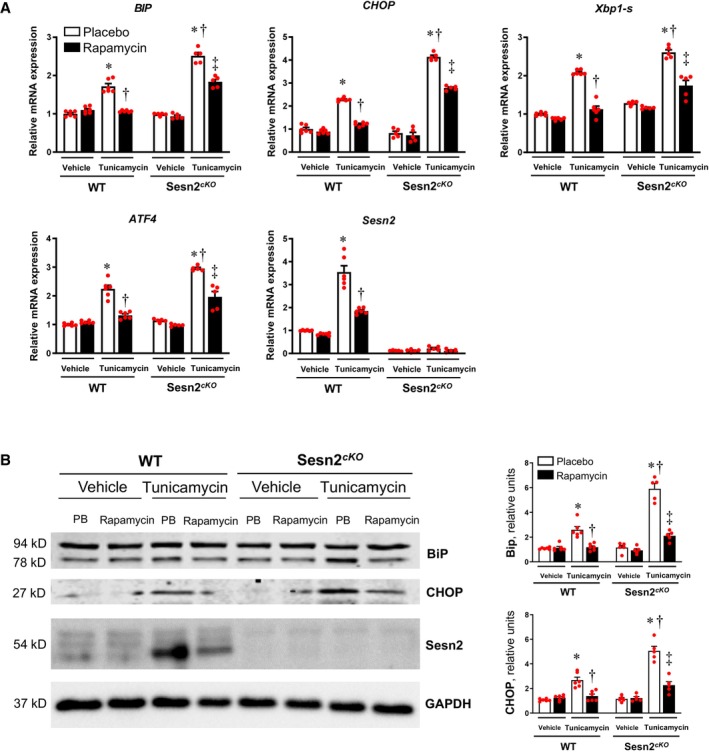
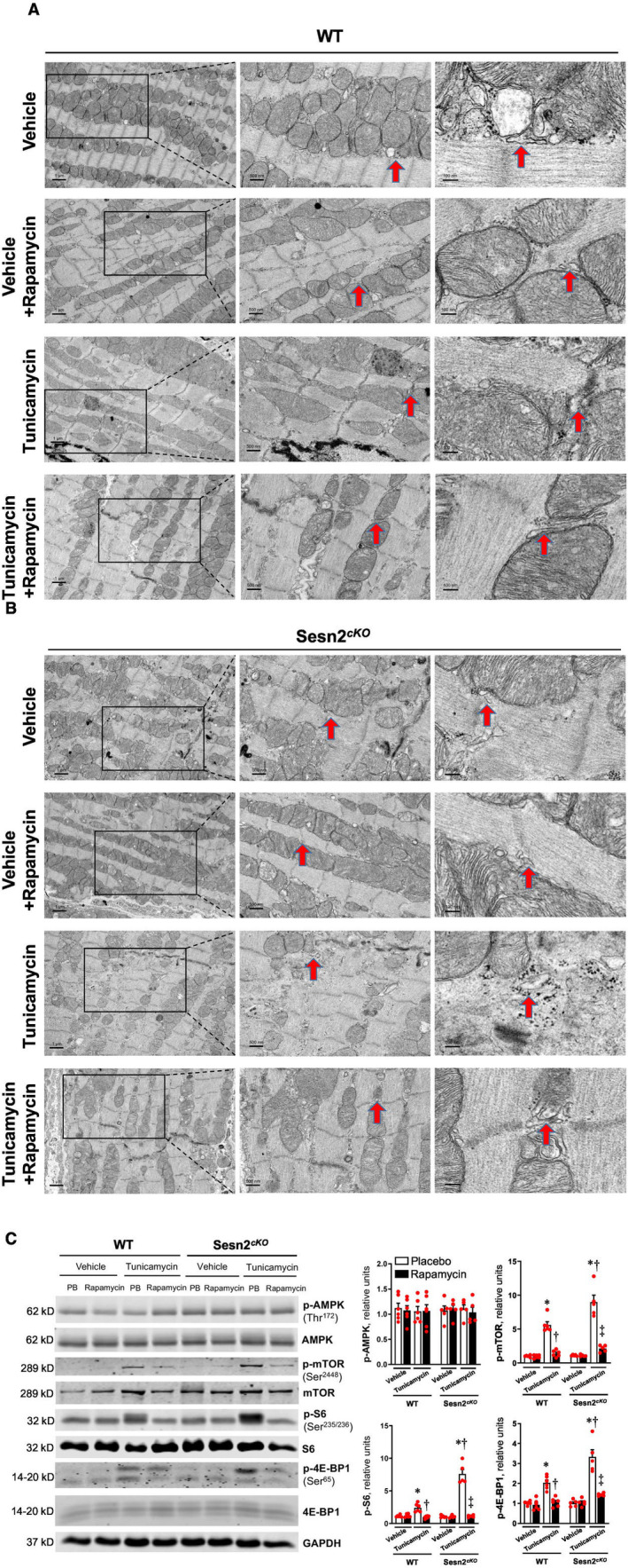
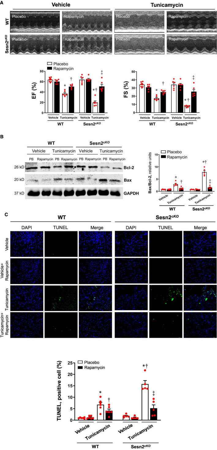
Methods and results: In vivo cardiac I/R was induced by ligation and subsequent release of the left anterior descending coronary artery in wild-type (WT) and cardiac-specific Sesn2 knockout (Sesn2cKO) mice. At 6 hours and 24 hours after reperfusion, cardiac function was evaluated, and heart samples were collected for analysis. I/R induced cardiac ER stress and upregulated Sesn2 mRNA and protein levels. Inhibiting ER stress with 4-phenylbutyric acid reduced infarct size by 37.5%, improved cardiac systolic function, and mitigated myocardial cell apoptosis post-I/R. Hearts from Sesn2cKO mice displayed increased susceptibility to ER stress during I/R compared with WT. Notably, cardiac mTOR signaling was further increased in Sesn2cKO hearts compared with WT hearts during I/R. In mice with cardiac Sesn2 deficiency, compared with WT, ER lumen was significantly expanded after tunicamycin-induced ER stress, as assessed by transmission electron microscopy. Additionally, pharmacological inhibition of mTOR signaling with rapamycin improved cardiac function after tunicamycin treatment and significantly attenuated the unfolded protein response and apoptosis in WT and Sesn2cKO mice.

Conclusions: Sesn2 attenuates cardiac ER stress post-I/R injury via regulation of mTOR signaling. Thus, modulation of the mTOR pathway by Sesn2 could be a critical factor for maintaining cardiac ER homeostasis control during myocardial I/R injury.

Keywords: cardiac injury; heart; mTOR signaling; unfolded protein response.


Figures

Figure 1
Figure 1. Ischemia/reperfusion induces endoplasmic reticulum (ER) stress and inhibiting ER stress protects the hearts from ischemia and reperfusion (I/R) injury.
A, Real‐time quantitative polymerase chain reaction results show the mRNA level of ER stress‐related biomarkers and Sesn2 (Sestrin2) in hearts of wild‐type (WT) mice under sham or ischemia 45 min/reperfusion 24‐hour condition treated with vehicle or 4‐phenylbutyric acid (4‐PBA). Values are mean±SEM, n=5–6, *P<0.05 vs sham vehicle; P<0.05 vs I/R vehicle (Dunn multiple comparison test). B, Left: Representative immunoblots showing protein levels of BiP, CHOP, Bcl‐2, Bax, and Sesn2 in hearts of WT mice under sham and I/R conditions treated with vehicle or 4‐PBA. Right: quantitative analysis of the relative protein levels in different groups. Values are mean±SEM, n=6, *P<0.05 vs sham vehicle; P<0.05 vs I/R vehicle (1‐way ANOVA followed by Tukey post hoc tests). C, Transmission electron microscopy (TEM) showing ER morphology in cardiomyocytes of WT mice under sham and I/R conditions. Middle images (Scale bar: 500 nm) are the higher amplification of the box areas in left images (scale bar: 1 μm). Right images (scale bar: 100 nm) are higher magnification of middle images. Red arrows point out representative ER. D, Left: Representative echocardiography of WT mice under sham and ischemia 45 min/reperfusion 24 h condition treated with vehicle or 4‐PBA. Right: Quantitative analysis of ejection fraction (EF) and fraction shortening (FS) of hearts of WT mice under sham and I/R conditions. Values are mean±SEM, n=6, *P<0.05 vs sham vehicle; P<0.05 vs I/R vehicle (1‐way ANOVA followed by Tukey post hoc tests). E, Upper: Representative sections of myocardial infarction staining with TTC and Evans Blue in hearts of WT mice treated with vehicle or 4‐PBA. Lower: The ratio of the area at the risk (AAR) to the myocardial area; the ratio of the infarction area (INF) to AAR in vehicle and 4‐PBA groups. Values are mean±SEM, n=5, *P<0.05 vs vehicle (Mann–Whitney test).
Figure 2
Figure 2. Cardiac deletion of Sesn2 exacerbated ischemia and reperfusion (I/R) injury.
A, Left: Representative echocardiography of wild‐type (WT) and cardiac‐specific Sestrin2 knockout (Sesn2 cKO ) mice under sham and ischemia 45 min/reperfusion 24‐hour condition. Right: Quantitative analysis of ejection fraction (EF) and fraction shortening (FS) of WT and Sesn2 cKO hearts under sham and ischemia 45 min/reperfusion 24‐hour conditions. Values are means±SEM, n=6, *P<0.05 vs sham, respectively; P<0.05 vs WT I/R (1‐way ANOVA followed by Tukey post hoc tests). The littermates, carrying Sesn2 flox/flox were considered as WT controls. B, Upper: representative sections of myocardial infarction staining with TTC and Evans Blue in WT and Sesn2 cKO hearts. Lower: The ratio of AAR to the myocardial area; the ratio of INF to AAR in WT and Sesn2 cKO groups. Values are mean±SEM, n=5, *P<0.05 vs WT (Mann–Whitney test). C, Immunoblotting result showed the protein level of Sesn2 in WT and Sesn2 cKO hearts. n=3.
Figure 3
Figure 3. Sesn2‐mTOR pathway is related to endoplasmic reticulum (ER) stress caused by ischemia and reperfusion (I/R).
A, Representative immunoblotting of wild‐type (WT) and cardiac‐specific Sestrin2 knockout (Sesn2 cKO ) hearts under sham and ischemia 45 min/reperfusion 6‐hour (I/R 6 hours) conditions showed mammalian target of rapamycin (mTOR) pathway. B, The immunoblotting measured the protein level of biomarkers of ER stress. C, The immunoblotting measured the protein level of Bcl‐2 and Bax in WT and Sesn2 cKO hearts under sham and I/R 6‐hour conditions. Values are means±SEM, n=5–6, *P<0.05 vs sham, respectively; P<0.05 vs WT I/R 6 hours (1‐way ANOVA followed by Tukey post hoc tests). The littermates, carrying Sesn2 flox/flox were considered as WT controls.
Figure 4
Figure 4. Tunicamycin induced more severe endoplasmic reticulum (ER) stress in cardiac‐specific Sestrin2 knockout (Sesn2 cKO ) mice and inhibiting mammalian target of rapamycin (mTOR) activity attenuated ER stress.
A, Real‐time quantitative polymerase chain reaction shows the mRNA level of ER stress related biomarkers and Sesn2 (Sestrin2). Values are means±SEM, n=5–6, *P<0.05 vs vehicle (no tunicamycin, no rapamycin), respectively; P<0.05 vs wild‐type (WT)+tunicamycin, P<0.05 vs Sesn2 cKO +tunicamycin (Dunn multiple comparison test). B, Left: Protein levels of BiP, CHOP, and Sestrin2 were measured by immunoblotting. Right: quantitative analysis of different proteins in different groups. Values are means±SEM, n=5 to 6, *P<0.05 vs vehicle (no tunicamycin, no rapamycin), respectively; P<0.05 vs WT+tunicamycin, P <0.05 vs Sesn2 cKO +tunicamycin (Dunn multiple comparison test). The littermates, carrying Sesn2 flox/flox were considered as WT controls.
Figure 5
Figure 5. Inhibiting mammalian target of rapamycin (mTOR) activity attenuated changes of endoplasmic reticulum (ER) morphology.
A and B, ER morphology of wild‐type (WT) and cardiac‐specific Sestrin2 knockout (Sesn2 cKO ) hearts under different conditions. Middle images (Scale bar: 500 nm) are the higher amplification of the box areas in left images (scale bar: 1 μm). Right images (scale bar: 100 nm) are higher magnification of middle images. Red arrows pointed out representative ER. C, The mTORC1 pathway was excessively activated in Sesn2 cKO group after tunicamycin treatment. Values are mean±SEM, n=5–6, *P<0.05 vs vehicle (no tunicamycin, no rapamycin), respectively; P<0.05 vs WT+tunicamycin, P<0.05 vs Sesn2 cKO +tunicamycin (Dunn multiple comparison test). The littermates, carrying Sesn2 flox/flox were considered as WT controls.
Figure 6
Figure 6. Mammalian target of rapamycin complex 1 (mTORC1) pathway regulated endoplasmic reticulum (ER)‐stress related cardiac dysfunction and myocardial apoptosis.
A, Upper: Representative echocardiography of wild‐type (WT) and cardiac‐specific Sestrin2 knockout (Sesn2 cKO ) mice treated with tunicamycin and/or rapamycin. Lower: Quantitative analysis of ejection fraction (EF) and fraction shortening (FS) of WT and Sesn2 cKO hearts under different conditions. Values are mean±SEM, n=6 to 8, *P<0.05 vs vehicle (no tunicamycin, no rapamycin), respectively; P<0.05 vs WT+tunicamycin, P<0.05 vs Sesn2 cKO +tunicamycin (1‐way ANOVA followed by Tukey post hoc tests). The littermates, carrying Sesn2 flox/flox were considered as WT controls. B, Immunoblotting levels of Bcl‐2, Bax, and GAPDH of WT and Sesn2 cKO hearts under different conditions. Values are mean±SEM, n=5 to 6, *P<0.05 vs vehicle (no tunicamycin, no rapamycin), respectively; P<0.05 vs WT+tunicamycin, P<0.05 vs Sesn2 cKO +tunicamycin (Dunn multiple comparison test). C, Upper: TUNEL and DAPI staining of WT and Sesn2 cKO hearts. Lower: Quantitative analysis of TUNEL positive cells in WT and Sesn2 cKO hearts. Values are mean±SEM, n=10 to 25 sections from 5 to 6 mice, *P<0.05 vs vehicle (no tunicamycin, no rapamycin), respectively; P<0.05 vs WT+tunicamycin, P<0.05 vs Sesn2 cKO +tunicamycin (Dunn multiple comparison test).

Similar articles

Cited by

References

    1. Mozaffarian D, Benjamin EJ, Go AS, Arnett DK, Blaha MJ, Cushman M, de Ferranti S, Despres JP, Fullerton HJ, Howard VJ, et al. Heart disease and stroke statistics—2015 update: a report from the American Heart Association. Circulation. 2015;131:e29–e322. doi: 10.1161/CIR.0000000000000152 - DOI - PubMed
    1. Yellon DM, Hausenloy DJ. Myocardial reperfusion injury. N Engl J Med. 2007;357:1121–1135. doi: 10.1056/NEJMra071667 - DOI - PubMed
    1. Moran AE, Forouzanfar MH, Roth GA, Mensah GA, Ezzati M, Flaxman A, Murray CJ, Naghavi M. The global burden of ischemic heart disease in 1990 and 2010: the global burden of disease 2010 study. Circulation. 2014;129:1493–1501. doi: 10.1161/CIRCULATIONAHA.113.004046 - DOI - PMC - PubMed
    1. Pagliaro P, Moro F, Tullio F, Perrelli MG, Penna C. Cardioprotective pathways during reperfusion: focus on redox signaling and other modalities of cell signaling. Antioxid Redox Signal. 2011;14:833–850. doi: 10.1089/ars.2010.3245 - DOI - PubMed
    1. Groenendyk J, Agellon LB, Michalak M. Coping with endoplasmic reticulum stress in the cardiovascular system. Annu Rev Physiol. 2013;75:49–67. doi: 10.1146/annurev-physiol-030212-183707 - DOI - PubMed

MeSH terms

LinkOut - more resources