. 2024 Dec 15;213(12):1811-1824.
doi: 10.4049/jimmunol.2400042.

ISG15 Drives Immune Pathology and Respiratory Failure during Systemic Lymphocytic Choriomeningitis Virus Infection

Affiliations

ISG15 Drives Immune Pathology and Respiratory Failure during Systemic Lymphocytic Choriomeningitis Virus Infection

Namir Shaabani et al. J Immunol. .

Abstract

ISG15, an IFN-stimulated gene, plays a crucial role in modulating immune responses during viral infections. Its upregulation is part of the host's defense mechanism against viruses, contributing to the antiviral state of cells. However, altered ISG15 expression can also lead to immune dysregulation and pathological outcomes, particularly during persistent viral infections. Understanding the balance of ISG15 in promoting antiviral immunity while avoiding immune-mediated pathology is essential for developing targeted therapeutic interventions against viral diseases. In this article, using Usp18-deficient, USP18 enzymatic-inactive and Isg15-deficient mouse models, we report that a lack of USP18 enzymatic function during persistent viral infection leads to severe immune pathology characterized by hematological disruptions described by reductions in platelets, total WBCs, and lymphocyte counts; pulmonary cytokine amplification; lung vascular leakage; and death. The lack of Usp18 in myeloid cells mimicked the pathological manifestations observed in Usp18-/- mice and required Isg15. Mechanistically, interrupting the enzymes that conjugate/deconjugate ISG15, using Uba7-/- or Usp18C61A mice, respectively, led to accumulation of ISG15 that was accompanied by inflammatory neutrophil accumulation, lung pathology, and death similar to that observed in Usp18-deficient mice. Moreover, myeloid cell depletion reversed pathological manifestations, morbidity, and mortality in Usp18C61A mice. Our results suggest that dysregulated ISG15 production and signaling during persistent lymphocytic choriomeningitis virus infection can produce lethal immune pathology and could serve as a therapeutic target during severe viral infections with pulmonary pathological manifestations.


Conflict of interest statement

Competing interests

The authors declare that they have no competing interests.

Figures

Figure 1.
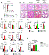
Figure 1.. Lack of USP18 isopeptidase domain leads to lung immunopathology and acute respiratory distress-like syndrome (ARDLS).
(A) WT and Usp18–/– mice were infected with 2×10⁶ PFU LCMV-Cl13. Mice survival was monitored (10 mice/group). (B) WT and Usp18–/– mice were infected with 2×10⁶ PFU of LCMV- Cl13. On day 5, White blood cells (WBC), platelets and lymphocytes were measured in the blood (n = 7–8 mice/group). (C) WT and Usp18–/– mice were infected with 2×10⁶ PFU of LCMV- Cl13. 6 days post infection, lung sections were stained with hematoxylin and eosin (n = 3 mice/group). (D-E) WT and Usp18–/– mice were infected with 2×10⁶ PFU of LCMV- Cl13. 6 days post infection, (D) Indicated cytokines were quantified by multiplex assay and (E) total protein and LDH were quantified in the BALF by BCA and ELISA assays (n = 5–7 mice/group). n.s., not significant; *P < 0.05 and **P < 0.01.
Figure 2.
Figure 2.. Deletion of Usp18 in LysM+ cells replicates inflammatory pathology, cytokine amplification and mortality observed in Usp18−/− mice.
(A) Usp18fl/fl and Usp18fl/fl x CD4-Cre were infected with 2 ×10⁶ PFU LCMV-Cl13. Mice survival was monitored (n = 5–10 mice/group). (B) Usp18fl/fl x LysM-Cre and WT mice were infected with 2 ×10⁶ PFU LCMV-Cl13, survival was monitored (n=7 mice/group). (C) Lung sections of Usp18fl/fl x LysM-Cre and WT mice infected with 2 ×10⁶ PFU LCMV-Cl13 for 7 days were stained with hematoxylin and eosin (n = 3 mice/group). (D-E) Usp18fl/fl x LysM-Cre treated with anti-CD8 or isotype antibodies at day −1 and 3 and WT mice were infected with 2 ×10⁶ PFU LCMV-Cl13. On day 6, (D) indicated cytokines (E) total protein, LDH and IgM were measured in the BALF (n = 8–9 mice/group). n.s. not significant, * P < 0.05, ** P < 0.01, *** P < 0.001 or **** P < 0.0001.
Figure 3.
Figure 3.. Lack of free ISG15 rescues Usp18C61A mice during Cl3 infection
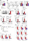
(A) WT and Usp18C61A, Usp18C61A x Uba7−/− and Usp18C61A x Isg15–/– mice were infected with 2 ×10⁶ PFU LCMV-Cl13. Mice survival was monitored. (n= 9–11 mice/group). (B-E) WT, Usp18C61A, Usp18C61A x Uba7−/− and Usp18C61A x Isg15–/– mice were infected with 2 ×10⁶ PFU LCMV-Cl13. (B) 8 days post clone-13 infection lung sections were stained with hematoxylin and eosin (n = 3 mice/group). (C) Indicated cytokines were analyzed in the BALF by multiplex assay (n = 4–5 mice/group). (D) Total protein, IgM and LDH were measured by BCA or ELISA assays from BALF fluid (n = 4–5 mice/group). (E) ISG15 levels were measured in the BALFs 8 days post infection by ELISA assay (n = 4–10 mice/group). n.s., not significant; *P < 0.05; **P < 0.01; ***P < 0.001 and ****P < 0.0001.
Figure 4.
Figure 4.. IFN-γ produced by T cells triggers inflammatory pathology, cytokine amplification and mortality observed in Usp18C61A mice.
(A) Usp18C61A mice were treated with anti-CD8, anti- IFN-γ antibodies or left untreated. Mice were infected with 2 ×10⁶ PFU LCMV-Cl13 and survival was monitored (n = 8–16 mice/group). (B-D) Usp18C61A mice were treated with anti-CD8 or isotype antibodies. Mice were infected with 2 ×10⁶ PFU LCMV-Cl13 and 8 days post infection, (B) ISG15 levels (C) indicated cytokines were measured in the BALF fluid (n = 8–10 mice/group). (D) total protein, LDH and IgM were measured by BCA or ELISA assays (n = 8–10 mice/group). n.s., not significant; *P < 0.05; **P < 0.01; ***P < 0.001 and ****P < 0.0001.
Figure 5.
Figure 5.. Transcriptome analysis of lung-infiltrating cells.
(A-F) WT and Usp18C61A mice were infected with Cl13 and treated with α-CD8 or isotype control. 7 days post infection, CD11b+ cells were purified from lung single cell suspension by flow cytometry, RNA isolated and sequenced using a ribo-depletion library preparation protocol. (A) Principal component analysis of total transcriptomes, top 2 components shown; (B) number of differentially expressed genes per pairwise group comparison, padj<0.05; (C) heatmap of relative expression of cytokine-encoding genes; (D) top 20 genes contributing to principal component 1 comprising 10 lowest and 10 highest loading value genes, neutrophil-enriched and neutrophil-specific genes marked in red; (E) expression of selected neutrophil markers per genotype, normalized counts shown; (F) expression pattern of selected neutrophil markers in the Immgen dataset (GSE109125), cell type abbreviations as defined in GSE109125; GN, granulocyte neutrophils; (G-N) WT, Usp18C61A and Usp18C61A x Isg15−/− mice were infected with Cl13, 7 days post infection, lungs collected infiltrating cells enriched for live singlets by flow cytometry, then subjected to single-cell transcriptome sequencing. (G) Dimensional reduction and clustering of cell transcriptomes; (H) contribution of sample cells to each cluster; (I) comparisons of relative cluster fractions in each sample group; (J) expression of the Gene Ontology gene set Response to interferon alpha per cluster (left panel); percentage change in cluster share of total cells in sample with respect to WT versus median expression of GO: response to interferon alpha gene set per cluster, mean values of biological replicates are shown, lines show linear regression for WT vs Usp18C61A, and WT vs Usp18C61A x Isg15−/−; (K) heatmap of relative expression of neutrophil genes within the neutrophil clusters, tSNE coordinates as in Fig. 5G; (L) heatmap of expression of gene signature distinguishing bone marrow neutrophils from splenic and peritoneal neutrophils (derived from GSE109125); (M) differentiation trajectory within neutrophil clusters rooted at cluster 6, heatmap shows relative expression of neutrophil cluster marker genes along trajectory pseudotime; (N) density of cells along trajectory, biological replicates pooled by genotype, values normalized for total number of cells per genotype. Two biological replicates were used per genotype; for further statistical details see Materials and methods.
Figure 6.
Figure 6.. Lack of deISGylation modulates neutrophil composition and function in lung leading to lethal immunopathology.
(A-B) WT, Usp18C61A and Usp18C61A x Isg15–/– mice were infected with 2 ×10⁶ PFU LCMV-Cl13. (A) Total numbers of CD11b+Ly6G+ neutrophils in the lung day 8 post-infection. (B) Flow cytometry staining of neutrophils in the lung on day 8 post-infection, frequencies and total numbers of c-kit+CXCR4+ neutrophils in the lung 8 days post-infection (n = 5 mice /group). (C) WT and Usp18C61A mice were infected with 2 ×10⁶ PFU LCMV-Cl13, myeloperoxidase (MPO) was assessed in the lung homogenates by western blot or (D) MPO and MMP9 were quantified by ELISA assay in BALF (n = 5 mice/group). (E-F) Usp18C61A mice were treated with anti-CD8, anti-IFN-γ or isotype antibodies. Mice in addition to WT were infected with 2 ×10⁶ PFU LCMV-Cl13 and 8 days post infection, (E) frequencies and total numbers of c-kit+CXCR4+ neutrophils in the lung 8 days post-infection (n = 4–5 mice /group). (F) MPO and MMP9 levels in BALF of WT, Usp18C61A and Usp18C61A treated with anti- CD8 or anti-IFN-γ or isotype antibody were quantified by ELISA assay. (G) Usp18C61A mice were infected with 2 ×10⁶ PFU LCMV-Cl13 and treated with anti-Ly6G (RB6–8C5) or isotype antibodies at day 1 and 4 post-infection and monitored for survival (n = 5 Iso and 10 anti-Ly6G). (H-I) Mice were infected with 2 ×10⁶ PFU LCMV-Cl13 and 8 days post infection (H) indicated cytokines measured by multiplex assay and (I) total protein, LDH and IgM were measured in BALF by BCA and ELISA assay, respectively (n = 5 mice/group). n.s., not significant; *P < 0.05; **P < 0.01; ***P < 0.001 and ****P < 0.0001.

Similar articles

References

    1. McNab F, Mayer-Barber K, Sher A, Wack A, O’Garra A, Type I interferons in infectious disease. Nat Rev Immunol 15, 87–103 (2015). - PMC - PubMed
    1. Shaabani N et al. , The probacterial effect of type I interferon signaling requires its own negative regulator USP18. Sci Immunol 3 (2018). - PMC - PubMed
    1. Teijaro JR et al. , Persistent LCMV infection is controlled by blockade of type I interferon signaling. Science 340, 207–211 (2013). - PMC - PubMed
    1. Ng CT et al. , Blockade of interferon Beta, but not interferon alpha, signaling controls persistent viral infection. Cell Host Microbe 17, 653–661 (2015). - PMC - PubMed
    1. Wilson EB et al. , Blockade of chronic type I interferon signaling to control persistent LCMV infection. Science 340, 202–207 (2013). - PMC - PubMed

LinkOut - more resources