. 2025 Jan 23;68(2):1092-1112.
doi: 10.1021/acs.jmedchem.4c02010. Epub 2024 Nov 4.

A Target Class Ligandability Evaluation of WD40 Repeat-Containing Proteins

Suzanne Ackloo  1 Fengling Li  1 Magda Szewczyk  1 Almagul Seitova  1 Peter Loppnau  1 Hong Zeng  1 Jin Xu  2 Shabbir Ahmad  1 Yelena A Arnautova  3 A J Baghaie  3 Serap Beldar  1 Albina Bolotokova  1 Paolo A Centrella  4 Irene Chau  1 Matthew A Clark  4 John W Cuozzo  3   5 Saba Dehghani-Tafti  1 Jeremy S Disch  3   5 Aiping Dong  1 Antoine Dumas  3 Jianwen A Feng  2 Pegah Ghiabi  1 Elisa Gibson  1 Justin Gilmer  2 Brian Goldman  3   5 Stuart R Green  1 Marie-Aude Guié  4 John P Guilinger  4 Nathan Harms  3 Oleksandra Herasymenko  1 Scott Houliston  6 Ashley Hutchinson  1 Steven Kearnes  2   5 Anthony D Keefe  4 Serah W Kimani  1 Trevor Kramer  3   5 Maria Kutera  1 Haejin A Kwak  1 Cristina Lento  7 Yanjun Li  1 Jenny Liu  4 Joachim Loup  8 Raquel A C Machado  1 Christopher J Mulhern  3   4 Sumera Perveen  1 Germanna L Righetto  1 Patrick Riley  2   5 Suman Shrestha  1 Eric A Sigel  3 Madhushika Silva  1 Michael D Sintchak  9 Belinda L Slakman  3   4   5 Rhys D Taylor  4 James Thompson  2 Wen Torng  2 Carl Underkoffler  3   5 Moritz von Rechenberg  3   5 Ryan T Walsh  4 Ian Watson  2 Derek J Wilson  7 Esther Wolf  7 Manisha Yadav  1 Aliakbar K Yazdi  1 Junyi Zhang  3   5 Ying Zhang  4 Vijayaratnam Santhakumar  1 Aled M Edwards  1 Dalia Barsyte-Lovejoy  1   10 Matthieu Schapira  1   10 Peter J Brown  1 Levon Halabelian  1   10 Cheryl H Arrowsmith  1   6
Affiliations

A Target Class Ligandability Evaluation of WD40 Repeat-Containing Proteins

Suzanne Ackloo et al. J Med Chem. .

Abstract

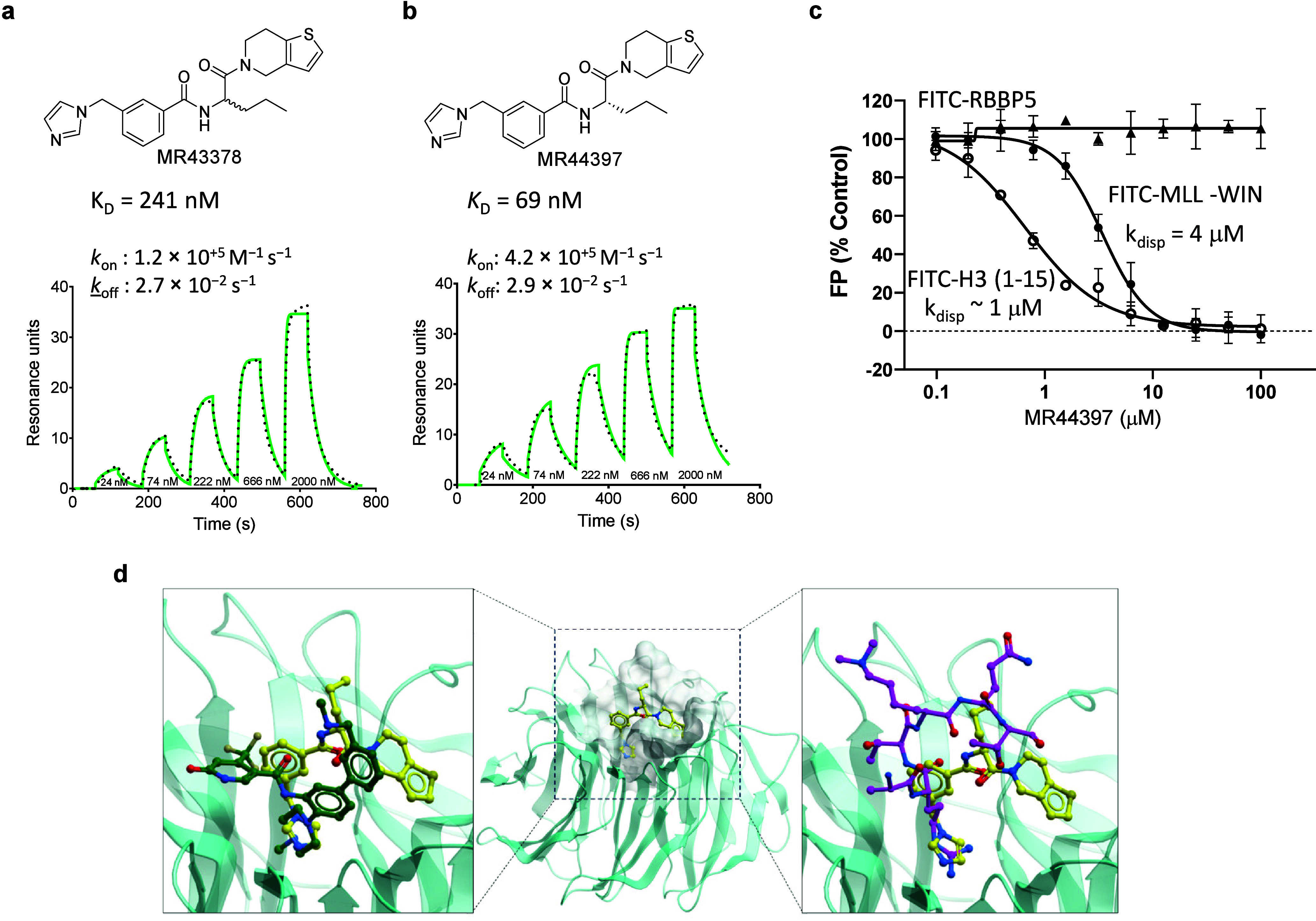
Target class-focused drug discovery has a strong track record in pharmaceutical research, yet public domain data indicate that many members of protein families remain unliganded. Here we present a systematic approach to scale up the discovery and characterization of small molecule ligands for the WD40 repeat (WDR) protein family. We developed a comprehensive suite of protocols for protein production, crystallography, and biophysical, biochemical, and cellular assays. A pilot hit-finding campaign using DNA-encoded chemical library selection followed by machine learning (DEL-ML) to predict ligands from virtual libraries yielded first-in-class, drug-like ligands for 7 of the 16 WDR domains screened, thus demonstrating the broader ligandability of WDRs. This study establishes a template for evaluation of protein family wide ligandability and provides an extensive resource of WDR protein biochemical and chemical tools, knowledge, and protocols to discover potential therapeutics for this highly disease-relevant, but underexplored target class.


Conflict of interest statement

The authors declare the following competing financial interest(s): BLS, BG, JSD, JZ, JWC, MvR, PR, SK, and TK are current or past employees and shareholders of Relay Therapeutics.

Figures

Figure 1
Figure 1
A roadmap for developing target-class focused pharmacological tools. (a) Structure of a canonical 7-bladed WDR protein (PDB ID: 7KQQ) is shown in ribbon and surface representations. The triangle highlights one WD40 repeat, and the arrow points to the central pocket. (b) A phylogenetic tree of the WDR protein family with annotations for targets as follows: cloned (grey), purified (green), new 3D structures (blue), prior 3D structures (yellow), cellular target engagement assays (cyan), and subjected to hit-finding (pink) in this study. (c) The hit-finding funnel encompassing DEL selections (Table S9) to ligand binding assays (Table S4). (d) Ligandability of the central pocket in AlphaFold-generated structures evaluated with the drug-like density index (DLID) calculated with ICM (Molsoft, San Diego) across all human WDR containing proteins where pockets predicted to be ligandable have a DLID > 0; details in Table S8. The WDRs are color-coded based on the most potent hit discovered by DEL-ML. (e) The number of primary predicted ligands tested, and (f) the number of follow-up compounds tested. The follow-up compounds were selected and prioritized using the same GCNN model that was used to predict the primary hits. Follow-up compounds were ordered for DCAF1, LRRK2, WDR12, RFWD3, and WDR91. (WDR12 is annotated with an light blue in (f) because MR44915 (KD value of 4 μM) was discovered by medicinal chemistry optimization of MR40903.) In (d–f): targets with a hit having KD value ≤ 10 μM are indicated with green, targets with hits having KD values between 11 and 20 μM are indicated with light blue, targets with hits having KD values > 20 μM are indicated with orange, and targets with no hits are indicated with no color.
Figure 2
Figure 2
Characterization of a novel WDR5 ligand. (a) Chemical structure of primary hit (racemate) MR43378, and an SPR sensorgram showing a dose–response titration for binding to WDR5 with a kinetic fit (green line) to a 1:1 binding model. (b) Chemical structure of MR44397 (the (S)-enantiomer of MR43378), and an SPR sensorgram showing a dose–response titration for binding to WDR5 with a kinetic fit (green line) to a 1:1 binding model. (c) FP-based displacement assays (with WDR5 protein at 5 μM) shows that MR44397 displaces the FITC-H3 (○ ARTKQTARKSTGGKA) with Kdisp = 1 μM, and the FITC-MLL-WIN peptide (● GSARAEVHLRKS) with Kdisp = 4 μM, but does not displace FITC-RBBP5 peptide (▲ EDEEVDVTSV) which binds at a different site. (d) (Center) Co-crystal structure of WDR5 (cyan) in complex with MR44397 (yellow sticks, PDB ID: 8T5I); (left) a close up view of the MR44397 binding site showing superimposed structures of WDR5 (cyan) in complex with MR44397 (yellow sticks) and WDR5 in complex with the potent WDR5 antagonist OICR-9429 (green sticks, PDB ID: 4QL1); and (right) superimposed structures of WDR5 (cyan) in complex with MR44397 (yellow sticks) and WDR5 in complex with a histone H3K4 peptide (magenta sticks, PDB ID: 2O9K). DSF and BLI data for MR44397 are in Figure S5. The SPR dose–response titration is done from a top concentration of 2 μM with 3-fold dilutions to 24 nM.
Figure 3
Figure 3
A novel ligand for WDR12 appears to bind near the central pocket. (a) Chemical structure of MR40903 (DEL-ML hit) and its chlorine analog MR44915 (Table S11). (b) (Left) SPR sensorgram showing a dose–response titration (from 90 μM with 3-fold dilutions to 1.1 μM) for MR40903 binding to WDR12, and (right) response vs concentration plot using a steady state affinity fit to a 1:1 binding model, KD = 18 μM. (c) Dose–response, ligand observed 19F-NMR for 10 μM of the primary hit MR40903 binding to WDR12, (d) (Left) SPR sensorgram showing a dose–response titration (from 100 μM with 3-fold dilutions to 1.2 μM) for MR44915 binding to WDR12, and (right) response vs concentration plot using a 1:1 steady state affinity fit to a 1:1 binding model, KD = 4 μM. (e) A cartoon representation of apo WDR12 (PDB: 6N31) with shading representing deuterium uptake in the HDX-MS experiment. The regions shaded in blue have the lowest rate of deuterium uptake. There was no sequence coverage for the regions shaded in black (with additional details in Figure S6).
Figure 4
Figure 4
| Two unique chemotypes were discovered for the WDR domain of LRRK2. (a) (Left) SPR sensorgram showing a dose–response titration for DR02380 binding to LRRK2, and (right) response vs concentration plot using a steady state affinity fit to a 1:1 binding model (red, dashed lines), KD = 4 μM. (b) (Ligand observed) 19F-NMR spectrum of 10 μM DR02380 in the absence (black) and presence (red) of 20 μM LRRK2 WDR domain, demonstrating a significant change (in peak height and broadening) in the chemical environment of the trifluoromethyl group. (c) (Left) SPR sensorgram showing a dose–response titration for DR02034 binding to LRRK2, and (right) response vs concentration plot using a steady state affinity fit to a 1:1 binding model (red, dashed lines), KD = 11 μM. (d) Chemical structures of DR02034 and a fluorine analog DR02244. (e) (Left) SPR sensorgram showing a dose–response titration for DR02244 binding to LRRK2, and (right) response vs concentration plot using a steady state affinity fit to a 1:1 binding model, KD = 20 μM. (f) (Ligand observed) 19F-NMR spectrum of 10 μM DR02244 in the absence (black) and presence (red) of 20 μM LRRK2, demonstrating a small but noticeable change (in intensity and splitting) in the chemical environment of the fluorine atom. The SPR dose–response experiments start at a top concentration of 50 μM with 3-fold dilutions to 0.6 μM.
Figure 5
Figure 5
A novel chemotype for DNMT3A is confirmed by NMR. (a) Chemical structure of MT34329 and a dose–response SPR sensorgram with a kinetic fit to a 1:1 binding model () yielded KD = 4 μM. (b) Indole proton region of the 1H-15N HSQC spectra of 100 μM 15N-labeled DNMT3A-PWWP (●) and 15N-labeled DNMT3A-PWWP with 500 μM of MT34329 (●). (c) Chemical structure of MT34335 and a dose–response SPR sensorgram with a kinetic fit to a 1:1 binding model () yielded KD = 10 μM. (d) Indole proton region of the 1H-15N HSQC spectra of 100 μM 15N-lablelled DNMT3A-PWWP (●) and 15N-lablelled DNMT3A-PWWP with 500 μM of MT34335 (●). In the presence of MT34329 and MT34335 we observe general broadening and/or chemical shift perturbations of a common subset of PWWP resonances. The full HSQC spectra are in Figure S7.
Figure 6
Figure 6
A novel peptide-competitive ligand of SETDB1. (a) Chemical structures of racemates MR40983 and MR43625. (b) (Right) Co-crystal structure of SETDB1-TTD (cyan) in complex with MR46747 (yellow sticks, PDB ID: 8UWP), and (left) superimposed crystal structures of SETDB1-TTD (cyan) in complex with MR46747 (yellow sticks, PDB ID: 8UWP), SETDB1-TTD in complex with H3K9me2K14Ac (blue, PDB ID: 6BHD), and SETDB1-TTD in complex with (R, R)-59 (magenta, PDB ID: 7CJT). The three Tudor domains are indicated as TD1–3. (c) (Left) SPR sensorgram showing a dose–response titration for MR46747 binding to SETDB1-TTD, and (right) the chemical structure of MR46747 (the (S)-enantiomer of MR43625) and a response vs concentration plot using a steady state affinity fit (●) and a 1:1 binding model, KD = 4 μM. (d) MR46747 displaces FITC-H3K9me2K14ac (ARTKQTARK(me2)STGGK(ac)APRKQLATKAA) with Kdisp of 36 μM in an FP-based assay.
Figure 7
Figure 7
MR44397 cellular WDR5 target engagement. (a) Schematic representation of CETSA. (b) MR44397 and the WDR5 chemical probe OICR-9429 (positive control) stabilize WDR5 in HEK293T cells. Cells were transfected with N-terminally HiBiT (HB) tagged WDR5 and incubated with 50 μM MR44397 and 20 μM OICR-9429 for 1 h. After heating for 3 min in an indicated temperature gradient, cells were lysed and incubated with LgBIT. The results are MEAN± SD of NanoLuc bioluminescence signal (n = 4). (c) Schematic representation of fluorescent tracer nanoBRET assay. (d) Dose–response of OICR-9429 and MR44397 competition with a fluorescent tracer compound indicated by decreased nanoBRET ratio of WDR5 and tracer interaction in HEK293T cells. Cells were transfected with N-terminally NL-tagged WDR5 for 24 h and incubated with 1 μM fluorescent tracer and indicated compound concentrations for 2 h MEAN± SD (n = 4). (e) Schematic representation of a nanoBRET-based PPI assay. (f) In contrast to WDR5 chemical probe OICR-9429 (positive control), MR44397 does not decrease the nanoBRET ratio between WDR5 and histone H3 in HEK293T cells. Cells were cotransfected with C-terminally NanoLuc (NL) tagged WDR5 and C-terminally HaloTag (HT) tagged histone H3 for 24 h and incubated with indicated compound concentrations for 4 h. The results are MEAN± SD (n = 4).

Comment in

References

    1. Song R.; Wang Z. D.; Schapira M. Disease Association and Druggability of WD40 Repeat Proteins. Journal of proteome research 2017, 16 (10), 3766–3773. 10.1021/acs.jproteome.7b00451. - DOI - PubMed
    1. Wang J.; Yazdani S.; Han A.; Schapira M. Structure-based view of the druggable genome. Drug Discov Today 2020, 25 (3), 561–567. 10.1016/j.drudis.2020.02.006. - DOI - PubMed
    1. Schapira M.; Tyers M.; Torrent M.; Arrowsmith C. H. WD40 repeat domain proteins: a novel target class?. Nature reviews.Drug discovery 2017, 16 (11), 773–786. 10.1038/nrd.2017.179. - DOI - PMC - PubMed
    1. Xu C.; Min J. Structure and function of WD40 domain proteins. Protein Cell 2011, 2 (3), 202–14. 10.1007/s13238-011-1018-1. - DOI - PMC - PubMed
    1. Yeh J. I.; Levine A. S.; Du S.; Chinte U.; Ghodke H.; Wang H.; Shi H.; Hsieh C. L.; Conway J. F.; Van Houten B.; Rapic-Otrin V. Damaged DNA induced UV-damaged DNA-binding protein (UV-DDB) dimerization and its roles in chromatinized DNA repair. Proc. Natl. Acad. Sci. U.S.A. 2012, 109 (41), E2737-4610.1073/pnas.1110067109. - DOI - PMC - PubMed

Publication types

LinkOut - more resources