. 2024 Nov 4;51(1):1116.
doi: 10.1007/s11033-024-10027-4.

Branched-chain amino acids and their metabolites decrease human and rat hepatic stellate cell activation

Affiliations

Branched-chain amino acids and their metabolites decrease human and rat hepatic stellate cell activation

Maria Camila Trillos-Almanza et al. Mol Biol Rep. .

Abstract

Background: End-stage liver diseases (ESLDs) are a significant global health challenge due to their high prevalence and severe health impacts. Despite the severe outcomes associated with ESLDs, therapeutic options remain limited. Targeting the activation of hepatic stellate cells (HSCs), key drivers of extracellular matrix accumulation during liver injury presents a novel therapeutic approach. In ESLDs patients, branched-chain amino acids (BCAAs, leucine, isoleucine and valine) levels are decreased, and supplementation has been proposed to attenuate liver fibrosis and improve regeneration. However, their effects on HSCs require further investigation.

Objective: To evaluate the efficacy of BCAAs and their metabolites, branched-chain α-keto acids (BCKAs), in modulating HSCs activation in human and rat models.

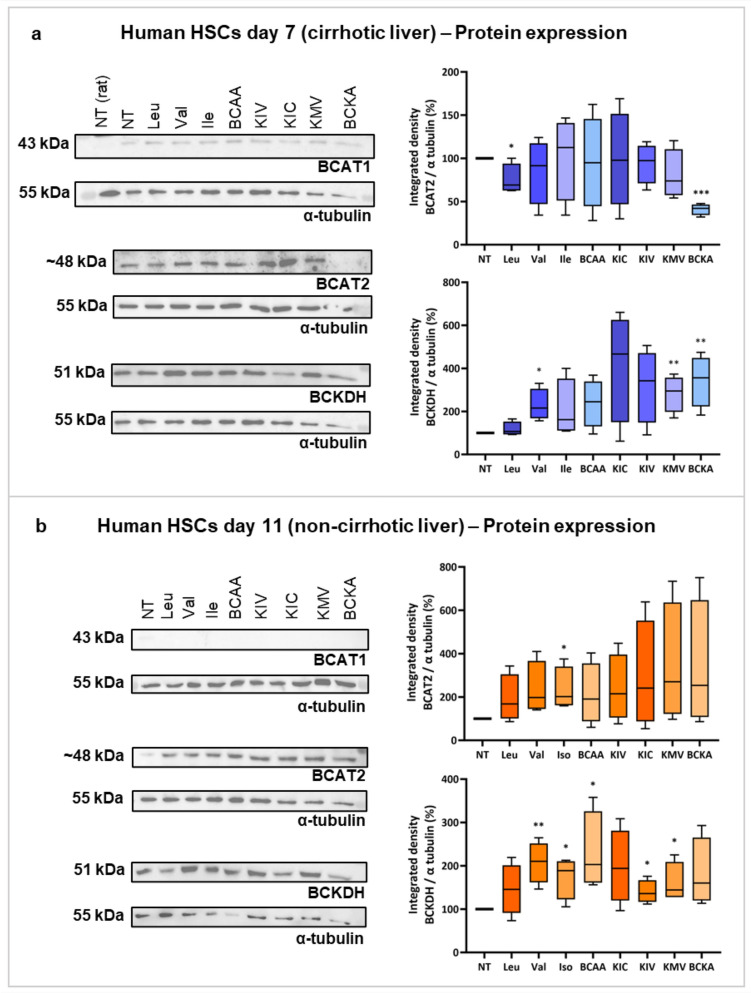
Methods: Primary HSCs from rats and cirrhotic and non-cirrhotic human livers, were cultured and treated with BCAAs or BCKAs to assess their effects on both preventing (from day 1 of isolation) and reversing (from day 7 of isolation) HSCs activation.

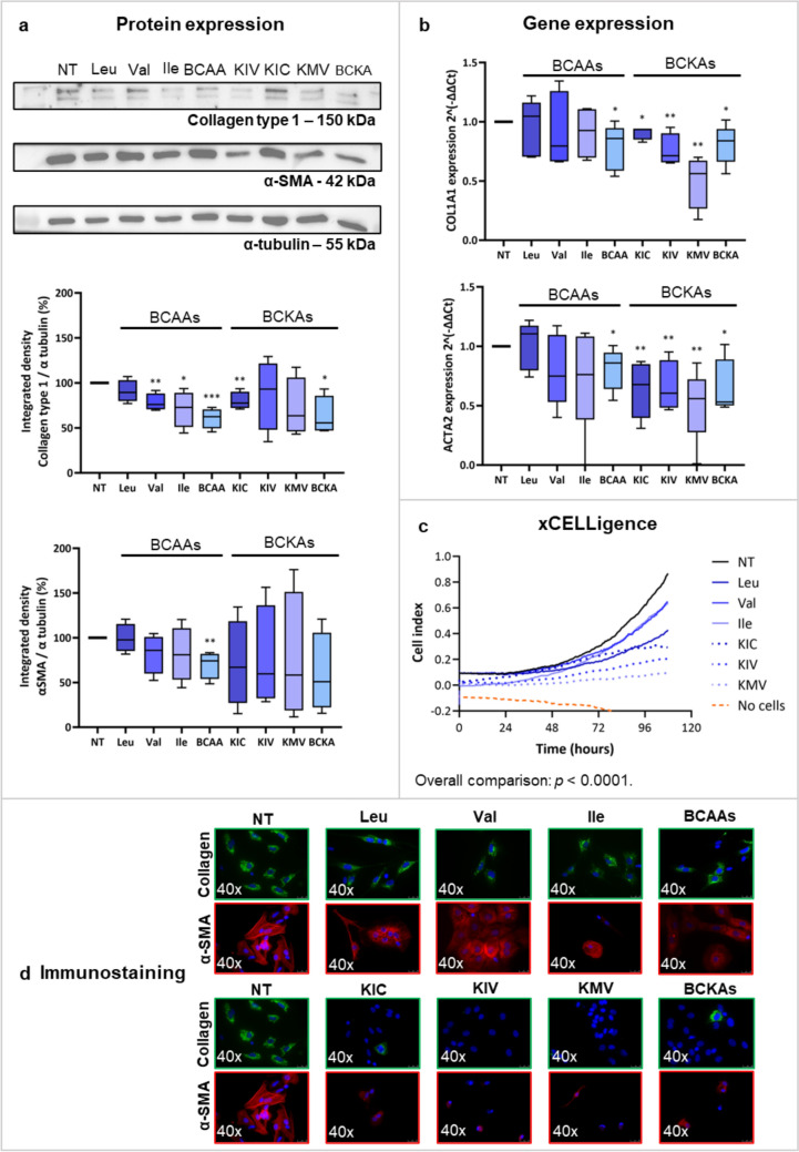
Results: In rat HSCs, leucine and BCKAs significantly reduced fibrotic markers and cell proliferation. In human HSCs, the metabolite of isoleucine decreased cell proliferation around 85% and increased the expression of branched-chain ketoacid dehydrogenase. The other metabolites also showed antifibrotic effects in HSCs from non-cirrhotic human livers.

Conclusion: BCAAs and their respective metabolites inhibit HSC activation with species-specific responses. Further research is needed to understand how BCAAs influence liver fibrogenesis. BCKAs supplementation could be a strategic approach for managing ESLDs, considering the nutritional status and amino acid profiles of patients.

Keywords: BCAAs; BCKAs; Branched-chain amino acids; Branched-chain keto acids; End-stage liver disease; Fibrosis; Hepatic stellate cells; Liver cirrhosis.


Conflict of interest statement

The authors declare no competing interests.

Figures

Fig. 1
Fig. 1
Methodology. HSCs were isolated from rat (A) and human (B) livers. Quiescent HSCs from rats were cultured in a medium enriched with BCAAs or BCKAs for 7 days, or cultured in a flask with standard IMDM medium for 6 days to achieve activation. Afterward, they were treated for 4 days with BCAAs- or BCKAs-enriched medium. For humans, HSCs were isolated from both cirrhotic and non-cirrhotic livers. HSCs from cirrhotic livers were cultured in a BCAAs- or BCKAs-enriched medium for 7 days, and HSCs from non-cirrhotic livers were cultured in a flask with standard IMDM medium for 6 days to achieve complete activation, and then treated for 4 days with a BCAAs- or BCKAs-enriched medium. aHSCs activated hepatic stellate cells, BCAAs branched-chain amino acids, BCKAs branched-chain keto acids, ESLD end-stage liver disease, qHSCs quiescent hepatic stellate cells. Figure created with Biorender.com
Fig. 2
Fig. 2
Expression of BCAT and BCKDH genes and proteins in HSCs and skeletal muscle tissues from rats. Representative Western blots and protein expression of BCAT2 and BCKDH in HSCs on day 7 (a) and day 11 (b) after treatment with leucine, valine, isoleucine, or a combination of the three BCAAs. Panel c shows the gene expression of BCAT1, BCAT2, and BCKDH in HSCs from rats, indicated as delta-delta Ct, at day 7 (blue bars) and day 11 (orange bars) of treatment with BCAAs as indicated. Panel d illustrates the gene expression levels, indicated as delta-delta Ct value of BCAT1, BCAT2, and BCKDH in skeletal muscle cells from rats after various culture times. BCAAs branched-chain amino acids, BCAT branched-chain amino acid transaminase, BCKDH branched-chain keto acid dehydrogenase, Ile Isoleucine, Leu Leucine, Val Valine. *p < 0.05; *** p < 0.001. Figure created with Biorender.com
Fig. 3
Fig. 3
Effect of BCAAs and BCKAs on activation markers in rHSCs cultured with BCAAs from day 1 to 7. Panel A shows the protein expression of Collagen type 1 and α-SMA, given as the integrated density of the protein band divided by the integrated density of α-tubulin (used as a housekeeping protein), and then normalized to the control sample, in samples from rHSCs at day 7 after treatment with BCAAs and BCKAs as indicated. Panel B corresponds to the gene expression levels of Col1a1 and Acta2, as delta-delta Ct with the housekeeping gene 36B4 in rHSCs at day 7. Panel C illustrates the cell proliferation in rHSCs treated with leucine, valine, isoleucine, KIC, KIV, KMV (blue lines), without any treatment (black line), or the control without cells (orange line). Panel D shows a representative immunofluorescence staining of Collagen type 1 (green) and α-SMA (red) under the indicated conditions. α-SMA alpha-smooth muscle actin, BCAAs branched-chain amino acids, BCKAs Branched-chain Keto Acids, Ile Isoleucine, KIC α-Ketoisocaproate, KIV α-Ketoisovalerate, KMV α-Keto-β-methylvalerate, Leu Leucine, NT Non-treatment, Val Valine. *p < 0.05; **p < 0.01; ***p < 0.001; ****p < 0.0001. Figure created with Biorender.com
Fig. 4
Fig. 4
Effects of BCAAs and BCKAs on activation markers in rHSCs cultured with BCAAs from day 7 to 11. Panel A shows the protein expression of Collagen type 1 and α-SMA, given as the integrated density of the protein band divided by the integrated density of α-tubulin (used as a housekeeping protein), and then normalized to the control sample, in samples from rHSCs at day 11 after 4 days of treatment with BCAAs and BCKAs as indicated. Panel B shows the gene expression levels of Col1a1 and Acta2, indicated as delta-delta Ct with the housekeeping gene 36b4, under the indicated treatment conditions in rHSCs at day 11. Panel C indicates a representative immunofluorescence staining of Collagen type 1 (green) and α-SMA (red) under the indicated conditions. α-SMA alpha-smooth muscle actin, BCAAs Branched-chain Amino Acids, BCKAs Branched-chain Keto Acids, Ile Isoleucine, KIC α-Ketoisocaproate, KIV α-Ketoisovalerate, KMV α-Keto-β-methylvalerate, Leu Leucine, NT non-treatment, Val Valine. *p < 0.05; **p < 0.01; ***p < 0.001; ****p < 0.0001. Figure created with Biorender.com
Fig. 5
Fig. 5
Protein expression of BCAT and BCKDH in human HSCs from cirrhotic and non-cirrhotic livers. Panel A illustrates the protein expression of BCAT and BCKDH in human HSCs isolated from cirrhotic livers (blue bars), treated with BCAAs or BCKAs as indicated. Panel B shows the protein expression of BCAT and BCKDH in human HSCs from non-cirrhotic livers (orange bars), also treated with BCAAs or BCKAs as indicated. BCAAs branched-chain amino acids, BCAT branched-chain amino acid transaminase, BCKAs branched-chain keto acids, BCKDH branched-chain keto acid dehydrogenase, Ile isoleucine, KIC α-ketoisocaproate, KIV α-ketoisovalerate, KMV α-keto-β-methylvalerate, Leu leucine, Val valine. *p < 0.05; **p < 0.01; ***p < 0.001. Figure created with Biorender.com
Fig. 6
Fig. 6
Effect of BCAAs and BCKAs on activation markers in hHSCs from cirrhotic tissue, cultured with BCAAs and BCKAs from day 1 to 7. Panel A shows the protein expression of Collagen type 1 and α-SMA, given as the integrated density of the protein band divided by the integrated density of α-tubulin (used as a housekeeping protein), and then normalized to the control sample when treating the cells with BCAAs and BCKAs as indicated. Panel B shows the gene expression levels of COL1A1 and ACTA2, indicated as delta-delta Ct with the housekeeping gene 18S, under the indicated treatment conditions. Panel C indicates the cell proliferation in hHSCs treated with leucine, valine, or isoleucine (continued blue lines), KIC, KIV, or KMV (intermittent blue lines), without any treatment (black line), or the controls without cells (orange line). Panel D shows the immunofluorescence staining of Collagen type 1 (green) and α-SMA (red) under the indicated conditions. α-SMA alpha-smooth muscle actin, BCAAs branched-chain amino acids, BCKAs branched-chain keto acids, Ile isoleucine; KIC α-ketoisocaproate, KIV α-ketoisovalerate, KMV α-keto-β-methylvalerate, Leu leucine, NT non-treatment, Val valine. *p < 0.05; **p < 0.01; ***p < 0.001. Figure created with Biorender.com
Fig. 7
Fig. 7
Effect of BCAAs and BCKAs on activation markers in hHSCs from non-cirrhotic tissue, cultured with BCAAs and BCKAs from day 7 to 11. Panel A shows the protein expression of Collagen type 1 and α-SMA, given as the integrated density of the protein band divided by the integrated density of α-tubulin (used as a housekeeping protein), and then normalized to the control sample, when treating the cells with BCAAs and BCKAs as indicated. Panel B shows the gene expression levels of COL1A1 and ACTA2, indicated as delta-delta Ct with the housekeeping gene 18S, under the indicated treatment conditions. Panel C indicates the immunofluorescence staining of Collagen type 1 (green) and α-SMA (red) under the indicated conditions. α-SMA alpha-smooth muscle actin, BCAAs branched-chain amino acids, BCKAs branched-chain keto acids, Ile isoleucine, KIC α-ketoisocaproate, KIV α-ketoisovalerate, KMV α-keto-β-methylvalerate, Leu leucine, NT non-treatment, Val valine. *p < 0.05; **p < 0.01. Figure created with Biorender.com

References

    1. Huang DQ, Terrault NA, Tacke F, Gluud LL, Arrese M, Bugianesi E, Loomba R (2023) Global epidemiology of cirrhosis—aetiology, trends and predictions. Nat Rev Gastroenterol Hepatol 20:388–398. 10.1038/S41575-023-00759-2 - PMC - PubMed
    1. Yao L, Hu X, Dai K, Yuan M, Liu P, Zhang Q, Jiang Y (2022) Mesenchymal stromal cells: promising treatment for liver cirrhosis. Stem Cell Res Ther. 10.1186/S13287-022-03001-Z - PMC - PubMed
    1. Ginès P, Krag A, Abraldes JG, Solà E, Fabrellas N, Kamath PS (2021) Liver cirrhosis. Lancet 398:1359–1376. 10.1016/S0140-6736(21)01374-X - PubMed
    1. Zhang CY, Yuan WG, He P, Lei JH, Wang CX (2016) Liver fibrosis and hepatic stellate cells: etiology, pathological hallmarks and therapeutic targets. World J Gastroenterol 22:10512. 10.3748/WJG.V22.I48.10512 - PMC - PubMed
    1. Dimou A, Tsimihodimos V, Bairaktari E (2022) The critical role of the Branched Chain Amino Acids (BCAAs) Catabolism-Regulating Enzymes, Branched-Chain Aminotransferase (BCAT) and Branched-Chain α-Keto Acid Dehydrogenase (BCKD), in human pathophysiology. Int J Mol Sci. 10.3390/IJMS23074022 - PMC - PubMed

MeSH terms

LinkOut - more resources