. 2024 Nov;28(21):e70187.
doi: 10.1111/jcmm.70187.

Exosomal miR-155-5p promote the occurrence of carotid atherosclerosis

Affiliations

Exosomal miR-155-5p promote the occurrence of carotid atherosclerosis

Wen-Wen Yang et al. J Cell Mol Med. 2024 Nov.

Abstract

Periodontitis is a significant independent risk factor for atherosclerosis. Yet, the exact mechanism of action is still not fully understood. In this study, we investigated the effect of exosomes-miR-155-5p derived from periodontal endothelial cells on atherosclerosis in vitro and in vivo. Higher expression of miR-155-5p was detected in the plasma exosomes of patients with chronic periodontitis (CP) and carotid atherosclerosis (CAS) compared to patients with CP. Also, the expression level of miR-155-5p was associated with the severity of CP. miR-155-5p-enriched exosomes from HUVECs increased the angiogenesis and permeability of HAECs and promoted the expression of angiogenesis, permeability, and inflammation genes. Along with the overexpression or inhibition of miR-155-5p, the biological effect of HUVECs-derived exosomes on HAECs changed correspondingly. In ApoE-/- mouse models, miR-155-5p-enriched exosomes promoted the occurrence of carotid atherosclerosis by increasing permeable and angiogenic activity. Collectively, these findings highlight a molecular mechanism of periodontitis in CAS, uncovering exosomal miR-155-5p derived periodontitis affecting carotid endothelial cells in an 'exosomecrine' manner. Exosomal miR-155-5p may be used as a biomarker and target for clinical intervention to control this intractable disease in future, and the graphic abstract was shown in Figure S1.

Keywords: LPS; carotid atherosclerosis; chronic periodontitis; exosome; miR‐155‐5p.


Conflict of interest statement

The authors declare that they have no competing interests.

Figures

FIGURE 1
FIGURE 1
Expression of miR‐155‐5p is upregulated in plasma exosomes from patients with CP and CAS. (A) Electron microscopy images of human plasma exosomes. Scale bar = 100 nm. (B) NTA was used to determine the particle size and concentration of human plasma exosomes. (C) Flow cytometric analysis of CD9 and CD63 (exosomal marker) expression in human plasma exosomes. (D) The heatmap of microRNAs expressed in plasma exosomes from patients with CP or patients with CP and CAS (n = 6 per group). (E, F) Plasma exosomes were isolated from patients with mild or moderate CP (n = 6), severe CP (n = 6), mild or moderate CP and CAS (n = 6) and severe CP and CAS (n = 7). Real‐time PCR was used to determine the miR‐155‐5p expression levels. (G) Real‐time PCR was used to determine the miR‐155‐5p expression levels in Normal (n = 5) and CAS (n = 5) tissue samples. All data are expressed as mean ± SD, *p < 0.05.
FIGURE 2
FIGURE 2
LPS is involved in the angiogenesis and permeability of endothelial cells. (A) CCK‐8 assays were used to assess HUVECs after incubation with or without 0.5 μg/mL LPS. (B) Migration assay for HUVECs after incubation with or without 0.5 μg/mL LPS. Scale bar = 200 μm. (C, D) Tube formation assays for HUVECs and HAECs after incubation with or without 0.5 μg/mL LPS. Scale bar = 500 μm. (E) Permeability assay for HUVECs after incubation with or without 0.5 μg/mL LPS. (F, G) qRT‐PCR analysis of relative VEGFA, IL‐8, VE‐cadherin, GM‐CSF, MCP‐1, RANTES, IL‐6 and IL‐1β mRNA expression levels in HUVECs cultured with 0.5 μg/mL LPS. All data are expressed as mean ± SD, *p < 0.05.
FIGURE 3
FIGURE 3
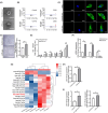
Exosomes derived from HUVECs cultured with 0.5 μg/mL LPS promote angiogenesis and permeability and increase the miR‐155‐5p level of HAECs. (A) Electron microscopy images of exosomes from HUVECs cultured with or without LPS (named LPS exo or Control exo). Scale bar = 100 nm. (B) Flow cytometric analysis of CD63 (exosomal marker) expression in Control exosomes and LPS exosomes. (C) The uptake of PKH26‐labelled exosomes (red) by HAECs was identified using fluorescence confocal microscopy. Scale bar = 10 μm. (D) Images of tube formation of HAECs after incubation with Control exosomes or LPS exosomes. Scale bar = 500 μm. (E) Permeability assay for HAECs after incubation with Control exosomes or LPS exosomes. (F) qRT‐PCR was used to assess the relative VEGFA, IL‐8 and VE‐cadherin expression levels in HAECs after stimulation with Control exosomes or LPS exosomes. (G) The heatmap and hierarchical clustering of microRNAs differentially expressed in Control exosomes or LPS exosomes. Red, high relative expression; blue, low relative expression. (H, I) qRT‐PCR analysis of relative miR‐155‐5p expression levels in Control exosomes and LPS exosomes, and miR‐155‐5p levels in HAECs after incubation with 0.5 μg/mL LPS, Control exosomes, or LPS exosomes. All data are expressed as mean ± SD, *p < 0.05.
FIGURE 4
FIGURE 4
miR‐155‐5p promotes endothelial cells' angiogenesis and permeability. (A) Fluorescence microscopy of HUVECs stably transfected with the control vector (Vector cells) or the miR‐155‐5p expression vector (OE cells). (B) Expression of miR‐155‐5p in Vector or OE cells. (C) Migration assays of Vector and OE cells. (D) Representative images of tube formation of Vector and OE cells. (E) Permeability assays for Vector and OE cells. (F) Expression of miR‐155‐5p in HUVECs transiently transfected with miR‐155‐5p inhibitor (Inhibitor cells) or negative control (NC cells). (G, H) Permeability and tube formation assays of NC and Inhibitor cells. (I) qRT‐PCR analysis of VEGFA, IL‐8, and VE‐cadherin expression levels in Vector, OE, NC or Inhibitor cells. (J) The binding sites of miR‐155‐5p and PIK3CA and the expression of PIK3CA in Vector, OE, NC or Inhibitor cells by qRT‐PCR. All data are expressed as mean ± SD, *p < 0.05.
FIGURE 5
FIGURE 5
miR‐155‐5p induces angiogenesis and permeability in HAECs through exosomes. (A) Electron microscopy images of exosomes from Vector, OE, NC or Inhibitor cells (Vector exo, OE exo, NC exo and Inhibitor exo, respectively) are shown. Scale bar = 100 nm. (B) Flow cytometric analysis of CD9 and CD63 expression in Vector exo, OE exo, NC exo and Inhibitor exo. (C) Confocal images of HAECs incubated with PKH26‐labelled Vector or OE exosomes. Scale bar = 10 μm. (D) qRT‐PCR of the relative miR‐155‐5p expression level in Vector and OE exosomes. (E, F) Tube formation and permeability assays for HAECs incubated with Vector or OE exosomes. Scale bar = 500 μm. (G) The expression level of miR‐155‐5p in NC and Inhibitor exosomes was determined by qRT‐PCR. (H, I) Tube formation and permeability assays for HAECs incubated with NC or Inhibitor exosomes. Scale bar = 500 μm. (J) The qRT‐PCR analysis was used to determine miR‐155‐5p, VEGFA, IL‐8 and VE‐cadherin expression of HAECs treated with Vector exosomes and OE exosomes, respectively. All data are expressed as mean ± SD, *p < 0.05.
FIGURE 6
FIGURE 6
Effects of exosomal miR‐155‐5p on atherosclerotic plaque formation in vivo. (A) In vivo bioluminescence images used to study the biodistribution of PHK67‐labelled Vector exosomes or OE exosomes in mice. (B) Vascular ultrasound analysis of plaques in the carotid artery and aorta from Vector or OE exosomes group. (C) Vascular vasodilatation of the carotid artery and aorta in mice is evaluated by ultrasound and presented as changes in vasodilation and vasoconstriction diameter. (D) The atherosclerotic lesion areas in the thoracic aorta of ApoE−/− mice stained with en face oil red O (left). Quantitative analysis of the plaque areas (right) is shown. (E) Representative images of oil‐red‐O‐stained aortic sections (left). Quantitative analysis of the plaque areas is shown. Scale bar = 200 μm. (F) Representative H&E‐stained images of carotid sections. Scale bar (left) = 200 μm, Scale bar (right) = 20 μm. (G) Plasma exosomes from mice in the Vector exosomes or OE exosomes group were isolated, and qRT‐PCR determined the miR‐155‐5p expression levels. (H) qRT‐PCR analysis of miR‐155‐5p, IL‐8, VE‐cadherin expression levels in the carotid artery and abdominal aorta tissues from mice. All data are expressed as mean ± SD, *p < 0.05.
FIGURE 7
FIGURE 7
Effects of LPS on atherosclerotic plaque formation in vivo. (A) MicroCT 2D image of mouse left maxilla. Green line: The cemento‐enamel junction, Red line: The alveolar bone crest. Scale bar = 1 mm (B) The atherosclerotic lesion areas in the thoracic aorta of ApoE−/− mice stained with en face oil red O (left). Quantitative analysis of the total area of plaque areas (right) is shown (n = 6 per group). (C) Haematoxylin–eosin (H&E) staining of tissue sections of carotid artery (n = 6 per group). Scale bar (left) = 200 μm, Scale bar (right) = 20 μm. (D) Plasma exosomes from mice in the Control or LPS group (n = 6) were isolated, and qRT‐PCR determined the miR‐155‐5p expression levels. (E, F) miR‐155‐5p, VEGFA, IL‐8, VE‐cadherin expression levels in the carotid artery and abdominal aorta tissues from mice (n = 6 per group) were analysed by qRT‐PCR. All data are expressed as mean ± SD, *p < 0.05.

References

    1. Saba L, Saam T, Jäger HR, et al. Imaging biomarkers of vulnerable carotid plaques for stroke risk prediction and their potential clinical implications. Lancet Neurol. 2019;18:559‐572. - PubMed
    1. Zhu X, Huang H, Zhao L. PAMPs and DAMPs as the bridge between periodontitis and atherosclerosis: the potential therapeutic targets. Front Cell Dev Biol. 2022;10:856118. - PMC - PubMed
    1. Patel UK, Malik P, Kodumuri N, et al. Chronic periodontitis is associated with cerebral atherosclerosis—A Nationwide Study. Cureus. 2020;12:e11373. - PMC - PubMed
    1. Sanz M, Marco Del Castillo A, Jepsen S, et al. Periodontitis and cardiovascular diseases: consensus report. J Clin Periodontol. 2020;47:268‐288. - PMC - PubMed
    1. Yamada S, Komiyama T, Ohi T, et al. Regular dental visits, periodontitis, tooth loss, and atherosclerosis: The Ohasama study. J Periodontal Res. 2022;57:615‐622. - PMC - PubMed

MeSH terms

LinkOut - more resources