

Effects of gestational age on blood cortisol and prolactin levels during pregnancy in malaria endemic area
- PMID: 39495748
- PMCID: PMC11534236
- DOI: 10.1371/journal.pone.0310372
Effects of gestational age on blood cortisol and prolactin levels during pregnancy in malaria endemic area
Abstract
Background: The hormonal shift occurring in pregnant women is crucial for the outcome of pregnancy. We conducted a study in pregnant women living in a malaria endemic area to determine the potential effect of gestational age on the modulation of the endocrine system by cortisol and prolactin production during pregnancy.
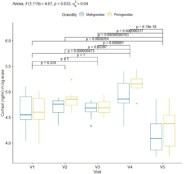
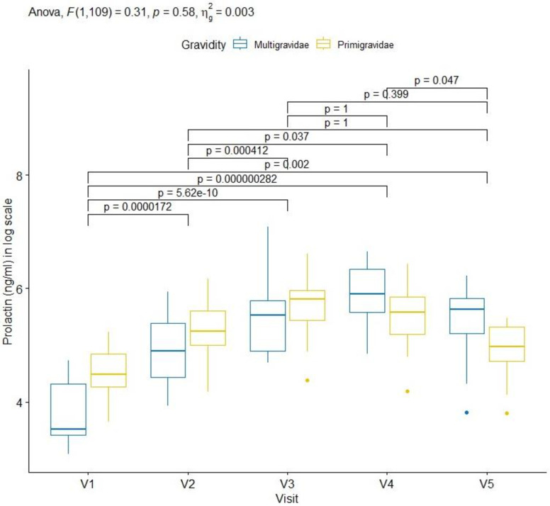
Methods: Primigravidae and multigravidae with a gestational age between 16-20 weeks were included in the study and followed up to delivery and 6-7 weeks thereafter. Venous blood was collected at scheduled visit: Visit 1 (V1; 16-20 weeks of amenorrhea), Visit 2 (V2; 28 ±1 weeks of pregnancy), Visit 3 (V3; 32 ±1 weeks of pregnancy), Visit4 (V4; delivery) and Visit5 (V5; 6-7 weeks after delivery). In addition, a cord blood sample was also collected during labour at delivery. Nulliparous and primiparous/multiparous non-pregnant women were enrolled in the control group. Cortisol and prolactin plasma concentrations were measured using ichroma II and i-chamber apparatus. Light microscopy was used to detect Plasmodium falciparum infections. A linear mixed-effects regression (LMER) model was used to assess the association between the variation of cortisol titres and prolactin levels during the pregnancy and the post-partum.
Results: Results showed that cortisol and prolactin levels in the peripheral blood were globally up-regulated during pregnancy. Concentrations of cortisol during follow-up was significantly higher in primigravidae than in multigravidae during the whole pregnancy (p<0.024). Moreover, the level of prolactin which was higher before delivery in primigravidae reversed at delivery and postpartum visit, but the difference was not statistically significant during the follow-up (V1 to V5) (p = 0.60). The cortisol level in peripheral blood at delivery was higher than that in the cord blood, and conversely for prolactin. Cortisol and prolactin levels decreased after delivery, though the level of prolactin was still higher than that at enrolment. An increase of one unit of prolactin was associated with the decrease of the average concentration of cortisol by 0.04 ng/ml (p = 0.009). However, when cortisol increases with one unit, the average concentration of prolactin decreases by 1.16 ng/ml (p = 0.013).
Conclusion: These results showed that the up-regulation effects of cortisol and prolactin are related to gestational age. A The downward regulation effect that both hormones have on each other during the pregnancy when each increase to 1 unit (1.0 ng/ml) was also reported.
Copyright: © 2024 Kiemde et al. This is an open access article distributed under the terms of the Creative Commons Attribution License, which permits unrestricted use, distribution, and reproduction in any medium, provided the original author and source are credited.
Conflict of interest statement
The authors have declared that no competing interests exist.
Figures
References
-
- NG SC, Gilman-sachs A, Beaman KD, Beer AE, Kwak-kim J. Expression of intracellular Th1 and Th2 cytokines in women with recurrent spontaneous abortion, implantation failures after IVF/ET or normal pregnancy. American Journal of Reproductive Immunology. 2002;48(2):77–86. doi: 10.1034/j.1600-0897.2002.01105.x - DOI - PubMed
-
- Kwak-Kim JYH, Gilman-Sachs A, Kim CE. T Helper 1 and 2 Immune Responses in Relationship to Pregnancy, Nonpregnancy, Recurrent Spontaneous Abortions and Infertility of Repeated Implantation Failures. In: Immunology of Gametes and Embryo Implantation. Basel: KARGER; 2005. p. 64–79. - PubMed
-
- Szekeres-Bartho J, Barakonyi A, Miko E, Polgar B, Palkovics T. The role ofγ / δT cells in the feto-maternal relationship. Semin Immunol. 2001. Aug;13(4):229–33. - PubMed
MeSH terms
Substances
LinkOut - more resources
Full Text Sources
