. 2024 Nov 12;121(46):e2405671121.
doi: 10.1073/pnas.2405671121. Epub 2024 Nov 4.

A conserved juxtamembrane motif in plant NFR5 receptors is essential for root nodule symbiosis

Affiliations

A conserved juxtamembrane motif in plant NFR5 receptors is essential for root nodule symbiosis

Simon Boje Hansen et al. Proc Natl Acad Sci U S A. .

Abstract

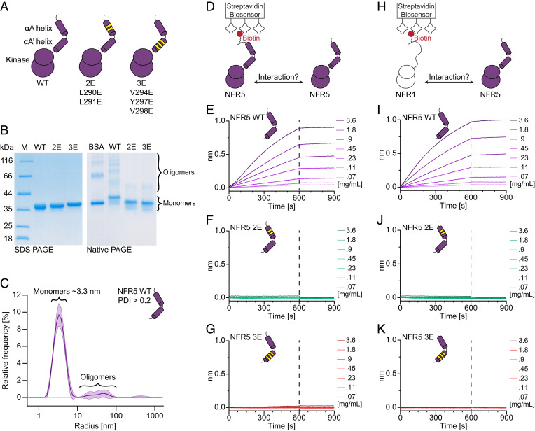
Establishment of root nodule symbiosis is initiated by the perception of bacterial Nod factor ligands by the plant LysM receptor kinases NFR1 and NFR5. Receptor signaling initiating the symbiotic pathway depends on the kinase activity of NFR1, while the signaling mechanism of the catalytically inactive NFR5 pseudokinase is unknown. Here, we present the crystal structure of the signaling-competent Lotus japonicus NFR5 intracellular domain, comprising the juxtamembrane region and pseudokinase domain. The juxtamembrane region is structurally well defined and forms two α-helices, αA and αA', which contain an exposed hydrophobic motif. We demonstrate that this "juxtamembrane motif" promotes NFR5-NFR5 and NFR1-NFR5 interactions and is essential for symbiotic signaling. Conservation analysis reveals that the juxtamembrane motif is present throughout NFR5-type receptors and is required for symbiosis signaling from barley RLK10, suggesting a conserved and broader function for this motif in plant-microbe symbioses.

Keywords: Nod factor receptors; kinases and pseudokinases; plant–microbe interactions; receptor signaling; root nodule symbiosis.


Conflict of interest statement

Competing interests statement:Some findings from this article are considered for a patent application.

Figures

Fig. 1.
Fig. 1.
Crystal structure of the signaling-competent NFR5 intracellular domain. (A) Phosphorylation assay showing catalytic activity of the NFR1 kinase and no activity of the NFR5 pseudokinase. (B) nfr5 complementation assay showing that the kinase domain of NFR5 is required for root nodule symbiosis. Number of nodules per plant on hairy roots expressing the indicated NFR5 constructs from the native Nfr5 promoter and terminator in nfr5 mutant plants, 6 wk after inoculation with M. loti R7A rhizobia. Transformation marker (tYFP) was used as the negative control. (C) NFR5-Nb200 structure model. N-lobe and C-lobe of NFR5 are highlighted. Nb200 binds to the C-lobe αG helix (see details in SI Appendix, Fig. S2 F and G). (D) Overview of the NFR5 intracellular domain. Secondary structure elements are highlighted. The juxtamembrane αA and αA′ helices precede the pseudokinase domain with the N- and C-lobe marked. An ATP molecule from the structure of PKA [PDB: 1ATP (41)] can be docked by superposition into the ATP binding pocket of NFR5 without steric clashes. The K339-E349 salt bridge, diverged DFG to “NFA” and HRD motifs are indicated. (E) The juxtamembrane region is marked along with the truncated glycine-rich and activation loops. The catalytic and regulatory spines are highlighted in transparent surface representations showing that the C-spine is broken due to the unoccupied ATP pocket, while the R-spine is fully formed. Highlighted residues A337, L440 in the C-spine and L353 in the R-spine were targeted in mutagenesis experiments in SI Appendix, Fig. S3. (F) Fusion of Nb200 as a C-terminal tag to the NFR1 receptor forces an artificial NFR1–NFR5 receptor complex that induces spontaneous nodulation in Lotus nfr1 mutant plants. Number of nodules per plant on hairy roots expressing the indicated NFR1 constructs from the native Nfr1 promoter and terminator in nfr1 mutant plants, 9 wk after transformation. Transformation marker (tYFP) was used as the negative control. (B and F) Lowercase letters indicate significant differences between samples [ANOVA (Kruskal–Wallis) and post hoc analysis (Dunn’s test), P < 0.05]. Circles represent individual plants, and the number of nodulating plants out of total plants is indicated below boxplots.
Fig. 2.
Fig. 2.
The αA and αA′ helices define a surface-exposed hydrophobic juxtamembrane motif in NFR5. (A) The NFR5 juxtamembrane region is highlighted, and the secondary structure elements are indicated. (B) Zoom of the juxtamembrane region showing the 2Fo-Fc electron density map (contour σ = 1, mesh representation), along with the atomic model (cartoon representation). Backbone and sidechains are well defined in the electron density and support modeling of two α-helices (see SI Appendix, Fig. S4 for stereo view). (C) Overview of the αA and αA′ helices. The αA residues L290 and L291 form the N-terminal part, while the αA′ residues V294, Y297, and V298 form the C-terminal part of the juxtamembrane motif. The helices are separated by a small linker region imposing a ~65° bend. S292 and S295 are positioned at the linker bend separating the αA and αA′ helices. (D) Electrostatic potential surface representation of the αA and αA′ helices, displaying the hydrophobicity and spatial continuity of the motif. ±5 kT/e, k = Boltzmann constant, T = temperature in Kelvin, e = elementary charge. (E) αA and αA′ alignment of Lotus NFR5 (S282-K300) and barley RLK10 (R314-K332) along with the sequence logo of the larger alignment shown in SI Appendix, Fig. S5. The juxtamembrane motif is highlighted below the sequence logo with red boxes. (F) Surface conservation analysis of the αA and αA′ helices shows that the juxtamembrane motif region is conserved throughout NFR5-type receptors.
Fig. 3.
Fig. 3.
The juxtamembrane motif mediates NFR5–NFR5 and NFR1–NFR5 complex formation. (A) Illustration of the NFR5 intracellular domain along with the αA 2E (L290E and L291E) and αA′ 3E (V294E, Y297E, and V298E) variants. (B) SDS-PAGE and native PAGE analysis of NFR5 WT, 2E and 3E samples. All three samples migrate as single bands to the expected molecular size in SDS-PAGE. NFR5 WT, but not 2E or 3E, show an oligomeric pattern resembling the pattern formed by the oligomer forms of BSA. (C) DLS assay of NFR5 WT revealing a PDI >0.2 and particle radii larger than the ~3.3 nm monomer form, indicating oligomer populations form in solution. (D) Illustration of the NFR5–NFR5 biolayer interferometry interaction experiment. An Avi-tagged version of NFR5 WT was purified (SI Appendix, Fig. S2A) and immobilized on streptavidin biosensors. NFR5 loaded biosensors were assayed against a concentration series (0.07 to 3.6 mg/mL) of NFR5 WT, 2E, and 3E. (E) A concentration-dependent response was observed for NFR5 WT, and no response was observed for 2E and 3E (F and G). (HK) Identical experiment as (DG) except Avi-tagged NFR1 (SI Appendix, Fig. S6D) was immobilized on streptavidin biosensors. NFR5 WT (I) and not 2E or 3E (J and K) interact in a concentration-dependent manner with NFR1.
Fig. 4.
Fig. 4.
The juxtamembrane motif in NFR5-type receptors is required for symbiotic signaling. (A) Illustration of the Lotus NFR5 receptor and the NFR5-RLK10 chimeric receptor assayed in the nodulation complementation experiments. (B) Number of nodules per plant on hairy roots expressing the indicated receptor constructs from the native Nfr5 promoter and terminator in nfr5 mutant plants, 6 wk after inoculation with M. loti R7A rhizobia. No nodules form on nfr5 when transformed with an empty vector containing only a triple yellow fluorescent protein (tYFP) transformation marker. NFR5 WT complements nfr5 and forms nodules in almost all 49 plants assayed. NFR5 2E (L290E and L291E) and NFR5 3E (V294E, Y297E, and V298E) fail to form any nodules. NFR5 (L290F and L291F) and (Y297I) (SI Appendix, Fig. S8) complement nfr5. The NFR5-RLK10 chimera complements nfr5 at NFR5 WT levels. RLK10 2E (L322E and L323E) and 3E (V326E, F329E, and I330E) variants are incapable of driving nodule formation in Lotus. Lowercase letters indicate significant differences between samples [ANOVA (Kruskal–Wallis) and post hoc analysis (Dunn’s test), P < 0.05]. Circles represent individual plants, and the number of nodulating plants out of total plants is indicated below boxplots. (C) Our data suggest that an NFR5–NFR5 complex is required for NFR1 interaction and symbiosis signaling. Complex formation is supported by the juxtamembrane motif spanning the αA and αA′ helices. Disruption of this complex by targeting the juxtamembrane motif leads to NFR5 receptors incapable of NFR5–NFR5 and NFR1–NFR5 interaction and signaling.

References

    1. Radutoiu S., et al. , Plant recognition of symbiotic bacteria requires two LysM receptor-like kinases. Science 425, 585–592 (2003). - PubMed
    1. Madsen E. B., et al. , A receptor kinase gene of the LysM type is involved in legume perception of rhizobial signals. Science 425, 637–640 (2003). - PubMed
    1. Radutoiu S., et al. , LysM domains mediate lipochitin-oligosaccharide recognition and Nfr genes extend the symbiotic host range. EMBO J. 26, 3923–3935 (2007). - PMC - PubMed
    1. Rübsam H., et al. , Nanobody-driven signaling reveals the core receptor complex in root nodule symbiosis. Science 379, 272–277 (2023). - PubMed
    1. Broghammer A., et al. , Legume receptors perceive the rhizobial lipochitin oligosaccharide signal molecules by direct binding. Proc. Natl. Acad. Sci. U.S.A. 109, 13859–13864 (2012). - PMC - PubMed

LinkOut - more resources