. 2024 Nov 13;11(11):ENEURO.0320-24.2024.
doi: 10.1523/ENEURO.0320-24.2024. Print 2024 Nov.

Distinct Strategies Regulate Correlated Ion Channel mRNAs and Ionic Currents in Continually versus Episodically Active Neurons

Affiliations

Distinct Strategies Regulate Correlated Ion Channel mRNAs and Ionic Currents in Continually versus Episodically Active Neurons

Jose A Viteri et al. eNeuro. .

Abstract

Relationships among membrane currents allow central pattern generator (CPG) neurons to reliably drive motor programs. We hypothesize that continually active CPG neurons utilize activity-dependent feedback to correlate expression of ion channel genes to balance essential membrane currents. However, episodically activated neurons experience absences of activity-dependent feedback and, thus, presumably employ other strategies to coregulate the balance of ionic currents necessary to generate appropriate output after periods of quiescence. To investigate this, we compared continually active pyloric dilator (PD) neurons with episodically active lateral gastric (LG) CPG neurons of the stomatogastric ganglion (STG) in male Cancer borealis crabs. After experimentally activating LG for 8 h, we measured three potassium currents and abundances of their corresponding channel mRNAs. We found that ionic current relationships were correlated in LG's silent state, but ion channel mRNA relationships were correlated in the active state. In continuously active PD neurons, ion channel mRNAs and ionic currents are simultaneously correlated. Therefore, two distinct relationships exist between channel mRNA abundance and the ionic current encoded in these cells: in PD, a direct correlation exists between Shal channel mRNA levels and the A-type potassium current it carries. Conversely, such channel mRNA-current relationships are not detected and appear to be temporally uncoupled in LG neurons. Our results suggest that ongoing feedback maintains membrane current and channel mRNA relationships in continually active PD neurons, while in LG neurons, episodic activity serves to establish channel mRNA relationships necessary to produce the ionic current profile necessary for the next bout of activity.

Keywords: central pattern generator; stomatogastric.


Conflict of interest statement

The authors declare no competing financial interests.

Figures

Figure 1.
Figure 1.
Activation of the lateral gastric (LG) neuron's active state via stimulation of descending inputs to the STG. A, Schematic of the stomatogastric nervous system from the crab Cancer borealis. All descending modulatory inputs were preserved: the oesophageal ganglion (OG) and the commissural ganglia (CoG) provide descending modulatory inputs to the STG via the stomatogastric nerve (stn). The lateral gastric (LG) neuron used in these experiments is highlighted in the STG. A stimulator (A-M Systems) provided the necessary voltage (10–15 V) to experimentally turn on the gastric mill rhythm. This was accomplished by surrounding the dorsal posterior oesophageal nerves (dpons) with a petroleum jelly well and stimulating the dpon nerves. This stimulates the ventral cardiac neurons which in turn activate MCN1 and CPN2 neurons in the CoG. This induces the release of the Cancer borealis tachykinin-related peptide Ia (CabTRP Ia) which converges to LG and initiates LG's active state. B, Representative recordings of LG's different states when the gastric mill is silent or active. Recordings of LG are taken from the same neuron before and after activation of the gastric mill rhythm. Recordings were taken extracellularly from the lvn and dgn nerves of the stomatogastric nervous system (STNS). Recordings from the lvn allow for visualization of the pyloric rhythm while recordings from the dgn allow for visualization of the gastric mill rhythm. LG axons run through the lvn and allow for confirmation of LG's active state during gastric mill activity. Calibration: 30 mV and 5 s. C, Ionic currents measured in silent and active LG neurons. Ionic current magnitudes in LG neurons were measured at 0 mV on an IV plot generated from the current traces. In silent LG neurons, currents were measured acutely. In active LG neurons, currents were measured after 8 h of activity. The A-type potassium current (IA) was measured by subtracting the high threshold potassium current (IHTK) from an IA TEVC protocol with a holding potential of −80 mV and 10 voltage steps from −60 to +30 mV (10 mV intervals). IHTK was measured by using a leak-subtracted TEVC protocol with a holding potential of −40 mV and 10 voltage steps from −60 to +30 mV (10 mV intervals). The calcium-activated potassium current (IKCA) was measured by subtracting postcadmium (250 µM CdCl2) IHTK current traces from precadmium IHTK current traces. The delayed rectifier potassium current (IKd) was measured by running the IHTK TEVC protocol after the application of cadmium (250 µM CdCl2) to block IKCa.
Figure 2.
Figure 2.
The active state of LG neurons induces changes in peak ionic current magnitudes and changes in corresponding ion channel mRNA abundances. A, Bar graphs (mean ± SD) displaying the peak ionic current magnitudes for three of the potassium currents that were measured. Each point corresponds to a measurement collected from a single LG neuron from two different conditions: silent (gray) LG neurons where their active state was not induced or active (pink) LG neurons where their active state was induced for 8 h. The active state of LG neurons (pink) induced significant changes in ionic current magnitudes in 3/3 potassium currents (Welch's independent two-sample t test, p < 0.05) when compared with the silent condition. B, Bar graphs (mean ± SD) displaying the corresponding mRNA abundances for the three potassium currents that were measured. Note that some ionic currents are encoded by more than one mRNA transcript. Each point corresponds to a measurement collected from a single LG neuron from two different conditions: silent (gray) LG neurons where their active state was not induced and active (pink) LG neurons where their active state was induced for 8 h. The active state of LG neurons (pink) induced significant changes in BKKCA (IKCa) and SHAKER (IA) when compared with the silent condition (Welch's independent two-sample t test, p < 0.05). ns, not significant. See also Extended Data Table 6-1. C, Bar graphs displaying the change in the coefficient of variation (COV) for membrane currents (left panel) and ion channel mRNA abundances (right panel) during LG's silent and active state. The active state of LG neurons induced a significant decrease in the COV of all membrane currents but had no effect on the COV of mRNA abundances (Levene's test, p < 0.05 and p > 0.05, respectively). See also Extended Data Tables 2-1 and 2-2. * = p < 0.05, ** = p < 0.001, *** = p < 0.0001.
Figure 3.
Figure 3.
Ion channel mRNA relationships are correlated only in active LG neurons, but ionic current relationships are correlated only in silent LG neurons. A, BKKCA versus SHAL and BKKCA versus SHAKER mRNAs were positively correlated only in active LG neurons (open pink circles: Spearman value > 0.6; p < 0.05). However, the corresponding ionic current relationship IKCa versus IA was negatively correlated only in silent LG neurons (solid gray circles: Pearson’s value < −0.6; p < 0.05). B, BKKCA versus SHAB mRNAs were positively correlated only in active LG neurons (open pink circles: Spearman value > 0.6; p < 0.05). However, the corresponding ionic current relationship IKCa versus IKd was negatively correlated only in silent LG neurons (solid gray circles: Pearson’s value greater than −0.6; p < 0.05). C, The SHAL versus SHAB and SHAKER versus SHAB relationships were positively correlated in active LG neurons (open pink circles: Pearson’s value > 0.6; p < 0.05). However, the corresponding ionic current relationship IA versus IKd was positively correlated only in silent LG neurons (solid gray circles: Pearson’s value > 0.6; p < 0.05). See also Extended Data Tables 3-1 and 3-2. *Pearson’s correlations denoted by “R.” *Spearman correlations denoted by “rho.”
Figure 4.
Figure 4.
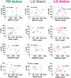
Both ion channel mRNA relationships and ionic current relationships are correlated in active PD neurons. A, The BKKCA versus SHAL mRNA relationship was positively correlated (open pink circles: Pearson’s value > 0.6; p < 0.05) but not BKKCA versus SHAKER in active PD neurons. The corresponding ionic current relationship IKCa versus IA was positively correlated (solid gray circles: Pearson’s value > 0.6; p < 0.05). B, The SHAL versus SHAB mRNA relationship was positively correlated (open pink circles: Pearson’s value  > 0.6; p < 0.05) but not BKKCA versus SHAB in active PD neurons. The corresponding ionic current relationship IA versus IKd was positively correlated (solid gray circles: Pearson’s value > 0.6; p < 0.05). C, The BKKCA versus SHAB mRNA relationship was not correlated in active PD neurons. The corresponding ionic current relationship IKCa versus IKd was positively correlated (solid gray circles: Pearson’s value > 0.6; p < 0.05). See also Extended Data Tables 4-1 and 4-2. *Pearson’s correlations denoted by “R.”
Figure 5.
Figure 5.
Ion channel mRNA transcripts are correlated with the ionic currents they encode in PD, but not LG neurons. Ion channel mRNAs were plotted against the ionic currents they encode across PD and LG neurons (silent and active state). The IA versus SHAL relationship is positively correlated in PD (open green circles: Pearson’s value > 0.6; p < 0.05). The IA versus SHAKER relationship is negatively correlated in PD (open green circles: Pearson’s value > 0.6; p < 0.05). However, none of these relationships were correlated in silent or active LG neurons (open gray and pink circles). See also Extended Data Table 5-1. *Pearson’s correlations denoted by “R.”
Figure 6.
Figure 6.
A model for temporally uncoupled regulation of channel mRNA and protein in episodically versus continually active neurons. A, In PD neurons (continually active), there is continuous activity- and modulator-dependent feedback that signals to maintain and tune both channel mRNA and protein relationships in a continually coregulated state, so that ongoing activity can continue throughout the lifetime of the animal without interruption. This ongoing regulation is revealed by artificially silencing PD neurons with tetrodotoxin (TTX): when PD neurons are experimentally turned OFF (silent), both mRNA and membrane current relationships that are correlated in the active state are no longer maintained (Temporal et al., 2012, 2014; Santin and Schulz, 2019). B, In LG neurons (episodically active), when the animal is (1) in the “No Feeding” state, the gastric mill is silent, and the LG neuron is in its OFF state with no activity. In this state, membrane currents are correlated and presumably balanced to generate appropriate cell type–specific output on demand. Concurrently, the mRNA relationships for these channels are not actively being maintained (hence not correlated). When (2) “Feeding” is initiated, the gastric mill—including LG—becomes active. This results in LG neurons receiving both activity- and modulator-dependent feedback. Our data indicate that these feedback pathways result in coregulated channel mRNAs, manifesting as correlated channel mRNA abundance (Viteri and Schulz, 2023). Meanwhile, measured membrane currents are no longer correlated. However, the variability of the magnitudes of these currents across individuals is significantly decreased during the active state of LG (Fig. 2C), suggesting that neuromodulation influences state-dependent relationships among these currents to ensure robust output (Marder and Bucher, 2007; Stein, 2009) In the (3) “Post Feeding” phase, we hypothesize that the correlated mRNAs are used as templates for coregulated translation and processing of ion channel proteins, which are then turned over in the membrane (solid blue arrow) to prepare the LG neurons for the next feeding cycle. These new channels ensure appropriate output is generated again on demand, tuned by the feedback received in the previous activity cycle.

References

    1. Beenhakker MP, Blitz D, Nusbaum M (2004) Long-lasting activation of rhythmic neuronal activity by a novel mechanosensory system in the crustacean stomatogastric nervous system. J Neurophysiol 91:78–91. 10.1152/jn.00741.2003 - DOI - PMC - PubMed
    1. Bhat VD, Jayaraj J, Babu K (2022) RNA and neuronal function: the importance of post-transcriptional regulation. Oxford Open Neurosci 1:1–19. 10.1093/oons/kvac011 - DOI - PMC - PubMed
    1. Blitz DM, Nusbaum MP (2012) Modulation of circuit feedback specifies motor circuit output. J Neurosci 32:9182–9193. 10.1523/JNEUROSCI.1461-12.2012 - DOI - PMC - PubMed
    1. Blitz DM, White RS, Saideman SR, Cook A, Christie AE, Nadim F, Nusbaum MP (2008) A newly identified extrinsic input triggers a distinct gastric mill rhythm via activation of modulatory projection neurons. J Exp Biol 211:1000–1011. 10.1242/jeb.015222 - DOI - PMC - PubMed
    1. Cazalets J, Border M, Clarac F (1996) The synaptic drive from the spinal locomotor network to motoneurons in the newborn rat. J Neurosci 16:298–306. 10.1523/JNEUROSCI.16-01-00298.1996 - DOI - PMC - PubMed

LinkOut - more resources