. 2025 Jan 15;45(3):e0478242024.
doi: 10.1523/JNEUROSCI.0478-24.2024.

Self-Awareness from Whole-Body Movements

Affiliations

Self-Awareness from Whole-Body Movements

Akila Kadambi et al. J Neurosci. .

Abstract

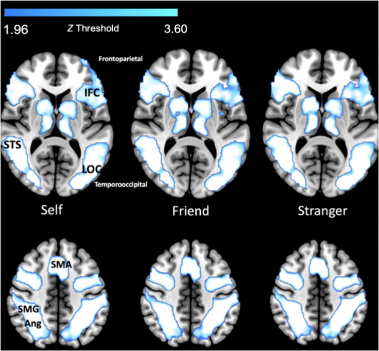
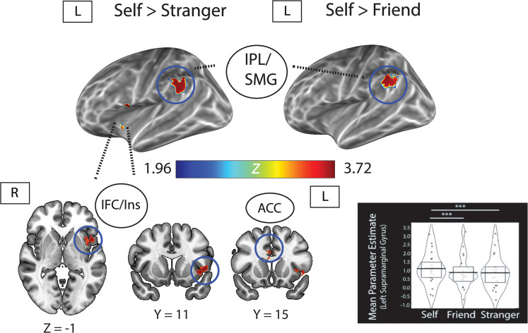
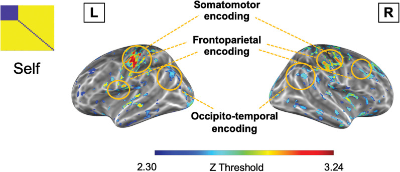
Humans can recognize their whole-body movements even when displayed as dynamic dot patterns. The sparse depiction of whole-body movements, coupled with a lack of visual experience watching ourselves in the world, has long implicated nonvisual mechanisms to self-action recognition. Using general linear modeling and multivariate analyses on human brain imaging data from male and female participants, we aimed to identify the neural systems for this ability. First, we found that cortical areas linked to motor processes, including frontoparietal and primary somatomotor cortices, exhibit greater engagement and functional connectivity when recognizing self-generated versus other-generated actions. Next, we show that these regions encode self-identity based on motor familiarity, even after regressing out idiosyncratic visual cues using multiple regression representational similarity analysis. Last, we found the reverse pattern for unfamiliar individuals: encoding localized to occipitotemporal visual regions. These findings suggest that self-awareness from actions emerges from the interplay of motor and visual processes.

Keywords: actions; motor; neuroimaging; self-awareness.


Conflict of interest statement

The authors declare no competing financial interests.

Figures

Figure 1.
Figure 1.
Trial structure including timing. Participants centrally attended to a white fixation cross until the action (self/friend/other) appeared for 5 s. On a subsequent screen, participants were then provided 2 s to make their identity judgment, followed by the variable ITI (mean centered at 5 s). The response order of self, friend, and other was counterbalanced in order to reduce any impact of motor order.
Figure 2.
Figure 2.
Left panel, Representational (dis)similarity matrices (RDMs) used for each RDA averaged across participants. RDMs reflect the Euclidean distance between identity and action categories for speed, movement distinctiveness, and body structure. For motor familiarity, identity was based on the degree of motor dissimilarity to oneself (self-generated actions, i.e., verbal instruction: zero dissimilarity; self-imitated actions, i.e., visual instruction: small dissimilarity, 0.3; friend actions: medium dissimilarity, 0.6; strangers: most dissimilarity, 1). Brighter colors for all RDMs indicate more dissimilarity. Top right panel, Upper triangular pairwise dissimilarity (1, Spearman's rho) between each of the group-level RDMs. Brighter colors indicate more dissimilarity. Bottom right panel, DTW figure showing movement trajectory of one joint from one actor's action time series (shown as red dots indicating locations) with lines measuring similarity to the corresponding joint in another actor's time series (shown as green dots) to find the optimal decrease in dissimilarity over time.
Figure 3.
Figure 3.
Behavioral results of identity recognition accuracy. Top, Self-recognition performance for different actions color coded by action type (verbal instruction, gray; visual instruction, blue). Light gray fill indicates bar plots for verbal instruction. Light blue fill indicates bar plot for visual instruction. Inference bands denote 95% Bayesian highest density interval with 1,000 iterations. Horizontal blue line indicates chance-level recognition accuracy (0.33). Bottom left panel, Confusion matrix for each identity. No significant misattributions were found for the self relative to other identities, though friend and stranger were more confused relative to the self (∼55% increase in misattributions for friend and strangers). Bottom right panel, Average recognition accuracy for each identity. All identities were recognized significantly above chance. Self actions were recognized significantly better than friend actions. Light gray fill indicates bar plots. Inference bands denote 95% Bayesian highest density interval with 1,000 iterations. Horizontal blue line indicates chance-level recognition accuracy (0.33). *p < 0.05, **p < 0.01, ***p < 0.001.
Figure 4.
Figure 4.
Group-level activity obtained using FSL's nonparametric permutation approach (randomise) with TFCE, p < 0.05. From left to right, Self versus baseline; friend versus baseline; and stranger versus baseline. +Large cluster sizes were obtained with TFCE due to the optimal cluster-defining threshold; hence cluster peaks are reported with visual interpolation using manual thresholding with a sliding scale. Abbreviations: IFC, inferior frontal cortex; STS, superior temporal sulcus; LOC, lateral occipital cortex; SMA, supplementary motor area; SMG, supramarginal gyrus; Ang, angular gyrus.
Figure 5.
Figure 5.
Univariate group-level activity for self > stranger (left) and self > friend (right) using the FSL randomise permutation approach, cluster corrected with TFCE (p < 0.05). Violin plot shows mean parameter estimates (PE) for the left posterior supramarginal gyrus (SMG) for all identities. The left SMG significantly discriminated contrasts of PE for both self versus stranger (p = 0.001) and self versus friend (p = 0.005), but not friend versus stranger (p = 0.821). Extended Data Figures 5-1 and 5-3 report the activity maps and peak clusters for both TFCE contrasts, as well as RFT cluster-corrected results (Extended Data Figs. 5-2 and 5-5). Abbreviations: IFC, inferior frontal cortex; Ins, insula; IPL, inferior parietal lobule; ACC, anterior cingulate cortex.
Figure 6.
Figure 6.
Task-modulated functional connectivity of left and right IPL. Left IPL (top panel) seed showed increased connectivity with bilateral occipitotemporal regions, bilateral superior and inferior parietal areas, and bilateral inferior frontal cortex during self > stranger. For self > friend, functional connectivity analysis revealed greater connectivity with the bilateral inferior frontal cortices and occipitotemporal regions. Task-modulated functional connectivity of the right IPL (bottom panel) showed a similar activity pattern to the left: strengthened frontoparietal and parieto-occipital connectivity for both contrasts. All activity cluster corrected at Z > 2.3, p < 0.01. Abbreviations: IPL, inferior parietal lobule; IPS, intraparietal sulcus; IFC, inferior frontal cortex; OT, occipitotemporal regions; EBA, extrastriate body area; STS, superior temporal sulcus.
Figure 7.
Figure 7.
Multiple regression searchlight RDA results for motor familiarity. This figure depicts the z-transformed activity map for significant correlations between the motor familiarity RDM and the neural RDM based on activity patterns for actions (self encoded as least dissimilar, with action separation to account for motor familiarity between action types; friend as medium dissimilarity, stranger as most), after accounting for speed and movement distinctiveness (DTW). Activation map reflects brain activity after 10,000 nonparametric Monte Carlo simulations, using TFCE and p < 0.01. Regions, bilateral somatomotor cortex: primary motor cortex, primary somatosensory cortex, superior parietal lobule; frontoparietal cortex: inferior parietal lobule, inferior frontal cortex, medial prefrontal cortex; occipitotemporal cortex: inferior temporal cortex, superior temporal sulcus and gyrus. All activity patterns are reported in Extended Data Table 7-1.
Figure 8.
Figure 8.
Multiple regression searchlight RDA results for each identity (self, friend, stranger). Activation maps reflect TFCE-corrected brain activity after 10,000 nonparametric Monte Carlo simulations, p< 0.01 for self and friend; p < 0.05 for stranger. Dissimilarity matrices reflect dissimilarity based on identity across all actions. Regions, Frontoparietal: inferior parietal lobule; superior frontal gyrus, lateral and medial prefrontal cortices. Somatomotor: primary motor cortex (M1), primary somatosensory cortex (S1). Occipitotemporal: superior temporal sulcus, middle temporal gyrus, extrastriate body area. Activity patterns are reported in Extended Data Tables 8-1–8-4 and Figure 8-1.
Figure 9.
Figure 9.
Multiple regression searchlight RDA results for self-identity, regressing out motor responses. Activation maps reflect TFCE-corrected brain activity after 10,000 nonparametric Monte Carlo simulations, p < 0.01 for self. Dissimilarity matrix reflects dissimilarity based on self-identity across all actions. Regions, frontoparietal: inferior parietal lobule; superior frontal gyrus, lateral and medial prefrontal cortices. Somatomotor: primary motor cortex (M1), primary somatosensory cortex (S1). Occipitotemporal: superior temporal sulcus, middle temporal gyrus, extrastriate body area. Activity patterns are reported in Extended Data Table 9-1.

Similar articles

References

    1. Abdulkarim Z, Guterstam A, Hayatou Z, Ehrsson HH (2023) Neural substrates of body ownership and agency during voluntary movement. J Neurosci 43:2362–2380. 10.1523/JNEUROSCI.1492-22.2023 - DOI - PMC - PubMed
    1. Apps MA, Tsakiris M (2014) The free-energy self: a predictive coding account of self-recognition. Neurosci Biobehav Rev 41:85–97. 10.1016/j.neubiorev.2013.01.029 - DOI - PMC - PubMed
    1. Arbib MA (1981) Perceptual structures and distributed motor control. In: Handbook of physiology, section I: the nervous system, vol. 2: motor control (Brooks VB, ed.), pp 1449–1480. Baltimore: Williams and Wilkins.
    1. Arbib MA (1992) Schema theory. In: The encyclopedia of artificial intelligence, Vol. 2, pp 1427–1443. New York, NY: Wiley Interscience.
    1. Asakage S, Nakano T (2023) The salience network is activated during self-recognition from both first-person and third-person perspectives. Hum Brain Mapp 44:559–570. 10.1002/hbm.26084 - DOI - PMC - PubMed

LinkOut - more resources