. 2025 Jan 1;45(1):e0606242024.
doi: 10.1523/JNEUROSCI.0606-24.2024.

Human iPSC-Derived MSCs Induce Neurotrophic Effects and Improve Metabolic Activity in Acute Neuronal Injury Models

Affiliations

Human iPSC-Derived MSCs Induce Neurotrophic Effects and Improve Metabolic Activity in Acute Neuronal Injury Models

Keiji Kawatani et al. J Neurosci. .

Abstract

Mesenchymal stromal cell (MSC) therapy has regenerative potentials to treat various pathological conditions including neurological diseases. MSCs isolated from various organs can differentiate into specific cell types to repair organ damages. However, their paracrine mechanisms are predicted to predominantly mediate their immunomodulatory, proangiogenic, and regenerative properties. While preclinical studies highlight the significant potential of MSC therapy in mitigating neurological damage from stroke and traumatic brain injury, the variability in clinical trial outcomes may stem from the inherent heterogeneity of somatic MSCs. Accumulating evidence has demonstrated that induced pluripotent stem cells (iPSCs) are an ideal alternative resource for the unlimited expansion and biomanufacturing of MSCs. Thus, we investigated how iPSC-derived MSCs (iMSCs) influence properties of iPSC-derived neurons. Our findings demonstrate that the secretome from iMSCs possesses neurotrophic effects, improving neuronal survival and promoting neuronal outgrowth and synaptic activity in vitro. Additionally, the iMSCs enhance metabolic activity via mitochondrial respiration in neurons, both in vitro and in mouse models. Glycolytic pathways also increased following the administration of iMSC secretome to iPSC-derived neurons. Consistently, in vivo experiments showed that intravenous administration of iMSCs compensated for the elevated energetic demand in male mice with irradiation-induced brain injury by restoring synaptic metabolic activity during acute brain damage. 18F-FDG PET imaging also detected an increase in brain glucose uptake following iMSC administration. Together, our results highlight the potential of iMSC-based therapy in treating neuronal damage in various neurological disorders, while paving the way for future research and potential clinical applications of iMSCs in regenerative medicine.

Keywords: MSC; iPSC; neuron; neurotrophic effect; stem cell therapy.


Conflict of interest statement

The authors declare no competing financial interests.

Figures

Figure 1.
Figure 1.
Conditioned medium from iMSCs induces neurotrophic effects on iPSC-derived neurons. A, Schematic representation of the differentiation timeline from iPSCs to iPSC-derived MSCs (iMSCs). B, Flow cytometry analysis showing that over 95% of iMSCs are positive for MSC markers CD105, CD73, and CD90. C, The iPSC-derived neurons were cultured in the presence of normal neuron media, serum-free DMEM/F12 media, or cocultured with iMSCs for 1 week in serum-free DMEM/F12 media using a trans-well chamber from 1 week after the differentiation from NPCs. The iPSC-derived neurons were immunostained for βIII-tubulin. Nuclei were stained with DAPI. Scale bar, 200 µm. D, The number of βIII-tublin-positive iPSC-derived neurons were quantified and normalized to cell number/field. Data represents mean ± SEM (D; n = 6 technical replicates/group). E, The iPSC-derived neurons were cultured in normal neuron media or iMSC conditioned media for 3 weeks after differentiation from NPCs. The iMSC conditioned media was used from 1 week after the differentiation. iPSC-derived neurons were immunostained for βIII-tubulin. Nuclei were stained with DAPI. Scale bar, 200 µm. Total axon length (F) and branch number (G) of iPSC-derived neurons were quantified. H, Sholl analysis was performed from the soma to 500 µm distance using a step size of 10 in the iPSC-derived neurons. Data represents mean ± SEM (F–H; n = 30 neurons/group) **p < 0.01, ****p < 0.0001 by one-way ANOVA with Tukey's correction (D), two-tailed Student's t test (F, G), or ****p <0.0001 by two-tailed Wilcoxon test (H).
Figure 2.
Figure 2.
Conditioned medium from iMSCs accelerates spontaneous electrical activity and synaptic network formation in iPSC-derived neurons. A, Spontaneous firing patterns in iPSC-derived neurons were measured 4 weeks after differentiation from NPCs in the presence of normal neuron media or iMSC conditioned media (iMSC CM). B, The frequency of spontaneous firing was monitored in the iPSC neurons for 5 weeks after differentiation (n = 11–12 technical replicates/group). C, Extracellular recordings of spontaneous firing including synchronized burst firing (SBF; red rectangles) in the iPSC neurons were measured 4 weeks after differentiation in the presence of normal neuron media or iMSC conditioned media. D, The incidence of SBF was monitored in the iPSC-derived neurons for 5 weeks after the differentiation. Data represents mean ± SEM (n = 11–12 technical replicates/group). *p < 0.05, ****p < 0.0001 by two-tailed Student's t test.
Figure 3.
Figure 3.
Conditioned medium from iMSCs activates mitochondrial respiration and glycolysis in iPSC-derived neurons. A, B, The oxygen consumption rate (OCR) in iPSC-derived neurons was measured in the presence of normal neuron media or iMSC conditioned media 6 weeks after differentiation from NPCs. The iMSC conditioned media (iMSC CM) was used from 1 week after the differentiation of iPSC-derived neurons. The OCR measurements were normalized with cell density determined by nuclear DNA staining with DAPI (n = 14 technical replicates/group). A.U., arbitrary unit. C, D, Glycolysis in the iPSC-derived neurons was measured in the presence of normal or iMSC conditioned media 6 weeks after the differentiation. The ECAR measurements were normalized with cell density determined by nuclear DNA staining with DAPI. Data represents mean ± SEM (n = 12 technical replicates/group). *p < 0.05, **p < 0.01, ****p < 0.0001 by two-tailed Student's t test.
Figure 4.
Figure 4.
Conditioned medium from iMSCs improves neuronal outgrowth in damaged iPSC-derived neurons. A, The iPSC-derived neurons were irradiated with a 1 Gy dose 1 week after the differentiation from NPCs in the presence of control media or iMSC conditioned medium (iMSC CM) for 5 d, followed by immunostaining for βIII-tubulin (green) and DAPI (blue). Scale bar, 50 µm. Total neurite length (B; n = 53–56 neurons/group), branch length (C; n = 35–36 neurons/group), and centroid–root distance (D; n = 53–56 neurons/group) were quantified in the irradiated iPSC-derived neurons treated with control media or iMSC CM. E, The iPSC-derived neurons were cultured with a 0.1 µM antimycin A 1 week after the differentiation from NPCs for 2 weeks in the presence of control media or iMSC conditioned medium (iMSC CM), followed by immunostaining for βIII-tubulin (green) and DAPI (blue). Scale bar, 50 µm. Total neurite length (F; n = 43–64 neurons/group), branch length (G; n = 36 neurons/group), and centroid–root distance (H; n = 43–64 neurons/group) were quantified in the iPSC-derived neurons treated with control media or iMSC CM. Data represent mean ± SEM. **p < 0.01, ****p < 0.0001 by two-tailed Student's t test.
Figure 5.
Figure 5.
iMSC secretome ameliorates IR-induced impairment of mitochondrial respiration in mouse synaptosomes. A, Male wild-type mice were treated with intravenous injection of saline (200 μl/each) or iMSCs (1 × 106 cells in 200 μl saline/each) 24 h after whole brain irradiation (IR) at 15 Gy. The synaptosomes were isolated from the mouse brains 7 d after IR and subjected to mitochondria respiration analysis. B, C, Oxygen consumption rate (OCR) of synaptosomes (15 μg/each) was measured by Mito Stress Test Kit through Seahorse XFe96 Extracellular Flux Analyzer. Synaptosomes extracted from mouse brains without IR administration were used as controls. Data represents mean ± SEM (n = 8 technical replicates from 4 mice/group). D, The synaptosomes were isolated from male wild-type mice, irradiated at 4 Gy in the presence of control media or iMSC conditioned medium (iMSC CM), and subjected to mitochondria respiration analysis. E, F, OCR of synaptosomes (15 μg/each) was measured 2 h after the IR. Data represents mean ± SEM (n = 6 technical replicates from 3 mice/group). A.U., arbitrary unit. *p < 0.05, **p < 0.01, ***p < 0.0001, ****p < 0.0001 by one-way ANOVA with Tukey's correction (C) or two-tailed Student's t test (F).
Figure 6.
Figure 6.
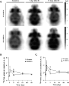
iMSC administration modulates brain glucose uptake after radiation-induced brain injury. Male wild-type mice were treated with intravenous injection of saline (200 μl/each) or iPSC-derived MSCs (iMSCs; 1 × 106 cells in 200 μl saline/each) 24 h after whole brain irradiation (IR) at 15 Gy. A, Representative 18F-FDG PET images of mouse brains before administration or IR (baseline) and at 1 and 7 d after irradiation were shown. These images demonstrate temporary increased 18F-FDG uptake at 24 h after irradiation (SUVmean of 3.1) with resolution to baseline at Day 7 (SUVmean of 2.2 and 2.5, respectively). Conversely, 18F-FDG PET in mouse brain treated with iMSC demonstrates persistently increased 18F-FDG at 24 and 7 d (SUVmean of 3.7 and 3.4, respectively) compared with baseline (SUVmean of 1.7). B, C, Summed 18F-FDG uptake in the cerebrum (B) and cerebellum (C) in the mice treated with saline or iMSC was measured at baseline and 1, 7, 21, and 35 d after the whole brain IR and normalized to that in liver at each time point. Data represents mean ± SEM (N = 5 mice/group). *p < 0.05 by two-tailed Student's t test.

References

    1. Ankrum JA, Ong JF, Karp JM (2014) Mesenchymal stem cells: immune evasive, not immune privileged. Nat Biotechnol 32:252–260. 10.1038/nbt.2816 - DOI - PMC - PubMed
    1. Arakawa M, et al. (2023) iPSC-derived mesenchymal stem cells attenuate cerebral ischemia-reperfusion injury by inhibiting inflammatory signaling and oxidative stress. Mol Ther Methods Clin Dev 30:333–349. 10.1016/j.omtm.2023.07.005 - DOI - PMC - PubMed
    1. Baak LM, et al. (2022) Feasibility and safety of intranasally administered mesenchymal stromal cells after perinatal arterial ischaemic stroke in the Netherlands (PASSIoN): a first-in-human, open-label intervention study. Lancet Neurol 21:528–536. 10.1016/S1474-4422(22)00117-X - DOI - PubMed
    1. Badyra B, Sulkowski M, Milczarek O, Majka M (2020) Mesenchymal stem cells as a multimodal treatment for nervous system diseases. Stem Cells Transl Med 9:1174–1189. 10.1002/sctm.19-0430 - DOI - PMC - PubMed
    1. Bloor AJC, et al. (2020) Production, safety and efficacy of iPSC-derived mesenchymal stromal cells in acute steroid-resistant graft versus host disease: a phase I, multicenter, open-label, dose-escalation study. Nat Med 26:1720–1725. 10.1038/s41591-020-1050-x - DOI - PubMed

Publication types

LinkOut - more resources