

Tumor growth and vascular redistribution contributes to the dosimetric preferential effect of microbeam radiotherapy: a Monte Carlo study
- PMID: 39496724
- PMCID: PMC11535247
- DOI: 10.1038/s41598-024-77415-5
Tumor growth and vascular redistribution contributes to the dosimetric preferential effect of microbeam radiotherapy: a Monte Carlo study
Abstract
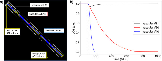
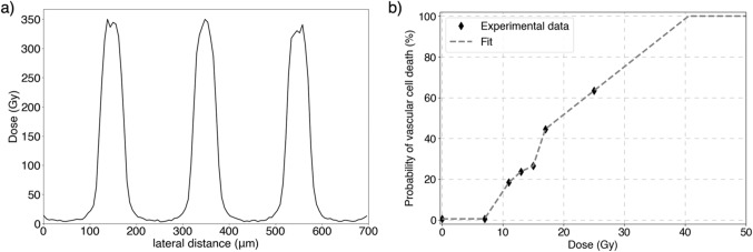
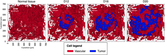
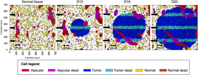
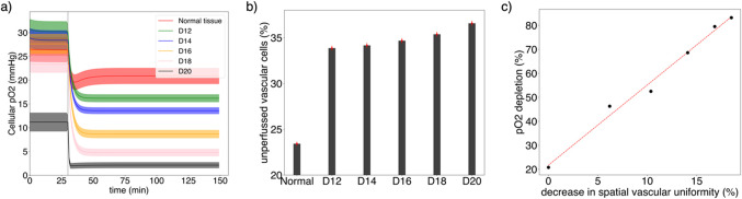
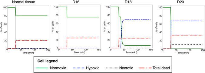
The radiobiological mechanisms behind the favorable response of tissues to microbeam radiation therapy (MRT) are not fully described yet. Among other factors, the differential action to tumor and normal tissue vasculature is considered to contribute to MRT efficacy. This computational study evaluates the relevance of tumor growth stage and associated vascular redistribution to this effect. A multiscale approach was employed with two simulation softwares: TOPAS and CompuCell3D. Segmentation images of the angioarchitecture of a non-bearing tumor mouse brain were used. The tumor vasculature at different tumor growth stages was obtained by simulating the tumor proliferation and spatial vascular redistribution. The radiation-induced damage to vascular cells and consequent change in oxygen perfusion were simulated for normal and tumor tissues. The multiscale model showed that oxygen perfusion to tissues and vessels decreased as a function of the tumor proliferation stage, and with the decrease in uniformity of the vasculature spatial distribution in the tumor tissue. This led to an increase in the fraction of hypoxic (up to 60%) and necrotic (10%) tumor cells at advanced tumor stages, whereas normal tissues remained normoxic. These results showed that tumor stage and spatial vascular distribution contribute to the preferential effect of MRT in tumors.
Keywords: Computational modeling; Microbeam radiotherapy; Spatially fractionated radiotherapy; Vasculature.
© 2024. The Author(s).
Conflict of interest statement
The authors declare no competing interests.
Figures
Similar articles
-
Microbeam radiation therapy alters vascular architecture and tumor oxygenation and is enhanced by a galectin-1 targeted anti-angiogenic peptide.Radiat Res. 2012 Jun;177(6):804-12. doi: 10.1667/rr2784.1. Epub 2012 May 18. Radiat Res. 2012. PMID: 22607585 Free PMC article.
-
A voxel-based multiscale model to simulate the radiation response of hypoxic tumors.Med Phys. 2015 Jan;42(1):90-102. doi: 10.1118/1.4903298. Med Phys. 2015. PMID: 25563250
-
Physics study of microbeam radiation therapy with PSI-version of Monte Carlo code GEANT as a new computational tool.Med Phys. 2000 Jul;27(7):1664-75. doi: 10.1118/1.599034. Med Phys. 2000. PMID: 10947271
-
Effects of pulsed, spatially fractionated, microscopic synchrotron X-ray beams on normal and tumoral brain tissue.Mutat Res. 2010 Apr-Jun;704(1-3):160-6. doi: 10.1016/j.mrrev.2009.12.003. Epub 2009 Dec 23. Mutat Res. 2010. PMID: 20034592 Review.
-
Effects of microbeam radiation therapy on normal and tumoral blood vessels.Phys Med. 2015 Sep;31(6):634-41. doi: 10.1016/j.ejmp.2015.04.014. Epub 2015 May 23. Phys Med. 2015. PMID: 26004351 Review.
Cited by
-
Simulation approach for common female cancers: a brief review.Front Oncol. 2025 Jul 9;15:1479225. doi: 10.3389/fonc.2025.1479225. eCollection 2025. Front Oncol. 2025. PMID: 40703544 Free PMC article. Review.
References
-
- Slatkin, D. N., Spanne, P., Dilmanian, F. A. & Sandborg, M. Microbeam radiation therapy. Med. Phys. 19, 1395–1400. 10.1118/1.596771 (1992). - PubMed
-
- Bräuer-Krisch, E. et al. Effects of pulsed, spatially fractionated, microscopic synchrotron X-ray beams on normal and tumoral brain tissue. Mutat. Res. 704, 1–3. 10.1016/j.mrrev.2009.12.003 (Jun. 2010). - PubMed
-
- Schültke, E. et al. Memory and survival after microbeam radiation therapy, Eur. J. Radiol. 68 (3 Suppl), S142-146 10.1016/j.ejrad.2008.04.051 (2008). - PubMed
-
- Serduc, R. et al. Characterization and quantification of cerebral edema induced by synchrotron x-ray microbeam radiation therapy. Phys. Med. Biol. 53 (5), 1153–1166. 10.1088/0031-9155/53/5/001 (2008). - PubMed
