. 2024 Nov 4;8(1):250.
doi: 10.1038/s41698-024-00736-1.

ERO1A levels are a prognostic indicator in EGFR mutated non small cell lung cancer

Affiliations

ERO1A levels are a prognostic indicator in EGFR mutated non small cell lung cancer

M A Voronkova et al. NPJ Precis Oncol. .

Abstract

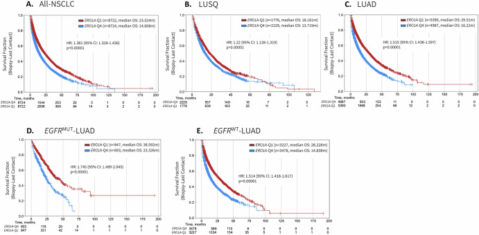
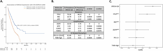
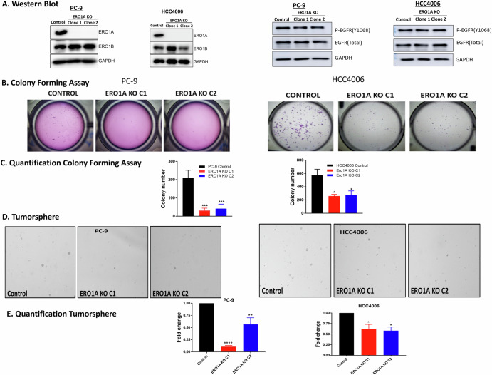
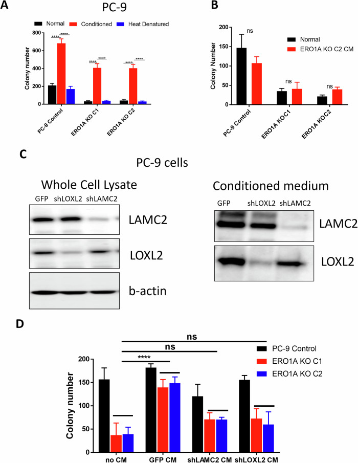
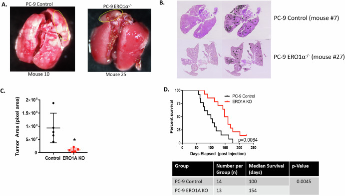
We have identified endoplasmic reticulum oxidoreductase 1 alpha (ERO1A) as a poor prognostic indicator in epidermal growth factor receptor (EGFR)-mutated non-small cell lung cancer (EGFRMUT-NSCLC). In addition, comparison of high versus low ERO1A expression among cohorts of EGFRMUT-NSCLC primary samples revealed that ERO1A expression correlated with increased expression of proteins that regulate secretion. Using the CPTAC proteomic data set in lung adenocarcinoma we found that high ERO1A protein expression correlated with both extracellular matrix and matrix modifying enzymes. In this report, we found that ablating ERO1A expression was a determinant of clonogenicity, tumor sphere formation, spheroid growth and growth in vivo, as well as response to Osimertinib. We validated that ERO1A-knockout EGFRMUT-LUAD cell lines demonstrated a reduction in secretion of both laminin gamma 2 (LAMC2) and the collagen modifying enzyme lysyl oxidase-like 2 (LOXL2). Our work supports the role of ERO1A in modulating the tumor microenvironment that is likely to contribute to tumor progression.


Conflict of interest statement

L.A.H. is co-founder of Modulation Therapeutics but this work is not related to the current pipeline.

Figures

Fig. 1
Fig. 1. ERO1A is associated with poor prognosis in NSCLC.
High expression of ERO1A is associated with poor prognosis in all-NSCLC(A), when considering squamous (B) and adenocarcinoma histology (C) and when accounting for EGFR mutation status (D, E). ERO1A quartiles are based on expression in all-NSCLC patients and were consistently applied across all subtypes of NSCLC.
Fig. 2
Fig. 2. Multivariate analysis to test the association of ERO1A expression with overall survival (OS).
ERO1A quartile cutoffs were determined based on the expression of ERO1A in EGFRmt-LUAD. A Patients with tumors expressing high ERO1A levels have increased survival. B Molecular alterations associated with ERO1A expression prevalent in either cohort at ≥2% and statistically significantly different are shown in the table. C Molecular alterations with a prevalence difference of at least 5% between the cohorts were incorporated as co-variates in a complete-case multivariate analysis. High ERO1A expression and TP53 mutations were independently associated with poor OS in EGFRmt-LUAD (n = 960).
Fig. 3
Fig. 3. ERO1A levels correlate with changes in protein secretion.
High expression of ERO1A was associated with an enrichment of a number of Hallmark Pathways as revealed by GSEA. The second most significant pathway was associated with regulating protein secretion (A). The association of ERO1A expression was established with the expression of genes involved in Hallmark protein secretion pathway. Genes were sorted based on their Spearman correlation with ERO1A and are listed to the right side of the heatmap (B). Subset of genes involved in vesicle trafficking from ER to Golgi as well as regulation of exosomes were investigated and observed to be significantly enriched in ERO1A-Q4 tumors (C). Correlation between ERO1A and levels of other proteins downloaded from CPTAC, a publicly available lung adenocarcinoma tumor proteomics dataset, were analyzed using LinkedOmics (http://linkedomics.org/data_download/CPTAC-LUAD/) (D). 7 out of 28 targets with highest correlation to ERO1A levels are extracellular matrix proteins (E).
Fig. 4
Fig. 4. Depletion of ERO1A does not alter EGFR activation, yet decreases clonogenicity and tumor sphere formation.
Shown is a representative image demonstrating CRISPR deletion of ERO1A in PC-9 and HCC006 cell line. ERO1B expression levels do not compensate for loss of ERO1A. Representative western blots demonstrate that ERO1A KO does not affect total levels of EGFR or p-EGFR in two EGFRMUT-NSCLC cell lines (A). ERO1A KO cells form smaller number of colonies in soft agar and on tissue culture plates. Representative fields of view from one independent experiment are shown. ***p = 0.0004 by one-way ANOVA, *p = 0.0111 by repeated measures ANOVA, n = 3–4 independent experiments performed in triplicates (B, C). ERO1A KO clones reveal a decrease in total tumor sphere formation in both PC-9 and HCC4006 cell lines. Representative fields of view from one independent experiment are shown. Shown is the means of 3-4 independent experiments performed in triplicates. Numbers are mean ± SEM. ****p < 0.0001, **p = 0.0072, *p < 0.05; Dunnett’s multiple comparisons test following one-way ANOVA (DE).
Fig. 5
Fig. 5. ERO1A protein levels correlate with levels of LOXL2 and LAMC2 protein levels.
Levels of LAMC2 and LOXL2 are dramatically reduced in conditioned medium collected from ERO1A KO cells. Representative western blots on whole cell lysates and conditioned media in PC-9 cell line are shown (A). Concentration of LAMC2 secreted into conditioned media by ERO1A KO cell lines is reduced compared to control cells. ELISA was performed in triplicate and the average ± S.D. of n = 3 combined results are shown. ****p < 0.0001; Dunnett’s multiple comparisons test following one-way ANOVA (BC). Representative images of immunofluorescence staining for LOXL2 and LAMC2 in HCC4006 spheroids, scale bar - 50 µm (DE). Quantification of LOXL2 and LAMC2 immunofluorescent staining in control and ERO1A KO HCC4006 cells cultured as tumor spheres. *p = 0.0209 by two-tailed unpaired t-test, n = 5, experiment was repeated 3 times. ns – not significant (F).
Fig. 6
Fig. 6. Tumorigenic properties of ERO1A are mediated in part by a secreted factor.
Soft agar colony formation assay using regular non-conditioned medium (Normal), medium conditioned by control cells (Conditioned) and conditioned medium from control cells after heat denaturation (Heat denatured), ****p < 0.0001, two-way ANOVA followed by Sidak’s multiple comparisons test, n = 3 (A). Conditioned medium generated by ERO1A KO cells does not rescue colony formation abilities of ERO1A KO cells (B). Representative immunoblot showing level of LAMC2 and LOXL2 knockdown in PC-9 cells. LAMC2 or LOXL2 knockdown dramatically decrease levels of secreted LAMC2 and LOXL2, accordingly (C). Conditioned medium generated by shLOXL2 and shLAMC2 PC-9 cells partially rescue colony formation abilities of ERO1A KO cells in soft agar assay. GFP—empty vector control. Data are means ± SD. ****p < 0.0001, Tukey’s multiple comparisons test following two-way ANOVA. ns – not significant (D).
Fig. 7
Fig. 7. ERO1A expression is associated with increased tumor burden and decreased overall survival in SCID-Beige mice bearing EGFRMUT-LUAD tumors.
Picture of tumors formed and H & E staining from the lungs of ERO1A replete and KO mice. H & E was performed on 5 random lungs from each group selected by a random number generator and tumor margins quantified using ImageJ (A, B). Each data point represents the average on three sections from each mouse. *p = 0.0110, unpaired t-test (C). Kaplan–Meier curve showing that ERO1A KO increased overall survival compared to the control group. p = 0.0064, log-rank (Mantel-Cox) test (D).

References

    1. Johnson, B. D., Geldenhuys, W. J. & Hazlehurst, L. A. The role of ERO1alpha in modulating cancer progression and immune escape. J. Cancer Immunol. (Wilmington)2, 103–115 (2020). - PMC - PubMed
    1. Hayes, K. E. et al. Inhibition of the FAD containing ER oxidoreductin 1 (Ero1) protein by EN-460 as a strategy for treatment of multiple myeloma. Bioorg. Med. Chem.27, 1479–1488 (2019). - PMC - PubMed
    1. Liu, L. et al. Ablation of ERO1A induces lethal endoplasmic reticulum stress responses and immunogenic cell death to activate anti-tumor immunity. Cell Rep. Med.4, 101206 (2023). - PMC - PubMed
    1. Gupta, N. et al. ERO1α promotes hypoxic tumor progression and is associated with poor prognosis in pancreatic cancer. Oncotarget10, 5970–5982 (2019). - PMC - PubMed
    1. Yang, S. et al. Endoplasmic reticulum resident oxidase ERO1-Lalpha promotes hepatocellular carcinoma metastasis and angiogenesis through the S1PR1/STAT3/VEGF-A pathway. Cell Death Dis.9, 1105 (2018). - PMC - PubMed

LinkOut - more resources