

Lethal versus surviving sepsis phenotypes displayed a partly differential regional expression of neurotransmitters and inflammation and did not modify the blood-brain barrier permeability in female CLP mice
- PMID: 39497013
- PMCID: PMC11535104
- DOI: 10.1186/s40635-024-00688-7
Lethal versus surviving sepsis phenotypes displayed a partly differential regional expression of neurotransmitters and inflammation and did not modify the blood-brain barrier permeability in female CLP mice
Abstract
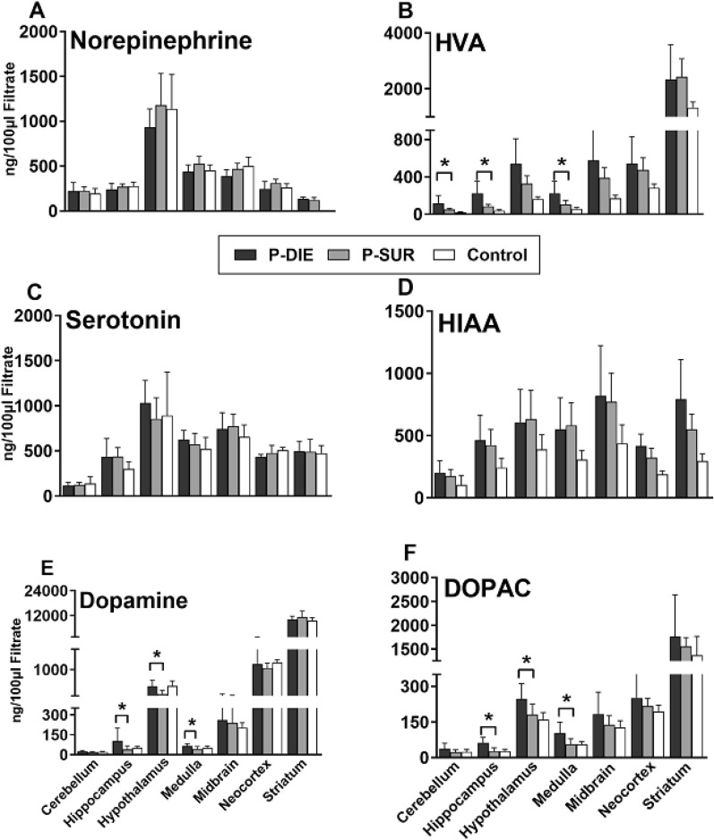
Background: Septic encephalopathy is frequent but its pathophysiology is enigmatic. We studied expression of neurotransmitters, inflammation and integrity of the blood-brain barrier (BBB) in several brain regions during abdominal sepsis. We compared mice with either lethal or surviving phenotype in the first 4 sepsis days. Mature CD-1 females underwent cecal ligation and puncture (CLP). Body temperature (BT) was measured daily and predicted-to-die (within 24 h) mice (for P-DIE; BT < 28 °C) were sacrificed together (1:1 ratio) with mice predicted-to-survive (P-SUR; BT > 35 °C), and healthy controls (CON). Brains were dissected into neocortex, cerebellum, midbrain, medulla, striatum, hypothalamus and hippocampus.
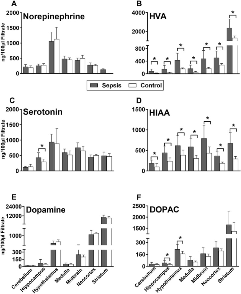
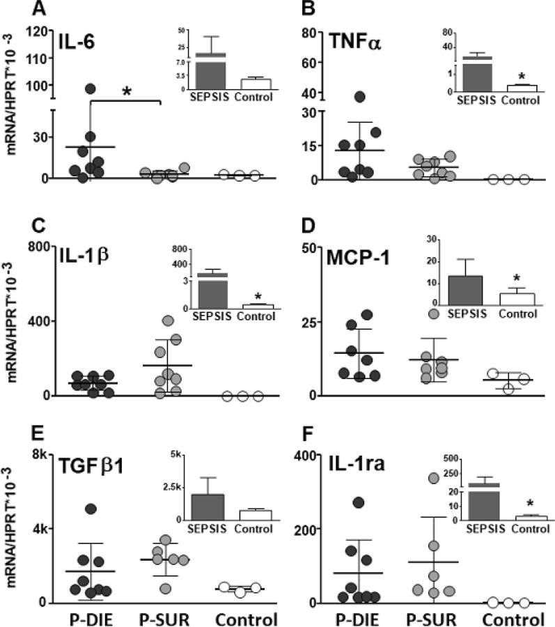
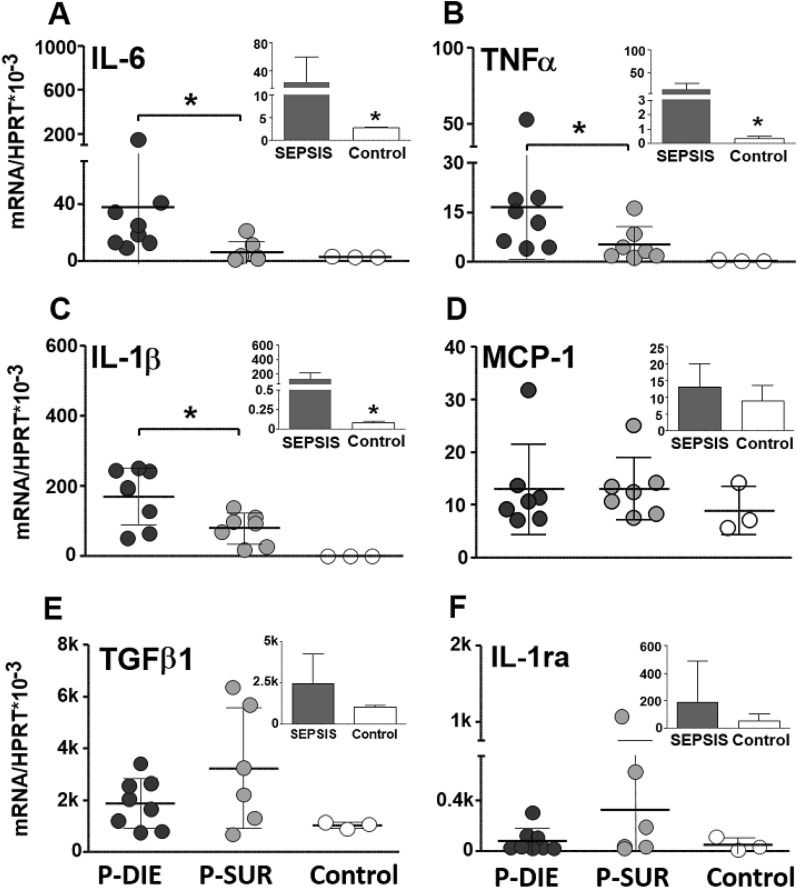
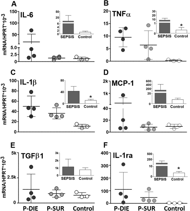
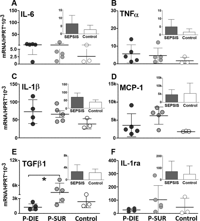
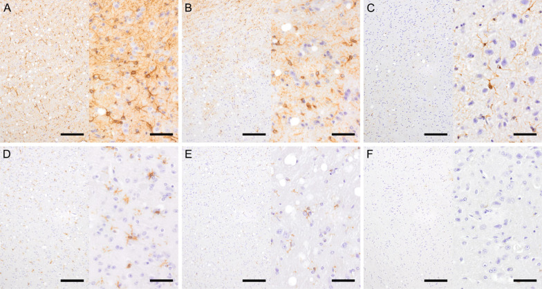
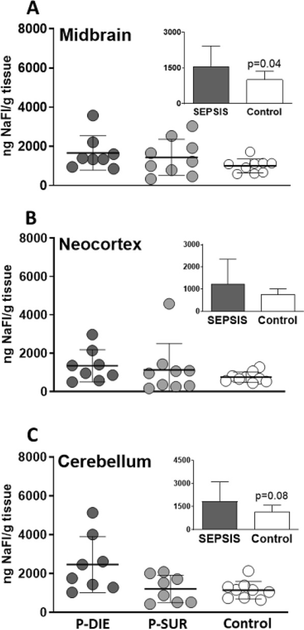
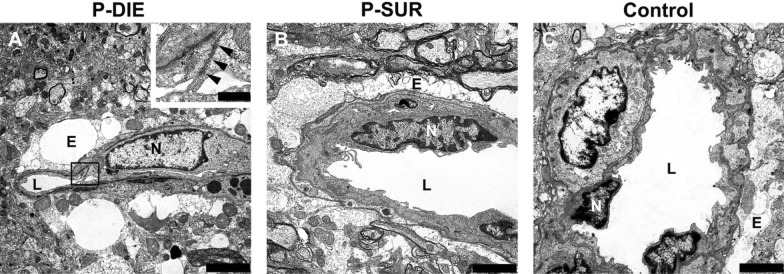
Results: CLP mice showed an up to threefold rise of serotonin in the hippocampus, 5-hydroxyindoleacetic and homovanillic acid (HVA) in nearly all regions vs. CON. Compared to P-SUR, P-DIE mice showed a 1.7 to twofold rise of HVA (386 ng/g of tissue), dopamine (265 ng/g) and 3,4-Dihydroxyphenylacetic acid (DOPAC; 140 ng/g) in the hippocampus, hypothalamus and medulla (174, 156, 82 ng/g of tissue, respectively). CLP increased expression of TNFα, IL-1β and IL-6 mRNA by several folds in the midbrain, cerebellum and hippocampus versus CON. The same cytokines were further elevated in P-DIE vs P-SUR in the midbrain and cerebellum. Activation of astrocytes and microglia was robust across regions but remained typically phenotype independent. There was a similar influx of sodium fluorescein across the BBB in both P-DIE and P-SUR mice.
Conclusions: Compared to survivors, the lethal phenotype induced a stronger deregulation of amine metabolism and cytokine expression in selected brain regions, but the BBB permeability remained similar regardless of the predicted outcome.
Keywords: Abdominal sepsis; Brain amines; Cytokines; Outcome prediction; Phenotyping.
© 2024. The Author(s).
Conflict of interest statement
The authors declare no competing interests.
Figures
References
-
- Gofton TE, Young GB (2012) Sepsis-associated encephalopathy. Nat Rev Neurol 8(10):557–566 - PubMed
-
- Papadopoulos MC, Davies DC, Moss RF, Tighe D, Bennett ED (2000) Pathophysiology of septic encephalopathy: a review. Crit Care Med 28(8):3019–3024 - PubMed
-
- Ebersoldt M, Sharshar T, Annane D (2007) Sepsis-associated delirium. Intensive Care Med 33:941–950 - PubMed
-
- Witt NJ, Zochodne DW, Bolton CF, Grand’Maison F, Wells G, Young GB, Sibbald WJ (1991) Peripheral nerve function in sepsis and multiple organ failure. Chest 99(1):176–184 - PubMed
Grants and funding
LinkOut - more resources
Full Text Sources
Miscellaneous
