. 2024 Nov 4;31(1):100.
doi: 10.1186/s12929-024-01088-5.

The molecular consequences of FOXF1 missense mutations associated with alveolar capillary dysplasia with misalignment of pulmonary veins

Affiliations

The molecular consequences of FOXF1 missense mutations associated with alveolar capillary dysplasia with misalignment of pulmonary veins

G G Edel et al. J Biomed Sci. .

Abstract

Background: Alveolar capillary dysplasia with misalignment of pulmonary veins (ACD/MPV) is a fatal congenital lung disorder strongly associated with genomic alterations in the Forkhead box F1 (FOXF1) gene and its regulatory region. However, little is known about how FOXF1 genomic alterations cause ACD/MPV and what molecular mechanisms are affected by these mutations. Therefore, the effect of ACD/MPV patient-specific mutations in the FOXF1 gene on the molecular function of FOXF1 was studied.

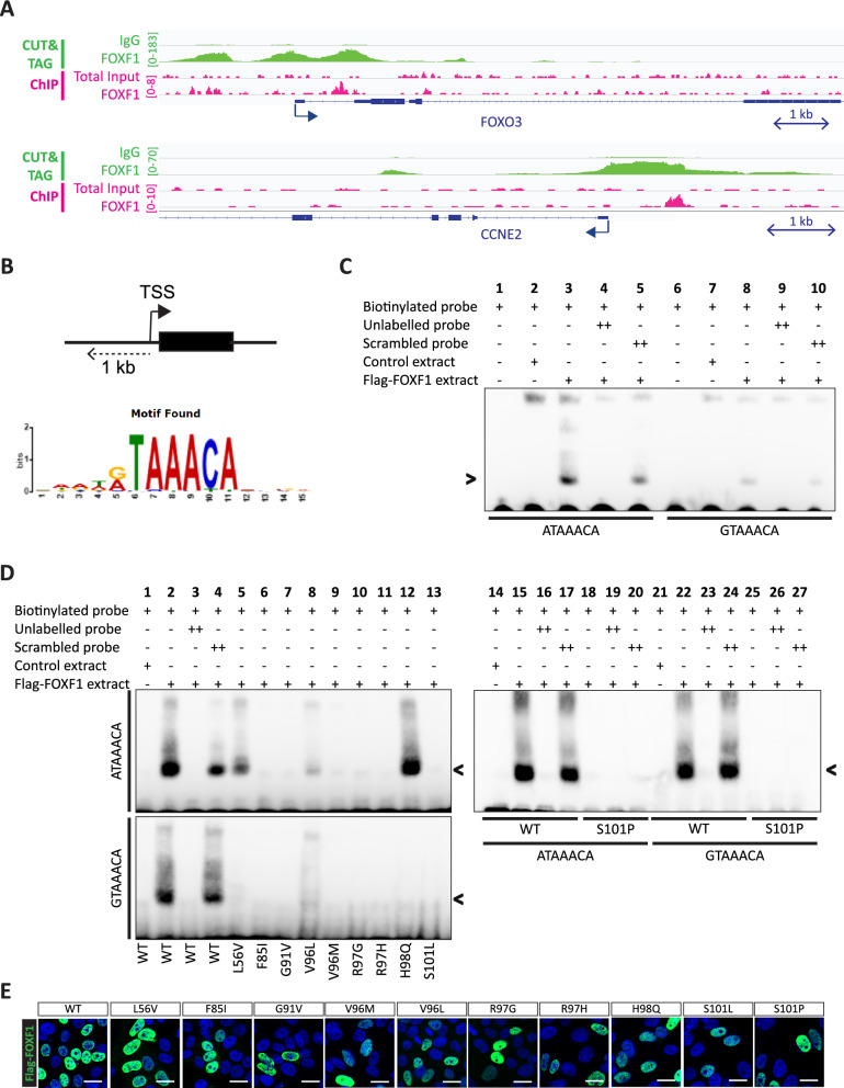
Methods: Epitope-tagged FOXF1 constructs containing one of the ACD/MPV-associated mutations were expressed in mammalian cell lines to study the effect of FOXF1 mutations on protein function. EMSA binding assays and luciferase assays were performed to study the effect on target gene binding and activation. Immunoprecipitation followed by SDS‒PAGE and western blotting were used to study protein‒protein interactions. Protein phosphorylation was studied using phos-tag western blotting.

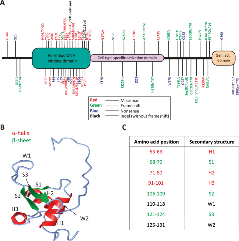
Results: An overview of the localization of ACD/MPV-associated FOXF1 mutations revealed that the G91-S101 region was frequently mutated. A three-dimensional model of the forkhead DNA-binding domain of FOXF1 showed that the G91-S101 region consists of an α-helix and is predicted to be important for DNA binding. We showed that FOXF1 missense mutations in this region differentially affect the DNA binding of the FOXF1 protein and influence the transcriptional regulation of target genes depending on the location of the mutation. Furthermore, we showed that some of these mutations can affect the FOXF1 protein at the posttranscriptional level, as shown by altered phosphorylation by MST1 and MST2 kinases.

Conclusion: Missense mutations in the coding region of the FOXF1 gene alter the molecular function of the FOXF1 protein at multiple levels, such as phosphorylation, DNA binding and target gene activation. These results indicate that FOXF1 molecular pathways may be differentially affected in ACD/MPV patients carrying missense mutations in the DNA-binding domain and may explain the phenotypic heterogeneity of ACD/MPV.

Keywords: ACD/MPV; Alveolar capillary dysplasia with misalignment of the pulmonary veins; DNA-binding domain; FOXF1; Missense mutations; Phosphorylation.


Conflict of interest statement

The authors declare that the research was conducted in the absence of any commercial or financial relationships that could be construed as potential conflicts of interest.

Figures

Fig. 1
Fig. 1
Distribution of FOXF1 ACD/MPV patient mutations in the different FOXF1 protein domains and predicted 3D model of the FOXF1 DNA binding domain. A Genomic ACD/MPV patient mutations in the coding region of the FOXF1 gene were translated to their corresponding amino acids to make an overview of the localization of the mutations in the FOXF1 protein. Forkhead DNA binding domain, cell-type specific activation domain and general activation domain (Gen. act. Domain) are shown in green, pink and orange, respectively. The locations of FOXF1 mutations are shown with lines and details of the mutations are shown next to them. Mutations were categorized in missense mutations (red), frameshifts (green), nonsense mutations (blue), insertion/deletions without a frameshift (Indel, black) and deletions (orange). B Predicted 3D model of the DNA binding domain of FOXF1, which consists of 2 wings (W), 3 α-helices (H) and 3 β-sheets (S). C Table of predicted secondary structures in the FOXF1 protein and their corresponding amino acid position in the FOXF1 protein
Fig. 2
Fig. 2
FLAG-tagged FOXF1 mutants have no or diminished binding to the FOXF1 binding motif. A FOXF1 ChIP-seq (pink) and CUT&TAG (green) analysis was performed in ECFCs and identified FOXF1 binding sites near transcriptional start sites of FOXO3 and CCNE2. Arrows indicate direction of transcription at transcriptional start site. B Motif analysis of FOXF1 ChIP-seq data in ECFCs identified a RTAAACA binding motif in promoter regions of FOXF1 target genes. ChIP-seq peaks located in 1 kb from transcription start site (TSS) were used for anaylsis as shown in the schematic. C EMSA-assay shows that WT FLAG-FOXF1 binds to the FOXO3 probe encoding the ATAAACA binding motif (A-motif) and to the CCNE2 probe containing the GTAAACA binding motif (G-motif). N = 3. D EMSA-assay shows that FLAG-L56V, FLAG-V96L and FLAG-H98Q have diminished binding to the FOXO3 probe. Other FOXF1 mutants show no binding. N = 3. WT: wild-type. Arrowhead indicates a band shift. E FLAG-tagged FOXF1 mutant proteins have no altered intracellular localization. Wild-type of mutant FLAG-tagged FOXF1 proteins were overexpressed in HepG2 cells and stained with immunofluorescence for the FLAG-tag to show intracellular localization of FOXF1. Scale bar = 20 µm
Fig. 3
Fig. 3
FOXF1 mutants show aberrant transcriptional activity. Luciferase assay to show transcriptional activity of FLAG-tagged WT FOXF1 or FOXF1 mutants to the binding motif containing minimal promoter of (A) FOXO3 (ATAAACA) gene (A-motif) and (B) CCNE2 (GTAAACA) gene (G-motif). One-way ANOVA (n = 3; *p < 0.05, **p < 0.01, ***p < 0.001 compared to scrambled). WT: wild-type
Fig. 4
Fig. 4
FOXF1 interacts with MST1/2. A Sites in the FOXF1 protein that are predicted to be phosphorylated by MST1/2 using GPS5.0. Below the table is a schematic representation of the FOXF1 protein and potential phosphorylated sites. B Western blot results of co-immunoprecipitation of transfected epitope-tagged FOXF1 and MST1/2 in HEK293T cells. Proteins were immunoprecipitated with antibodies against the indicated epitopes (MYC IP or FLAG IP) and Westerrn blots were labelled with antibodies against MYC, FLAG, FOXF1 or MST1/2. C Western blot results of co-immunoprecipitation experiments with transfected epitope-tagged FOXF1 and endogenous MST2. Immunoprecipitation was performed with antibodies against MST2 and blots were labelled with antibodies for MST2 and FOXF1. MST2 expression in TI was below detection level. TI: total input, IP: immunoprecipitation. U: unbound
Fig. 5
Fig. 5
Phosphorylation is affected in FOXF1 mutant proteins containing ACD/MPV-related mutations. A Results of phos-tag and SDS-PAGE western blot of transfected epitope-tagged FOXF1 and MST1 in HEK293T cells. Extracts were treated with phosphatase to demonstrate specificity of the phosphorylated FOXF1 band in the phos-tag results. Blots were labelled with antibodies against FLAG-tag or MST1/2 antibody mix. B Phos-tag (top) and SDS-page western blots (middle and bottom) results of co-transfected epitope-tagged FOXF1 wild-type (WT) or FOXF1 mutants with MST1/2 WT or catalytically inactive MST1/2 in HEK293T cells. Phos-tag blot was labeled with the anti-FLAG antibody to detect tagged FOXF1 proteins, and SDS-PAGE western blots were labelled with antibodies against MST1/2 (middle) or FLAG-tag (bottom) to evaluate protein levels. C Co-immunoprecipitation of extracts of HEK cells co-transfected with WT FLAG-FOXF1 and WT MST1/2 or kinase-dead MST1(K59R) and MST2 (K56R) proteins. The top shows the total input of the immunoprecipitated protein extracts. After immunoprecipitation with antibodies against the MYC (left)-or HA-tag (right), co-precipitated FLAG-FOXF1 is detected (bottom). Succesful precipitation was confirmed by labeling the blots with anti-MYC or anti-HA antibody (middle). MST2 wild-type and kinase-dead mutant could not be detected upon overexpression with FLAG-FOXF1 in total input samples, indicated by *. Nevertheless, MST2 was purified after immunoprecipitation, indicating that it was expressed, but below detection level in total input fraction

References

    1. Slot E, Edel G, Cutz E, van Heijst A, Post M, Schnater M, et al. Alveolar capillary dysplasia with misalignment of the pulmonary veins: clinical, histological, and genetic aspects. Pulm Circ. 2018;8(3):2045894018795143. - PMC - PubMed
    1. Bishop NB, Stankiewicz P, Steinhorn RH. Alveolar capillary dysplasia. Am J Respir Crit Care Med. 2011;184(2):172–9. - PMC - PubMed
    1. Sen P, Yang Y, Navarro C, Silva I, Szafranski P, Kolodziejska KE, et al. Novel FOXF1 mutations in sporadic and familial cases of alveolar capillary dysplasia with misaligned pulmonary veins imply a role for its DNA binding domain. Hum Mutat. 2013;34(6):801–11. - PMC - PubMed
    1. Stankiewicz P, Sen P, Bhatt SS, Storer M, Xia Z, Bejjani BA, et al. Genomic and genic deletions of the FOX gene cluster on 16q24.1 and inactivating mutations of FOXF1 cause alveolar capillary dysplasia and other malformations. Am J Hum Genet. 2009;84(6):780–91. - PMC - PubMed
    1. Steiner LA, Getman M, Schiralli Lester GM, Iqbal MA, Katzman P, Szafranski P, et al. Disruption of normal patterns of FOXF1 expression in a lethal disorder of lung development. J Med Genet. 2020;57(5):296–300. - PubMed

MeSH terms

Substances

Supplementary concepts