. 2024 Nov 4;21(1):286.
doi: 10.1186/s12974-024-03269-3.

Flow cytometry identifies changes in peripheral and intrathecal lymphocyte patterns in CNS autoimmune disorders and primary CNS malignancies

Affiliations

Flow cytometry identifies changes in peripheral and intrathecal lymphocyte patterns in CNS autoimmune disorders and primary CNS malignancies

Saskia Räuber et al. J Neuroinflammation. .

Abstract

Background: Immune dysregulation is a hallmark of autoimmune diseases of the central nervous system (CNS), characterized by an excessive immune response, and primary CNS tumors (pCNS-tumors) showing a highly immunosuppressive parenchymal microenvironment.

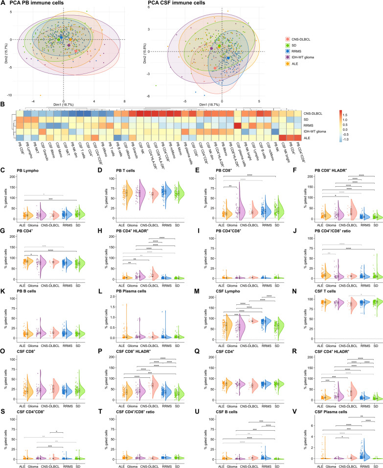
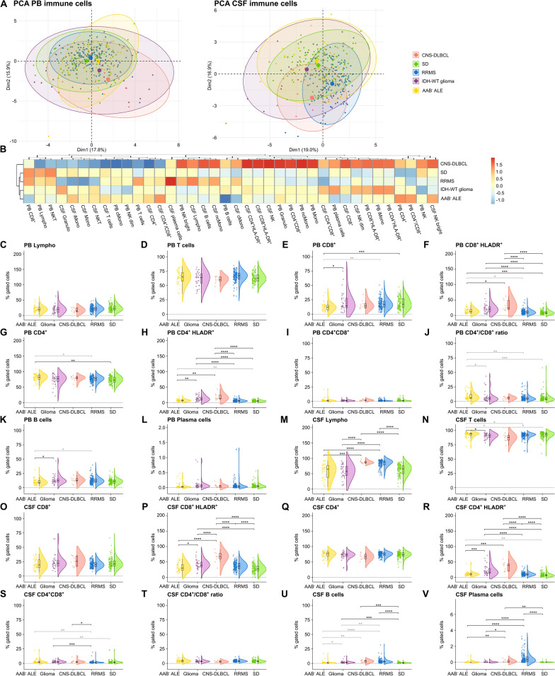
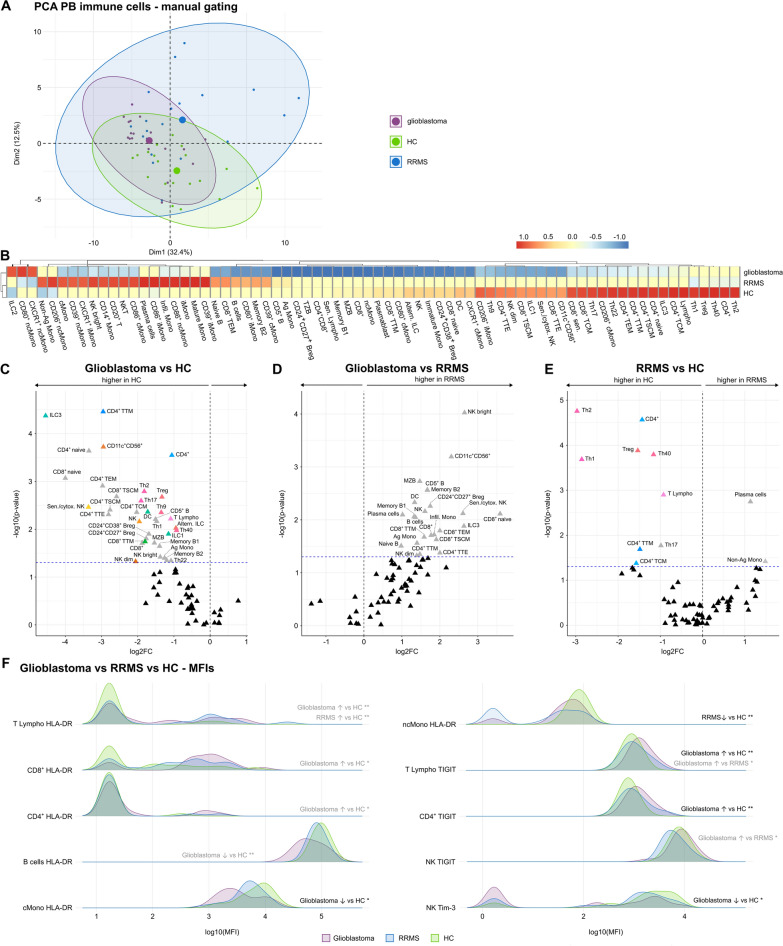
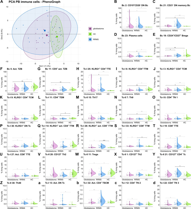
Methods: Aiming to provide novel insights into the pathogenesis of CNS autoimmunity and cerebral tumor immunity, we analyzed the peripheral blood (PB) and cerebrospinal fluid (CSF) of 81 autoimmune limbic encephalitis (ALE), 148 relapsing-remitting multiple sclerosis (RRMS), 33 IDH-wildtype glioma, 9 primary diffuse large B cell lymphoma of the CNS (CNS-DLBCL), and 110 controls by flow cytometry (FC). Additionally, an in-depth immunophenotyping of the PB from an independent cohort of 20 RRMS and 18 IDH-wildtype glioblastoma patients compared to 19 controls was performed by FC combined with unsupervised computational approaches.

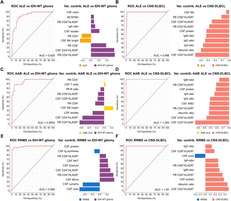
Results: We identified alterations in peripheral and intrathecal adaptive immunity, mainly affecting the T cell (Tc) but also the B cell (Bc) compartment in ALE, RRMS, and pCNS-tumors compared to controls. ALE, RRMS, and pCNS-tumors featured higher expression of the T cell activation marker HLA-DR, which was even more pronounced in pCNS-tumors than in ALE or RRMS. Glioblastoma patients showed signs of T cell exhaustion that were not visible in RRMS patients. In-depth characterization of the PB revealed differences mainly in the T effector and memory compartment between RRMS and glioblastoma patients and similar alterations in the Bc compartment, including atypical Bc, CD19+CD20- double negative Bc, and plasma cells. PB and CSF mFC together with CSF routine parameters could reliably differentiate ALE and RRMS from pCNS-tumors facilitating early diagnosis and treatment.

Conclusions: ALE, RRMS, and pCNS-tumors show distinct but partially overlapping changes mainly in HLA-DR+ Tc, memory Tc, exhausted Tc, and Bc subsets providing insights into disease pathogenesis. Moreover, mFC shows diagnostic potential facilitating early diagnosis and treatment.

Keywords: Autoimmune limbic encephalitis; Glioblastoma; Multidimensional flow cytometry; Primary diffuse large B cell lymphoma of the CNS; Relapsing–remitting multiple sclerosis.


Conflict of interest statement

SK reports funding from the IMF (Innovative medizinische Forschung; I-KO122003) and received research funding from Biogen and honoraria from Eisai, UCB and Jazz Pharma. The remaining authors declare no competing interests related to this study.

Figures

Fig. 1
Fig. 1
Pronounced adaptive immune response in ALE, IDH-wildtype glioma, and CNS-DLBCL compared to SD controls. A PCA including either PB mFC or CSF mFC parameters (% of gated cells) of ALE, RRMS, IDH-wildtype glioma, CNS-DLBCL, and SD patients. Every patient is displayed as a colored symbol. B Heatmap analysis of PB and CSF mFC parameters (% of gated cells) from ALE, RRMS, IDH-wildtype glioma, CNS-DLBCL, and SD patients: the median of each parameter was calculated, scaled, centered, and clustered hierarchically; CV Violin plots with overlaying box plots depicting the PB and CSF mFC parameters of ALE, RRMS, IDH-wildtype glioma, CNS-DLBCL, and SD patients. Boxes display the median as well as the 25th and 75th percentiles. The whiskers extend from the hinge to the largest and smallest values, respectively, but no further than 1.5 * IQR from the hinge. P-values were calculated by ANOVA with post-hoc Tukey HSD if normality could be assumed based on Shapiro–Wilk test, otherwise Kruskal Wallis test with Dunn post hoc test (p-adjustment method: Benjamini–Hochberg) was used. *p ≤ 0.05, **p ≤ 0.01, ***p ≤ 0.001, ****p ≤ 0.0001. ALE autoimmune limbic encephalitis, CD4+/CD8+ CD4+/CD8+ ratio, CNS Central nervous system, CSF cerebrospinal fluid, CNS-DLBCL diffuse large B cell lymphoma of the central nervous system, IDH isocitrate dehydrogenase, Lympho lymphocytes, mFC multidimensional flow cytometry, PB peripheral blood, PCA principal component analysis, RRMS relapsing remitting multiple sclerosis, SD somatic symptom disorder, WT wildtype
Fig. 2
Fig. 2
Antibody-negative ALE features similarities in adaptive immunity with IDH-wildtype glioma, and CNS-DLBCL. A PCA including either PB mFC or CSF mFC parameters (% of gated cells) of AAB ALE, RRMS, IDH-wildtype glioma, CNS-DLBCL, and SD patients. Every patient is displayed as a colored symbol. B Heatmap analysis of PB and CSF mFC parameters (% of gated cells) from AAB ALE, RRMS, IDH-wildtype glioma, CNS-DLBCL, and SD patients: the median of each parameter was calculated, scaled, centered, and clustered hierarchically; CV Violin plots with overlaying box plots depicting the PB and CSF mFC parameters of AAB ALE, RRMS, IDH-wildtype glioma, CNS-DLBCL, and SD patients. The whiskers extend from the hinge to the largest and smallest values, respectively, but no further than 1.5 * IQR from the hinge. P-values were calculated by ANOVA with post-hoc Tukey HSD if normality could be assumed based on Shapiro–Wilk test, otherwise Kruskal Wallis test with Dunn post hoc test (p-adjustment method: Benjamini–Hochberg) was used. *p ≤ 0.05, **p ≤ 0.01, ***p ≤ 0.001, ****p ≤ 0.0001. AAB ALE Antibody-negative autoimmune limbic encephalitis, CD4+/CD8+ CD4+/CD8+ ratio, CNS Central nervous system, CSF cerebrospinal fluid, CNS-DLBCL diffuse large B cell lymphoma of the central nervous system, IDH isocitrate dehydrogenase, Lympho lymphocytes; mFC multidimensional flow cytometry, PB peripheral blood, PCA principal component analysis, RRMS relapsing remitting multiple sclerosis, SD somatic symptom disorder, WT wildtype
Fig. 3
Fig. 3
CSF routine together with PB and CSF mFC parameters can reliably differentiate ALE and RRMS patients from patients with primary CNS tumors. AF ROC analyses of the classification results obtained from sPLS-DA including CSF routine, PB and CSF mFC parameters. ALE patients (A and B), antibody-negative ALE patients (C and D), or RRMS patients (E and F) were compared to either IDH-WT glioma or CNS-DLBCL. Loading plots visualize the top 10 variables contributing to latent component 1. Colors indicate the group in which the median is maximum. AAB ALE antibody-negative ALE, ALE autoimmune limbic encephalitis, AUC Area under the curve, BCSFBD blood-CSF barrier dysfunction, cMono classical monocytes, CSF cerebrospinal fluid, CNS-DLBCL diffuse large B cell lymphoma of the central nervous system, contrib contribution, IDH isocitrate dehydrogenase wildtype glioma, Granulo granulocytes, iMono intermediate monocytes, Lympho lymphocytes, mFC multidimensional flow cytometry, Mono monocytes, ncMono non-classical monocytes, NK natural killer cells, NKT Natural killer T cells, PB peripheral blood, ocbs oligoclonal bands, Q ratio, ROC receiver operating characteristic, RRMS relapsing remitting multiple sclerosis, SD somatic symptom disorder, sPLS-DA Sparse Partial Least Squares Discriminant Analysis, Var variable WBC white blood cell count, WT wildtype
Fig. 4
Fig. 4
- Glioblastoma patients feature a reduction in innate and adaptive immune cell population in the PB and a higher abundance of cell activation and exhaustion markers. A PCA including PB mFC parameters (cell clusters as % of living cells identified by manual gating) of RRMS and glioblastoma patients as well as HC. Every patient is displayed as a colored symbol. B Heatmap analysis of PB mFC parameters (cell clusters as % of living cells identified by manual gating): the median of each parameter was calculated, scaled, centered, and clustered hierarchically. CD4+ senescent T cells are not visualized given the median of 0 in all groups; CE Volcano plots showing PB mFC parameters of patients with glioblastoma or RRMS, and HC. The fold change of each single parameter between two groups is plotted against the corresponding p-value calculated by ANOVA with post-hoc Tukey HSD, if normality could be assumed based on Shapiro–Wilk test. Otherwise, Kruskal Wallis test with Dunn post hoc test (p-adjustment method: Benjamini–Hochberg) was used. Only significant parameters are labeled. Non-significant parameters are shown as black triangles. Parameters that did not remain significant after correction for age and sex are colored in grey. Senescent CD4+ are not visualized given the median of 0 in all groups. F Comparison of MFIs (medians) of different cell surface markers between patients with glioblastoma or RRMS and HC. *p ≤ 0.05, **p ≤ 0.01. Ag antigen-presenting, Altern alternative, B B cells, Breg B regulatory cells, cMono classical monocytes, cytox cytotoxic, DC dendritic cells, HC healthy control, ILC innate lymphoid cells, iMono intermediate monocytes, Infil infiltrating, Lympho lymphocytes, mFC multidimensional flow cytometry, MFI mean fluorescence intensity, Mono monocytes, MZB Marginal zone like B cells, ncMono non-classical monocytes, NK natural killer cells, NKT Natural killer T cells, PCA principal component analysis, RRMS relapsing remitting multiple sclerosis, Sen senescent, T T cells, TCM Central memory T cells, TEM Effector memory T cells, Th T helper cells, Treg Regulatory T cells, TSCM Stem memory T cells, TTE terminal effector T cells, TZB Transitional B cells
Fig. 5
Fig. 5
Unsupervised clustering identifies differences in T effector and memory subsets and similarities in the B cell compartment between glioblastoma and RRMS patients. A PCA including PB mFC parameters (cell clusters as % of living cells identified by PhenoGraph) of RRMS, glioblastoma patients, and HC. Every patient is displayed as a colored symbol. Bd Violin plots with overlaying box plots depicting the PB mFC parameters (cell clusters as % of living cells identified by PhenoGraph) of RRMS, glioblastoma patients, and HC. The whiskers extend from the hinge to the largest and smallest values, respectively, but no further than 1.5 * IQR from the hinge. P-values were calculated by ANOVA with post-hoc Tukey HSD if normality could be assumed based on Shapiro–Wilk test, otherwise Kruskal Wallis test with Dunn post hoc test (p-adjustment method: Benjamini–Hochberg) was used. *p ≤ 0.05, **p ≤ 0.01, ***p ≤ 0.001, ****p ≤ 0.0001. Act. Activated, Bc—B cells, Bregs B regulatory cells; cMono—classical monocytes; DN Bc—double negative (CD27IgD) B cells, DN Tc double negative (CD4+CD8+) T cells, FC fold change, HC healthy control, mFC multidimensional flow cytometry, Mono monocytes, ncMono non-classical monocytes, PCA principal component analysis, RRMS relapsing remitting multiple sclerosis, Tc T cells, TCM central memory T cells, TEM effector memory T cells, Th T helper cells, TN naïve T cells, Tregs regulatory T cells, TSCM stem memory T cells, TTE T terminal effector T cells, TTM transitional memory T cells, TZB transitional B cells

References

    1. Dalmau J, Graus F. Antibody-mediated encephalitis. N Engl J Med. 2018;378:840–51. - PubMed
    1. Melzer N, Budde T, Stork O, Meuth SG. Limbic encephalitis: potential impact of adaptive autoimmune inflammation on neuronal circuits of the amygdala. Front Neurol. 2015;6:171. - PMC - PubMed
    1. Breese EH, Dalmau J, Lennon VA, Apiwattanakul M, Sokol DK. Anti-N-methyl-D-aspartate receptor encephalitis: early treatment is beneficial. Pediatr Neurol. 2010;42:213–4. - PubMed
    1. Byrne S, Walsh C, Hacohen Y, Muscal E, Jankovic J, Stocco A, et al. Earlier treatment of NMDAR antibody encephalitis in children results in a better outcome. Neurol Neuroimmunol Neuroinflamm. 2015;2: e130. - PMC - PubMed
    1. Kao Y-C, Lin M-I, Weng W-C, Lee W-T. Neuropsychiatric disorders due to limbic encephalitis: immunologic aspect. Int J Mol Sci. 2020;22:389. - PMC - PubMed

MeSH terms

LinkOut - more resources