. 2024 Oct 21:15:1493848.
doi: 10.3389/fimmu.2024.1493848. eCollection 2024.

Analysis of lipid uptake, storage, and fatty acid oxidation by group 2 innate lymphoid cells

Affiliations

Analysis of lipid uptake, storage, and fatty acid oxidation by group 2 innate lymphoid cells

Audrey Roy-Dorval et al. Front Immunol. .

Abstract

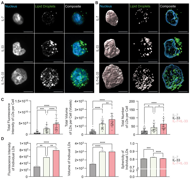
Group 2 Innate Lymphoid Cells (ILC2) are critical drivers of both innate and adaptive type 2 immune responses, known to orchestrate processes involved in tissue restoration and wound healing. In addition, ILC2 have been implicated in chronic inflammatory barrier disorders in type 2 immunopathologies such as allergic rhinitis and asthma. ILC2 in the context of allergen-driven airway inflammation have recently been shown to influence local and systemic metabolism, as well as being rich in lipid-storing organelles called lipid droplets. However, mechanisms of ILC2 lipid anabolism and catabolism remain largely unknown and the impact of these metabolic processes in regulating ILC2 phenotypes and effector functions has not been extensively characterized. ILC2 phenotypes and effector functions are shaped by their metabolic status, and determining the metabolic requirements of ILC2 is critical in understanding their role in type 2 immune responses and their associated pathophysiology. We detail here a novel experimental method of implementing flow cytometry for large scale analysis of fatty acid uptake, storage of neutral lipids, and fatty acid oxidation in primary murine ILC2 with complementary morphological analysis of lipid storage using confocal microscopy. By combining flow cytometry and confocal microscopy, we can identify the metabolic lipid requirements for ILC2 functions as well as characterize the phenotype of lipid storage in ILC2. Linking lipid metabolism pathways to ILC2 phenotypes and effector functions is critical for the assessment of novel pharmaceutical strategies to regulate ILC2 functions in type 2 immunopathologies.

Keywords: fatty acid oxidation (FAO); fatty acid uptake; flow cytometry; group 2 innate lymphoid cells (ILC2); immunometabolism; lipid droplets; microscopy; type 2 immunity.


Conflict of interest statement

The authors declare that the research was conducted in the absence of any commercial or financial relationships that could be construed as a potential conflict of interest. JHF declares that he was an editorial board member of Frontiers, at the time of submission. This had no impact on the peer review process and the final decision.

Figures

Figure 1
Figure 1
Schematic of experimental workflow. Female C57BL/6J wild-type mice aged 6-12 weeks were selected for either bone marrow or lung group 2 innate lymphoid cell (ILC2) isolation. Femurs and tibias were harvested to obtain bone marrow-derived ILC2 by flow cytometric cell sorting and were then cultured in vitro (dashed line) for expansion, followed by cytokine starvation and subsequent cytokine re-stimulation. Lung-derived ILC2 were obtained by flow cytometric cell sorting from naïve mice followed by in vitro cell culture expansion (dashed line). Alternatively, lung-derived ILC2 were obtained by flow cytometric cell sorting from animals that were challenged intranasally with PBS as control or IL-33 for three consecutive days (solid line). Bone marrow- and lung-derived ILC2 were used for the quantification of fatty acid uptake, neutral lipid storage, and fatty acid oxidation (FAO) by flow cytometry or microscopy. Created in BioRender.com. Deagle, R. (2024) BioRender.com/v17y111.
Figure 2
Figure 2
Flow cytometry gating strategies. After debris exclusion (FSC-A vs SSC-A) bone marrow- or lung-derived group 2 innate lymphoid cells (ILC2) were gated on singlets (FSC-A vs FSC-H) and on 7-AAD-negative live cells (7-AAD vs FSC-H). ILC2 were subsequently analyzed for lipid uptake (BODIPY FL C16), neutral lipid content (BODIPY 493/503) or fatty acid oxidation (FAO Blue).
Figure 3
Figure 3
Cytokine production and proliferation of murine ILC2 upon treatment with distinct cytokine combinations. Bone marrow-derived (A, B, E, F) or lung-derived (C, D) group 2 innate lymphoid cells (ILC2) were stimulated with either IL-7 only, IL-33 only, or a combination of IL-7 and IL-33 (A, C, E) or IL-2 only, IL-33 only, or a combination of IL-2 and IL-33 (B, D, F). All cytokines were applied at 10 ng/mL. (A–D) After 24 hours of stimulation, supernatants were harvested and analyzed for IL-5 content by ELISA. (E, F) Proliferation of ILC2 was assessed using CellTrace Yellow Cell Proliferation Kit after 3 days of incubation with respective cytokines. The data representing the IL-33 stimulation is the same for (A–F). Data are representative of three independent experiments with stimulations performed in duplicates. Data are shown as average ± standard deviation (SD). Statistical analysis was performed using one-way ANOVA followed by Tukey’s multiple comparisons test (p < 0.01 = ** and p < 0.0001 = ****); ND, not detectable.
Figure 4
Figure 4
ILC2 increase fatty acid uptake upon stimulation with activating cytokines. (A, B) Mice were treated intranasally with either PBS as control or with 250 ng of IL-33 for three consecutive days. Two days after the last treatment, lungs were collected and group 2 innate lymphoid cells (ILC2) were sort-purified and subsequently incubated with BODIPY FL C16 for one hour to assess capacity of lipid uptake by flow cytometric analysis (MFI, geometric mean fluorescence intensity). Bone marrow-derived (C–F) or lung-derived (G–J) ILC2 were stimulated with either IL-7 only, IL-33 only, or a combination of IL-7 and IL-33 (C, D, G, H) or IL-2 only, IL-33 only, or a combination of IL-2 and IL-33 (E, F, I, J). The data representing the IL-33 stimulation is the same for (C–J). All cytokines were applied at 10 ng/mL. BODIPY FL C16 was added after 23 hours of cytokine stimulation and incubated for one hour to assess capacity of lipid uptake by flow cytometric analysis. ILC2 that were not incubated with BODIPY FL C16 served as negative control (Unstained). Data are shown as average ± standard deviation (SD). Statistical analysis was performed using one-way ANOVA followed by Tukey’s multiple comparisons test (p < 0.05 = *, p < 0.01 = **, p < 0.001 = ***, p < 0.0001 = ****).
Figure 5
Figure 5
ILC2 increase neutral lipid storage upon stimulation with activating cytokines. (A, B) Mice were treated intranasally with either PBS as control or with 250 ng of IL-33 for three consecutive days. Two days after the last treatment, lungs were collected and group 2 innate lymphoid cells (ILC2) were sorted and subsequently incubated with BODIPY 493/503 for 20 minutes to assess capacity of neutral lipid storage by flow cytometric analysis (MFI, geometric mean fluorescence intensity). Bone marrow-derived (C–F) or lung-derived (G–J) ILC2 were stimulated with either IL-7 only, IL-33 only, or a combination of IL-7 and IL-33 (C, D, G, H) or IL-2 only, IL-33 only, or a combination of IL-2 and IL-33 (E, F, I, J). The data representing the IL-33 stimulation is the same for (C–J). All cytokines were applied at 10 ng/mL. BODIPY 493/503 was added after 24 hours of cytokine stimulation and incubated for 20 minutes to assess capacity of neutral lipid storage by flow cytometric analysis. ILC2 that were not incubated with BODIPY 493/503 served as negative control (Unstained). Data are shown as average ± standard deviation (SD). Statistical analysis was performed using one-way ANOVA followed by Tukey’s multiple comparisons test (p < 0.05 = *, p < 0.01 = **, p < 0.001 = ***, p < 0.0001 = ****).
Figure 6
Figure 6
ILC2 increase fatty acid oxidation upon stimulation with activating cytokines. (A, B) Mice were treated intranasally with either PBS as control or with 250 ng of IL-33 for three consecutive days. Two days after the last treatment, lungs were collected, and group 2 innate lymphoid cells (ILC2) were sorted and subsequently stained with FAO Blue for one hour to assess capacity of fatty acid oxidation by flow cytometric analysis (MFI, geometric mean fluorescence intensity). Bone marrow-derived (C–F) or lung-derived (G–J) ILC2 were stimulated with either IL-7 only, IL-33 only, or a combination of IL-7 and IL-33 (C, D, G, H) or IL-2 only, IL-33 only, or a combination of IL-2 and IL-33 (E, F, I, J). The data representing the IL-33 stimulation is the same for (C–J). All cytokines were applied at 10 ng/mL. After 24 hours of cytokine stimulation cells were incubated for one hour with FAO Blue and analyzed using flow cytometry. ILC2 that were not incubated with FAO Blue served as negative control (Unstained). Data are shown as average ± standard deviation (SD). Statistical analysis was performed using one-way ANOVA followed by Tukey’s multiple comparisons test (p < 0.05 = *, p < 0.01 = **, p < 0.001 = ***, p < 0.0001 = ****).
Figure 7
Figure 7
Allergic airway inflammation increases the lipid droplet number and size in pulmonary ILC2. Mice were treated intranasally with either PBS as control or with 250 ng of IL-33 for three consecutive days. Two days after the last treatment, lungs were collected, and group 2 innate lymphoid cells (ILC2) were sorted by flow cytometry and labelled with Hoechst 33342 to visualize the nucleus (blue) and BODIPY 493/503 to visualize neutral lipid storage (A, B). Confocal images (A) and 3D fluorescence reconstructions from Imaris (B) are shown (PBS - top row; IL-33 treatments - bottom row). Confocal images are displayed as a maximum intensity projection of approximately 10-20 frames (5-10 μm depth) produced in ImageJ. Images are representative from 3 independent experiments, scale bars represent 5 μm. (C) Quantification of the total fluorescence intensity (left), total volume (middle), and total number (right) of neutral lipid droplets cumulatively per cell. Fluorescence and morphology metrics were taken from individual neutral lipid droplets and were added together based on cell of origin and then by intranasal challenge. ILC2 that underwent PBS intranasal challenge are shown in blue (n = 30) and those treated with IL-33 are shown in off-white (n = 33). (D) Quantification of the total fluorescence intensity (left), total volume (middle), and sphericity (right) of individual neutral lipid droplets. Fluorescence and morphology metrics were taken from the same individual neutral lipid droplets shown in (C) and compiled based on intranasal challenge alone; PBS (n = 39) and IL-33 (n = 154). The data representing the IL-33 stimulation is the same for A-D. Data are represented as average ± standard deviation (SD) where n = number of cells (C) or average ± standard error of measurement (SEM) where n = number of neutral lipid droplets (D). Statistical significance (p-values) was calculated using unpaired two-tailed Student’s t-test (p < 0.0001 = ****).
Figure 8
Figure 8
In vitro stimulation of ILC2 with combinations of IL-7 and IL-33 changes the neutral lipid droplet size and number. Bone marrow-derived group 2 innate lymphoid cells (ILC2) were stimulated for 24 hours with either IL-7 only, IL-33 only, or a combination of IL-7 and IL-33 (all cytokines were applied at 10 ng/mL) and labelled with Hoechst 33342 to visualize the nucleus (blue) and BODIPY 493/503 to visualize neutral lipid storage (A, B). Confocal images (A) and 3D fluorescence reconstructions from Imaris (B) are shown. Confocal images are displayed as a maximum intensity projection of approximately 10-20 frames (5-10 μm depth) produced in ImageJ. Images are representative from 3 independent experiments, scale bars represent 5 μm. (C) Quantification of the total fluorescence intensity (left), total volume (middle), and total number (right) of neutral lipid droplets cumulatively per cell. Fluorescence and morphology metrics were taken from individual neutral lipid droplets and were added together based on cell of origin and then by cytokine treatment. ILC2 were treated with either IL-7 (grey, n = 30), IL-33 (off-white, n = 30), or IL-7+IL-33 (pink, n =30) for 24 hours prior to imaging. (D) Quantification of the total fluorescence intensity (left), total volume (middle), and sphericity (right) of individual neutral lipid droplets. Fluorescence and morphology metrics were taken from the same individual neutral lipid droplets shown in (C) and were compiled based on cytokine treatment alone; IL-7 (n = 637), IL-33 (n = 1,267), or IL-7+IL-33 (n = 1,569). The data representing the IL-33 stimulation is the same for (A–D). Data are represented as average ± standard deviation (SD) where n = the number of cells (C) or average ± standard error of measurement (SEM) where n = number of neutral lipid droplets (D). Statistical significance (p-values) was calculated using ordinary one-way ANOVA and post-hoc Tukey’s multiple comparison tests (p < 0.05 = *, p < 0.01 = **, p < 0.001 = ***, p < 0.0001 = ****).
Figure 9
Figure 9
In vitro stimulation of ILC2 with combinations of IL-2 and IL-33 changes the neutral lipid droplet volume. Bone marrow-derived group 2 innate lymphoid cells (ILC2) were stimulated for 24 hours with either IL-2 only, IL-33 only, or a combination of IL-2 and IL-33 (all cytokines were applied at 10 ng/mL) and labelled with Hoechst 33342 to visualize the nucleus (blue) and BODIPY 493/503 to visualize neutral lipid storage (A, B). Confocal images (A) and 3D fluorescence reconstructions from Imaris (B) are shown. Confocal images are displayed as a maximum intensity projection of approximately 10-20 frames (5-10 μm depth) produced in ImageJ. Images are representative from 3 independent experiments, scale bars represent 5 μm. (C) Quantification of the total fluorescence intensity (left), total volume (middle), and total number (right) of neutral lipid droplets cumulatively per cell. Fluorescence and morphology metrics were taken from individual neutral lipid droplets and were added together based on cell of origin and then by cytokine treatment. ILC2 were treated with either IL-2 (orange, n = 30), IL-33 (off-white, n = 30), or IL-2+IL-33 (teal, n = 30) for 24 hours prior to imaging. (D) Quantification of the total fluorescence intensity (left), total volume (middle), and sphericity (right) of individual neutral lipid droplets. Fluorescence and morphology metrics were taken from the same individual neutral lipid droplets in (C) and compiled based on cytokine treatment alone; IL-2 (n = 1,266), IL-33 (n = 1,267), or IL-2+IL-33 (n = 1,015). The data representing the IL-33 stimulation is the same for (A–D). Data are represented as average ± standard deviation (SD) where n = the number of cells (C) or average ± standard error of measurement (SEM) where n = number of neutral lipid droplets (D). Statistical significance (p-values) was calculated using ordinary one-way ANOVA and post-hoc Tukey’s multiple comparison tests (p < 0.05 = *, p < 0.001 = ***, p < 0.0001 = ****).
Figure 10
Figure 10
ILC2 increase in cell volume upon stimulation with activating cytokines. Quantification of the total cell volume using cytosolic BODIPY (493/503) fluorescence from confocal images. (A) group 2 innate lymphoid cells (ILC2) that underwent PBS intranasal challenge for three consecutive days are shown in blue (n = 30) and those treated with 250 ng/mL of IL-33 are shown in off-white (n = 33). (B) ILC2 were treated with either IL-7 (grey, n = 30), IL-33 (off-white, n = 30), or IL-7+IL-33 (pink, n = 30) for 24 hours prior to imaging. (C) ILC2 were treated with either IL-2 (orange, n = 30), IL-33 (off-white, n = 30), or IL-2+IL-33 (teal, n = 30) for 24 hours prior to imaging. All cytokines were administered at 10 ng/mL. Data are represented as average ± standard deviation (SD) where n = number of cells. The data representing the IL-33 stimulation is the same for (B, C). (A) Statistical significance (p-values) was calculated using unpaired two-tailed Student’s t-test. (B, C) Statistical significance (p-values) was determined by applying ordinary one-way ANOVA and post-hoc Tukey’s multiple comparison tests (p < 0.01 = ** and p < 0.0001 = ****).

References

    1. Helfrich S, Mindt BC, Fritz JH, Duerr CU. Group 2 innate lymphoid cells in respiratory allergic inflammation. Front Immunol. (2019) 10:930. doi: 10.3389/fimmu.2019.00930 - DOI - PMC - PubMed
    1. Kobayashi T, Motomura Y, Moro K. The discovery of group 2 innate lymphoid cells has changed the concept of type 2 immune diseases. Int Immunol. (2021) 33:705–9. doi: 10.1093/intimm/dxab063 - DOI - PMC - PubMed
    1. Zaiss DMW, Pearce EJ, Artis D, McKenzie ANJ, Klose CSN. Cooperation of ilc2s and T(H)2 cells in the expulsion of intestinal helminth parasites. Nat Rev Immunol. (2024) 24:294–302. doi: 10.1038/s41577-023-00942-1 - DOI - PubMed
    1. Starkey MR, McKenzie AN, Belz GT, Hansbro PM. Pulmonary group 2 innate lymphoid cells: surprises and challenges. Mucosal Immunol. (2019) 12:299–311. doi: 10.1038/s41385-018-0130-4 - DOI - PMC - PubMed
    1. Ricardo-Gonzalez RR, Molofsky AB, Locksley RM. Ilc2s - development, divergence, dispersal. Curr Opin Immunol. (2022) 75:102168. doi: 10.1016/j.coi.2022.102168 - DOI - PMC - PubMed

LinkOut - more resources