Review
. 2024 Oct 18;10(20):e39489.
doi: 10.1016/j.heliyon.2024.e39489. eCollection 2024 Oct 30.

Deciphering the multifaceted role of microRNAs in hepatocellular carcinoma: Integrating literature review and bioinformatics analysis for therapeutic insights

Affiliations
Review

Deciphering the multifaceted role of microRNAs in hepatocellular carcinoma: Integrating literature review and bioinformatics analysis for therapeutic insights

Fereshteh Rahdan et al. Heliyon. .

Abstract

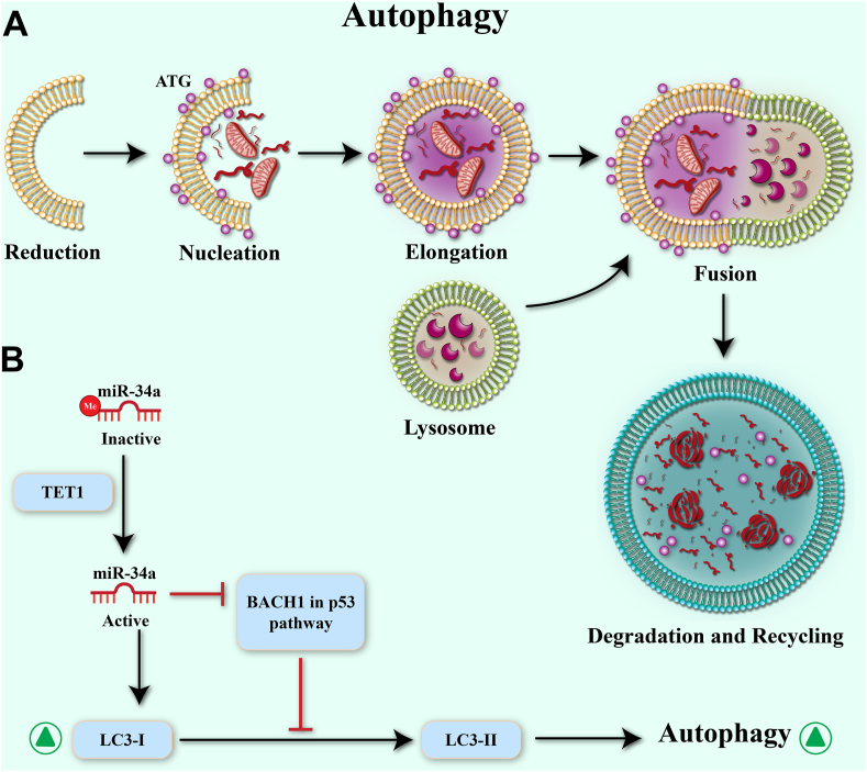
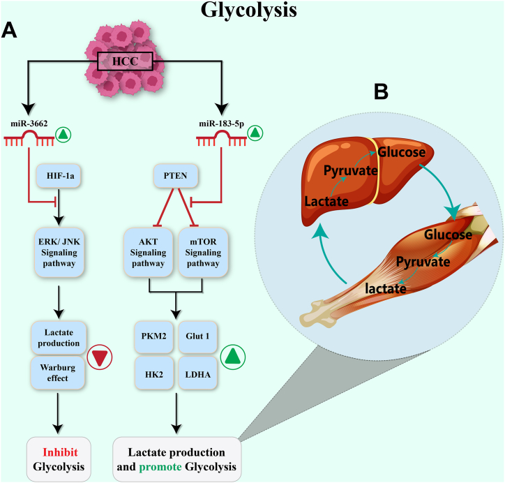
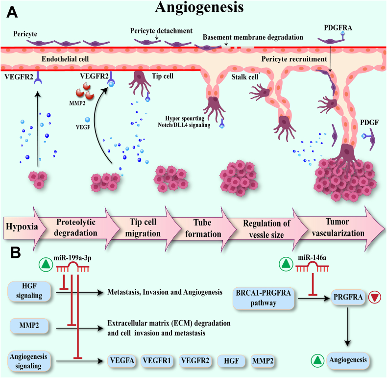
Hepatocellular carcinoma (HCC) poses a significant global health challenge, necessitating innovative therapeutic strategies. MicroRNAs (miRNAs) have emerged as pivotal regulators of HCC pathogenesis, influencing key processes such as self-renewal, angiogenesis, glycolysis, autophagy, and metastasis. This article integrates findings from a comprehensive literature review and bioinformatics analysis to elucidate the role of miRNAs in HCC. We discuss how dysregulation of miRNAs can drive HCC initiation, progression, and metastasis by modulating various signaling pathways and target genes. Moreover, leveraging high-throughput technology and bioinformatics tools, we identify key miRNAs involved in multiple cancer hallmarks, offering insights into potential combinatorial therapeutic strategies. Through our analysis considering p-values and signaling pathways associated with key features, we unveil miRNAs with simultaneous roles across critical cancer characteristics, providing a basis for the development of high-performance biomarkers. The microRNAs, miR-34a-5p, miR-373-3p, miR-21-5p, miR-214-5p, miR-195-5p, miR-139-5p were identified to be shared microRNAs in stemness, angiogenesis, glycolysis, autophagy, EMT, and metastasis of HCC. However, challenges such as miRNA stability and delivery hinder the translation of miRNA-based therapeutics into clinical practice. This review underscores the importance of further research to overcome existing barriers and realize the full potential of miRNA-based interventions for HCC management.

Keywords: Angiogenesis; Cancer metabolism; Epithelial-mesenchymal transition (EMT); Hepatocellular carcinoma (HCC); In silico analysis; Metastasis; Stemness; miRNA therapeutics.


Conflict of interest statement

The authors declare that they have no known competing financial interests or personal relationships that could have appeared to influence the work reported in this paper.

Figures

Fig. 1
Fig. 1
Transcription and processing of microRNA.
Fig. 2
Fig. 2
Functions of miRNAs in HCC Autophagy A. Autophagy maintains cellular homeostasis by degrading misfolded or damaged proteins as well as removing aged or malfunctioning organelles. Following the induction of autophagy, the pre-autophagosome occurs, which is a cup-shaped structure separated from the ER membrane and formed. The next step, the nuclear ATG complex, consists of a set of autophagy-related proteins that gradually begin to engulf damaged cellular proteins and components. The next stage is elongation, which starts the complete separation of membrane and autophagosome formation. After the formation of the autophagosome, it merges with the lysosome, and the contents inside the autophagosome are destroyed by lysosomal hydrolases. B. MicroRNAs are key regulators of autophagy. TET1 activates miR-34 through demethylation. miR-34 regulates autophagy through the miR-34a/BACH1/p53 axis. BACH1 is the target of the miR-34a gene in the p53 pathway, inhibiting BACH1 expression and increasing LC3-I expression leads to the cytoplasmic form of I3-LC being cleaved to the autophagic membrane form LC3- II is converted and then it leads to stimulation of autophagy formation steps.
Fig. 3
Fig. 3
MicroRNAs in HCC apoptosis. A. The miR-644a, as a promoter of apoptosis, suppresses HSF1 expression by binding to the 3′-UTR of HSF1, and the suppression of HSF1 expression leads to increased expression of BH3-only family proteins including BID, BAD, BIM, SMAC, Apaf-1 and cleaved caspases-3 and -9. B. miR-224 targets genes whose products are involved in cell apoptosis, and therefore, increased expression of miR-224 downregulates apoptosis-promoting genes (CDC42, CDH1, and PAK2) and increases the expression of the anti-apoptotic gene BCL-2.
Fig. 4
Fig. 4
MicroRNAs in EMT process. A. In the lower parts of epithelial and endothelial tissues, there is a thin layer of extracellular matrix called basement membrane (BM). Cancer cells must invade the basement membrane of the extracellular matrix to metastasize. Laminins are the main components of the ECM, which are involved in the epithelial-mesenchymal transition (EMT) of the basement membrane (BM) of cancer cells. Cells with (EMT) characteristics are degraded through the matrix metalloproteinase (MMP)-associated pathway. The increased expression of MMPs leads to increased destruction of the basement membrane and the entry of cells with EMT characteristics into the blood circulation, which leads to invasion and then metastasis. B.Increasing miR-345 by targeting IRF1 mRNA and inhibiting the activation of the mTOR/STAT3/AKT triple pathway and reducing the expression of its downstream targets such as Slug, Snail, and Twist reduce EMT in HCC cells. Up-regulation of miR-1296 inhibits hypoxia-promoting effects on metastasis and EMT of HCC cells by targeting SRPK1 gene expression and inhibiting PI3K/AKT signaling pathway activation which both miR-345 and miR-1296 could be used to control the EMT progression.
Fig. 5
Fig. 5
Role of miRNAs in metastasis of HCC. . 1. To create metastasis, first, cancer cells are separated from the tissue mass with the origin of cancer by ADAM proteins, a family of secretory and membrane metalloproteinases, and SRPK1, which is a splicing factor of SRSF protein kinase 1. They cause the movement and migration of cells removed from tumor tissue in the tumor microenvironment of blood vessels 2. Tumor cells pass through the vessels reach the surrounding healthy tissues and create the characteristics of cancer cells in healthy tissues. 3. Metastasis occurs in distant and nearby tissues. Up-regulation of miR-1296 inhibits hypoxia-promoting effects on metastasis and EMT of HCC cells by targeting SRPK1 gene expression and inhibiting PI3K/AKT signaling pathway activation. Also, Mir-655 inhibits metastasis in HCC cells by directly targeting ADAM10 inhibiting the β-catenin pathway, and downregulating E-cadherin protein expression.
Fig. 6
Fig. 6
Role of miRNAs regulating glycolysis in HCC. A. Tumor cells use glucose as their metabolic source to meet their biosynthetic and bioenergetic needs. By inactivating pyruvate dehydrogenase and increasing the expression of lactate dehydrogenase A, pyruvate is converted to lactate, and lactate increases to a large level. Increased levels of lactate become the main energy fuel in tumors. B.The increase of miR-3662 levels by directly targeting the hypoxia-inducible factor 1α (HIF-1α) by inhibiting the activation of ERK and JNK signaling pathways and by reducing lactate production and reducing the Warburg effect inhibits coagulation in cancer cells. Increasing the level of miR-183-5p by reducing the expression of the PTEN gene in the Akt, p-Akt, and mTOR pathway and increasing the expression of PKM2, HK2, LDHA, GLUT1 genes with lactate production leads to an increase in glycolysis in HCC cells.
Fig. 7
Fig. 7
Role of miRNAs regulating angiogenesis in HCC. A. Pericytes line the vascular endothelium throughout the body. Pericytes cover vascular endothelium throughout the body, platelet-derived growth factor receptors, and vascular endothelial growth factor receptors are located on these vascular surfaces. tip/stalk cells are a group of endothelial cells. Tumor cells widely secrete pro-angiogenic factors and create tumor tissue with poor perfusion. After that, a hypoxic microenvironment is created, which is one of the main drivers of angiogenesis in tumor tissue. PDGF by tip cells is secreted, and the high level of VEGF secretion by tumor cells blocks PDGFRb signaling through the receptor complex consisting of PDGFRb and VEGFR2. VEGF-stimulated tip/stalk cells tend to migrate and proteolytic enzymes such as MMP, elastase or trypsin are secreted by cancer cells. These enzymes destroy cell junctions. The released cells gradually mimic the behavior of blood vessel cells and form channel-like structures similar to vessels. B.By increasing the expression of miR-199a-3p, the signaling pathways of HGF, and MMP2 are blocked, and at the same time, the expression of VEGFR1 and VEGFR2, HGF, MMP2, VEGFA genes is inhibited, and then angiogenesis is inhibited. Increased miR-146 increases angiogenesis by inhibiting PDGFRA activation in the BRCA1–PDGFRA pathway.
Fig. 8
Fig. 8
Alluvial diagrams show the interaction between miRNAs- Targets in Apoptosis (a1 down. a2 up), autophagy (b1 down, b2 up), angiogenesis (c1 down, c2 up), and glycolysis (d1 down, d2 up).
Fig. 9
Fig. 9
Alluvial diagrams show the interaction between miRNAs-Targets in EMT (a1 down, a2 up) and metastasis (b1 down and b2 up).

References

    1. Samant H., Amiri H.S., Zibari G.B. Addressing the worldwide hepatocellular carcinoma: epidemiology, prevention and management. J. Gastrointest. Oncol. 2021;12(Suppl 2):S361. - PMC - PubMed
    1. Belghiti J., Kianmanesh R. Surgical treatment of hepatocellular carcinoma. HPB (Oxford) 2005;7(1):42–49. - PMC - PubMed
    1. Chen C.P. Role of radiotherapy in the treatment of hepatocellular carcinoma. J Clin Transl Hepatol. 2019;7(2):183–190. - PMC - PubMed
    1. Li S., et al. Low-dose radiotherapy combined with dual PD-L1 and VEGFA blockade elicits antitumor response in hepatocellular carcinoma mediated by activated intratumoral CD8(+) exhausted-like T cells. Nat. Commun. 2023;14(1):7709. - PMC - PubMed
    1. Yang Y.Q., et al. Current status and prospect of treatments for recurrent hepatocellular carcinoma. World J. Hepatol. 2023;15(2):129–150. - PMC - PubMed

LinkOut - more resources