. 2024 Dec;60(11):6799-6811.
doi: 10.1111/ejn.16589. Epub 2024 Nov 5.

Central amygdala-to-pre-Bötzinger complex neurotransmission is direct and inhibitory

Affiliations

Central amygdala-to-pre-Bötzinger complex neurotransmission is direct and inhibitory

Jeffrey Gu et al. Eur J Neurosci. 2024 Dec.

Abstract

Breathing behaviour is subject to emotional regulation, but the underlying mechanisms remain unclear. Here, we demonstrate a direct relationship between the central amygdala, a major output hub of the limbic system associated with emotional brain function, and the brainstem pre-Bötzinger complex, which generates the fundamental rhythm and pattern for breathing. The connection between these two sites is monosynaptic and inhibitory, involving GABAergic central amygdala neurons whose axonal projections act predominantly via ionotropic GABAA receptors to produce inhibitory postsynaptic currents in pre-Bötzinger neurons. This pathway may provide a mechanism to inhibit breathing in the context of freezing to assess threats and plan defensive action. The existence of this pathway may further explain how epileptic seizures invading the amygdala cause long-lasting apnea, which can be fatal. Although their ultimate importance awaits further behavioural tests, these results elucidate a link between emotional brain function and breathing, which underlies survival-related behaviour in mammals and pertains to human anxiety disorders.

Keywords: SUDEP; anxiety; central amygdala; defense; pre‐Bötzinger complex; respiration.


Conflict of interest statement

The authors have no financial or other conflicts of interest to declare.

Figures

FIGURE 1
FIGURE 1
Neuroanatomical tracing of projections from central amygdala (CeA) to pre‐Bötzinger complex (preBötC). (a) Parasagittal view of mouse brain with an arrow indicating cholera toxin B (CTb)‐Alexa488 injection in the right preBötC. Dotted vertical lines show midbrain sections in the CeA where CTb‐labelled neurons are shown in (d). This experiment produced the data in (b–d). (b) Transverse section showing the preBötC site of injection. (c) Percentage of CTb‐labelled neurons in the CeA (n = 3). (d) 10× magnification (left and upper panels) and 40× magnification (lower panels) confocal images of right CeA showing CTb‐Alexa488‐labelled neurons following preBötC injections. Images were converted into maximum intensity z‐projections, and their contrast was adjusted. Numerals 1–4 indicate corresponding sections at different levels of magnification. d1–d4 show no colocalization of CTb (cyan) and protein kinase C‐delta (PKCδ) (magenta) in CeA sections as indicated by distance from Bregma. (e) Parasagittal view of mouse brain with an arrow indicating the AAV‐ChR2(H134R)‐EYFP injection in the CeA. (f) Transverse section showing enhanced yellow fluorescent protein (EYFP) at the CeA injection site. (g) Recording site in the preBötC; ‘P’ refers to the patch‐recording pipette. (h) EYFP expression in the preBötC under high magnification. Dorsal and lateral directions are shown for orientation and those orientations apply to all images in the CeA and preBötC.
FIGURE 2
FIGURE 2
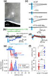
Synaptic projections from CeA to preBötC are monosynaptic and inhibitory. (a) Whole‐cell patch‐clamp recording from a preBötC neuron in the enhanced yellow fluorescent protein (EYFP)‐expressing region of the preBötC. ‘P’ indicates the location of the patch pipette. This neuron produced the data in (c) and (e). (b) Average waveform of 30 traces (black) triggered by randomly generated pulses (indicated by green arrowhead) with 95% confidence limit (grey shaded area) and light‐triggered average waveform (n = 30, blue). The blue rectangular bar indicates the duration of 470 nm photostimulation. (c) Light‐evoked inhibitory postsynaptic currents under different pharmacological conditions including standard artificial cerebrospinal fluid (ACSF) and in the presence of sequentially added TTX (1 μM), 4‐AP (200 μM), and bicuculline (10 μM). (d) Light‐evoked postsynaptic outward currents, rather than inward currents seen in 2c. Red line indicates the average waveform triggered by light stimulation. (e) Raw traces and histograms of the latency of light‐evoked postsynaptic currents in control (ACSF) and in the presence of TTX + 4AP. (f) Group data showing mean latency and its coefficient of variation (CV).
FIGURE 3
FIGURE 3
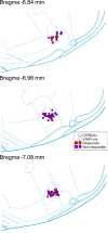
All the recorded neurons with measurable light‐evoked inhibitory postsynaptic currents, that is, responders and nonresponders, were located within the preBötC.

References

    1. Adke, A. P. , Khan, A. , Ahn, H. S. , Becker, J. J. , Wilson, T. D. , Valdivia, S. , Sugimura, Y. K. , Martinez Gonzalez, S. , & Carrasquillo, Y. (2021). Cell‐type specificity of neuronal excitability and morphology in the Central Amygdala. eNeuro, 8(1), ENEURO.0402‐20.2020. 10.1523/ENEURO.0402-20.2020 - DOI - PMC - PubMed
    1. Adolphs, R. (2010). What does the amygdala contribute to social cognition? Annals of the new York Academy of Sciences, 1191, 42–61. 10.1111/j.1749-6632.2010.05445.x - DOI - PMC - PubMed
    1. Arima, Y. , Yokota, S. , & Fujitani, M. (2019). Lateral parabrachial neurons innervate orexin neurons projecting to brainstem arousal areas in the rat. Scientific Reports, 9, 2830. 10.1038/s41598-019-39063-y - DOI - PMC - PubMed
    1. Ashhad, S. , Kam, K. , Del Negro, C. A. , & Feldman, J. L. (2022). Breathing rhythm and pattern and their influence on emotion. Annual Review of Neuroscience, 45, 223–247. 10.1146/annurev-neuro-090121-014424 - DOI - PMC - PubMed
    1. Bagur, S. , Lefort, J. M. , Lacroix, M. M. , de Lavilléon, G. , Herry, C. , Chouvaeff, M. , Billand, C. , Geoffroy, H. , & Benchenane, K. (2021). Breathing‐driven prefrontal oscillations regulate maintenance of conditioned‐fear evoked freezing independently of initiation. Nature Communications, 12, 2605. 10.1038/s41467-021-22798-6 - DOI - PMC - PubMed

Substances

LinkOut - more resources