. 2024 Nov;13(11):e12519.
doi: 10.1002/jev2.12519.

Extracellular vesicles from human-induced pluripotent stem cell-derived neural stem cells alleviate proinflammatory cascades within disease-associated microglia in Alzheimer's disease

Affiliations

Extracellular vesicles from human-induced pluripotent stem cell-derived neural stem cells alleviate proinflammatory cascades within disease-associated microglia in Alzheimer's disease

Leelavathi N Madhu et al. J Extracell Vesicles. 2024 Nov.

Abstract

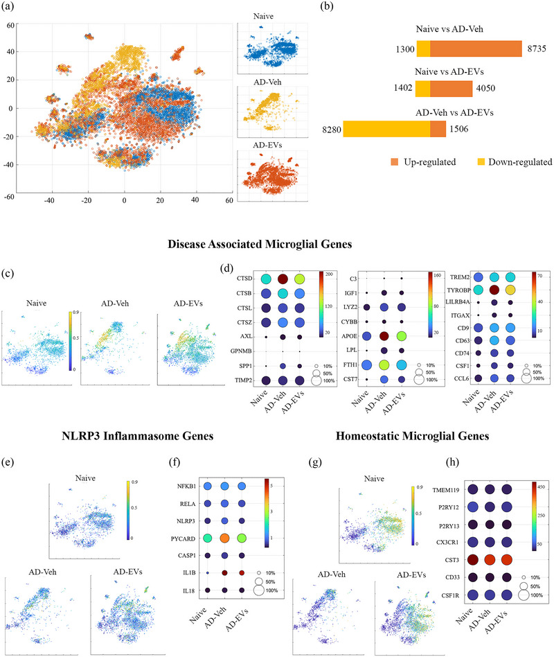
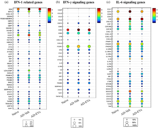
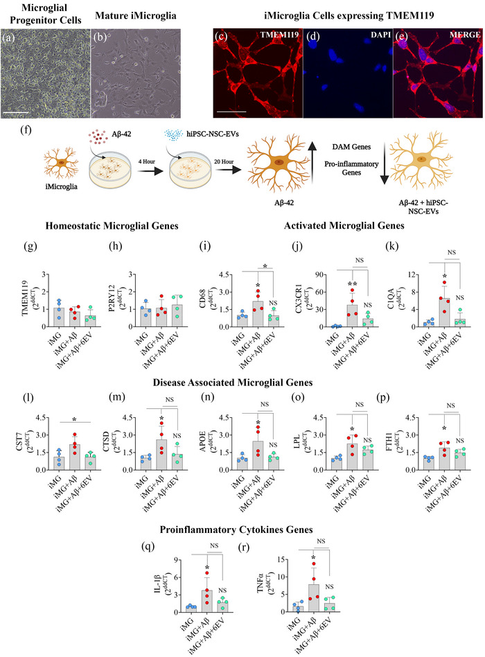
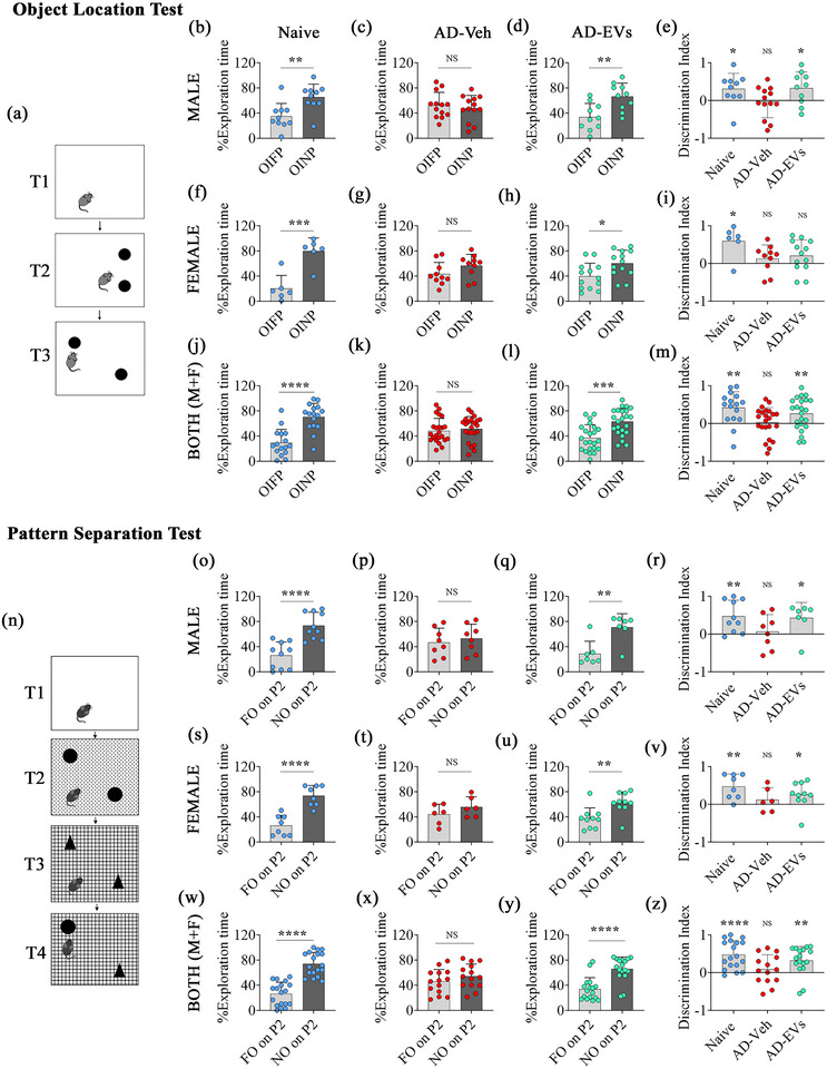
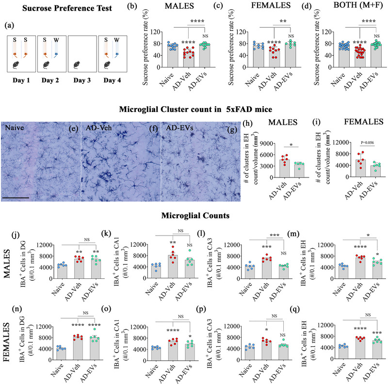
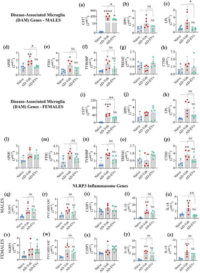
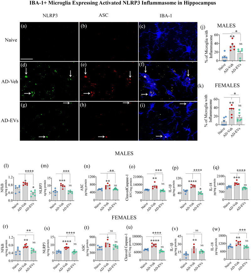
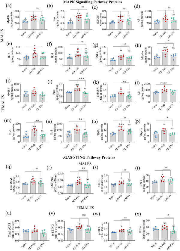
As current treatments for Alzheimer's disease (AD) lack disease-modifying interventions, novel therapies capable of restraining AD progression and maintaining better brain function have great significance. Anti-inflammatory extracellular vesicles (EVs) derived from human induced pluripotent stem cell (hiPSC)-derived neural stem cells (NSCs) hold promise as a disease-modifying biologic for AD. This study directly addressed this issue by examining the effects of intranasal (IN) administrations of hiPSC-NSC-EVs in 3-month-old 5xFAD mice. IN administered hiPSC-NSC-EVs incorporated into microglia, including plaque-associated microglia, and encountered astrocyte soma and processes in the brain. Single-cell RNA sequencing revealed transcriptomic changes indicative of diminished activation of microglia and astrocytes. Multiple genes linked to disease-associated microglia, NOD-, LRR-, and pyrin domain-containing protein 3 (NLRP3)-inflammasome and interferon-1 (IFN-1) signalling displayed reduced expression in microglia. Adding hiPSC-NSC-EVs to cultured human microglia challenged with amyloid-beta oligomers resulted in similar effects. Astrocytes also displayed reduced expression of genes linked to IFN-1 and interleukin-6 signalling. Furthermore, the modulatory effects of hiPSC-NSC-EVs on microglia in the hippocampus persisted 2 months post-EV treatment without impacting their phagocytosis function. Such effects were evidenced by reductions in microglial clusters and inflammasome complexes, concentrations of mediators, and end products of NLRP3 inflammasome activation, the expression of genes and/or proteins involved in the activation of p38/mitogen-activated protein kinase and IFN-1 signalling, and unaltered phagocytosis function. The extent of astrocyte hypertrophy, amyloid-beta plaques, and p-tau were also reduced in the hippocampus. Such modulatory effects of hiPSC-NSC-EVs also led to better cognitive and mood function. Thus, early hiPSC-NSC-EV intervention in AD can maintain better brain function by reducing adverse neuroinflammatory signalling cascades, amyloid-beta plaque load, and p-tau. These results reflect the first demonstration of the efficacy of hiPSC-NSC-EVs to restrain neuroinflammatory signalling cascades in an AD model by inducing transcriptomic changes in activated microglia and reactive astrocytes.

Keywords: Anti‐inflammatory effects; disease‐associated microglia; extracellular vesicles; human induced pluripotent stem cell‐derived neural stem cells; inflammasomes; interferon 1 signalling; mitogen‐activated protein kinase signalling.


Conflict of interest statement

The authors report no conflicts of interest.

Figures

FIGURE 1
FIGURE 1
Intranasal administration of extracellular vesicles from human induced pluripotent stem cell‐derived neural stem cells (hiPSC‐NSC‐EVs) altered the expression of genes linked to disease‐associated microglia (DAM) and NOD‐, LRR‐, and pyrin domain‐containing protein 3 (NLRP3) inflammasome activation in 5xFAD mice microglia when observed 72 h post‐EVs administration. Figure (a) illustrates the distinct t‐SNE plots of microglia in naïve, AD‐Veh, and AD‐EVs groups. (b) represents the total number of microglial genes upregulated and downregulated in the AD‐Veh and AD‐EVs groups compared to the naïve group. (c and d) represent the Ucell score scatter plot and dot plots of different DAM genes in microglia of naïve, AD‐Veh, and AD‐EVs groups. (e and f) represent the Ucell score scatter plot and dot plots of different NLRP3 inflammasome genes in microglia of different groups. (g and h) represent the Ucell score scatter plot and dot plots of different homeostatic microglial genes in microglia of different groups.
FIGURE 2
FIGURE 2
Intranasal administration of extracellular vesicles from human induced pluripotent stem cell‐derived neural stem cells (hiPSC‐NS‐EVs) altered the expression of genes linked to interferon‐1 (IFN‐1), interferon‐gamma (IFN‐γ) interleukin‐6 (IL‐6) signalling in 5xFAD mice microglia when observed 72 h post‐EVs administration. Dot plots in A‐C compare the expression of multiple genes linked to IFN‐1, IFN‐γ signalling, and IL‐6 signalling pathways between naive, AD‐Veh, and AD‐EVs groups. The expression of most genes was upregulated in the AD‐Veh group compared to the naive group but reduced in the AD‐EVs treatment group compared to the AD‐Veh group.
FIGURE 3
FIGURE 3
Intranasal administration of extracellular vesicles from human induced pluripotent stem cell‐derived neural stem cells (hiPSC‐NSC‐EVs) suppressed Aβ42‐induced activation of iMicroglia derived from hiPSCs. Images (a and b) show the progenitor and mature microglia from hiPSCs. (c‐e) images confirm TMEM119 expression in mature microglia. (f) is a diagrammatic representation of the experiment involving Aβ42 exposure to iMicroglia followed by hiPSC‐NSC‐EV treatment. Bar charts (g‐h), (i‐k), (l‐p), and (q‐r) respectively compare the expression of homeostatic genes (g‐h), activated microglia genes (i‐k), disease‐associated microglia (DAM) genes (l‐p), and proinflammatory cytokine genes (q‐r) in iMicroglia between control, Aβ42‐exposed, and Aβ42‐exposed and hiPSC‐NSC‐EVs treated cultures. Scale bar, A‐E = 100 µm; *, p < 0.05; **, p < 0.01; NS, not significant.
FIGURE 4
FIGURE 4
Intranasal administration of extracellular vesicles from human induced pluripotent stem cell‐derived neural stem cells (hiPSC‐NSC‐EVs) to 5xFAD mice preserved cognitive function. Cartoon A depicts different trials (t1‐t3) in an object location test (OLT). The bar charts in b‐d, f‐h, j‐l compare percentages of object exploration times spent with the object in the familiar place (OIFP) vis‐à‐vis the object in the novel place (OINP) in naïve (b, f, j), AD‐Veh (c, g, k), and AD‐EVs (d, h, l) in males (b‐d), females (f‐h), and when both males and females were considered together (j‐l). The bar charts e, i, m compare the OINP discrimination index (DI) for males (e), females (i), and both sexes (m) using a one‐sample t‐test. Cartoon N depicts different trials (t1‐t4) in a pattern separation test (PST). The bar charts in o‐q, s‐u, w‐y compare percentages of object exploration times spent with the familiar object on pattern 2 (FO on P2) vis‐à‐vis the novel object on P2 (NO on P2) in naïve (o, s, w), AD‐Veh (p, t, x), and AD‐EVs (q, u, y) in males (o‐q), females (s‐u) when both males and females were considered together (W‐Y) together. The bar charts R, V, Z compare the DI for the NO on P2 for males (r), females (v), and both sexes (z) using a one‐sample t‐test. *, p < 0.05; **, p < 0.01; ***, < 0.001; and ****, p < 0.0001; NS, not significant.
FIGURE 5
FIGURE 5
Intranasal administration of extracellular vesicles from human induced pluripotent stem cell‐derived neural stem cells (hiPSC‐NSC‐EVs) to 5xFAD mice prevented anhedonia and reduced microglial clusters and numbers. Cartoon (A) depicts the experimental design employed for the sucrose preference test. The bar charts (B‐D) compare the sucrose preference rate (SPR) in males (B), females (C) and males + females (D) across naïve, AD‐Veh and AD‐EVs groups. **, p < 0.01; ****, p < 0.0001; NS, not significant. Figure (E‐G) illustrates the representative images of microglial clusters in naïve (E), AD‐Veh (F), and AD‐EVs (G) groups. Bar charts (H‐I) compare the number of microglial clusters per mm3 unit area of the hippocampus in males (H) and females (I) between naive, AD‐Veh, and AD‐EV groups. Bar charts J‐Q compare numbers of microglia in males (J‐M) and females (N‐Q) per 0.1 mm3 area of the dentate gyrus (J, N), CA1 subfield (K, O), CA3 subfield (L, P), and the entire hippocampus (M, Q) between naive, AD‐Veh, and AD‐EVs groups. Scale bar, E‐G = 100 µm *, p < 0.05; **, p < 0.01; ***, p < 0.001; ****, p < 0.0001; NS, not significant.
FIGURE 6
FIGURE 6
Intranasal administration of extracellular vesicles from human induced pluripotent stem cell‐derived neural stem cells (hiPSC‐NSC‐EVs) to 5xFAD mice normalized the expression of genes linked to disease associated microglia (DAM) and inflammasome activation. The bar charts A‐P compare the expression of DAM genes (cst7, spp1, lpl, apoe, fth1, tyrobp, trem2, ctsd) in the hippocampus of males (A‐H) and females (I‐P) between naïve, AD‐Veh and AD‐EVs groups. The bar charts Q‐Z compare the expression of genes linked to NOD‐, LRR‐ and pyrin domain‐containing protein 3 (NLRP3) inflammasome activation (nlrp3, pycard, casp1, il‐1β, il‐18) in hippocampus of males (Q‐U) and females (V‐Z) between naïve, AD‐Veh and AD‐EVs groups. *, p < 0.05; **, p < 0.01; ***, p < 0.001; ****, p < 0.0001; NS, not significant.
FIGURE 7
FIGURE 7
Intranasal administration of extracellular vesicles from human induced pluripotent stem cell‐derived neural stem cells (hiPSC‐NSC‐EVs) to 5xFAD mice inhibited NOD‐, LRR‐ and pyrin domain‐containing protein 3 (NLRP3) inflammasome complex formation and activation. Figures (A‐I) illustrate examples of NLRP3 inflammasome complexes co‐expressing NLRP3 (green) and apoptosis‐associated speck‐like protein containing a CARD (ASC, red) in IBA‐1+ microglia (blue) from the CA3 subfield of the hippocampus in male mice from naïve (A‐C), AD‐Veh (D‐F), and AD‐EVs (G‐I) groups. The bar charts J‐K compare the percentages of microglia with inflammasome complexes in males (J) and females (K). The bar charts L‐W compare the concentrations of mediators of NLRP3 inflammasome activation (NF‐kB, NLRP3, ASC, and cleaved caspase‐1; L‐O, males and R‐U, females) and end products (IL‐1β, IL‐18; P‐Q, and V‐W) in males (L‐Q) and females (R‐W) between naive, AD‐Veh, and AD‐EVs groups. Scale bar, A‐I = 25 µm; *, p < 0.05; **, p < 0.01; ***, p < 0.001; ****, p < 0.0001; NS, not significant.
FIGURE 8
FIGURE 8
Intranasal administration of extracellular vesicles from human induced pluripotent stem cell‐derived neural stem cells (hiPSC‐NSC‐EVs) to 5xFAD mice thwarted the activation of p38 mitogen‐activated protein kinase and cyclic GMP‐AMP synthase (cGAS), and phosphorylated stimulator of interferon genes (p‐STING) signalling. The bar charts A‐P compare the concentrations of various components of p38/MAPK activation (MyD88, Ras, pMAPK, AP‐1; A‐D and I‐L) and end products (IL‐6, IL‐8, TNFα, Mip‐1α; E‐H and M‐P) in males (a‐h) and females (i‐p) between naive, AD‐Veh, and AD‐EVs groups. *, p < 0.05; **, p < 0.01; ***, p < 0.001; NS, not significant. The bar charts Q‐X compare total cyclic GMP‐AMP synthase (cGAS, q, u), stimulator of interferon genes (p‐STING, r, v), p‐interferon regulatory factor 3 (p‐IRF3, s, w), IFN‐α (t, x) across groups in males (q‐t) and females (u‐x). *, p < 0.05; **, p < 0.01; ***, p < 0.001; NS, not significant.
FIGURE 9
FIGURE 9
Intranasal administration of extracellular vesicles from human induced pluripotent stem cell‐derived neural stem cells (hiPSC‐NSC‐EVs) to 5xFAD mice reduced amyloid plaques and phosphorylated tau. Figures (A‐B) illustrate the distribution of amyloid plaques in the hippocampus from 5xFAD mice receiving the vehicle (Veh, A) or hiPSC‐NSC‐EVs (B). The bar charts compare area fraction (AF) of amyloid plaques (C, D), the concentration of soluble Aβ42 (E, F), and p‐tau (G, H) in males (C, E, G) and females (D, F, H) between AD‐Veh and AD‐EVs groups. Scale bar, A‐B = 500 µm; *, p < 0.05; **, p < 0.01; NS, not significant.

Update of

Similar articles

Cited by

References

    1. Abdi, S. , Javanmehr, N. , Ghasemi‐Kasman, M. , Bali, H. Y. , & Pirzadeh, M. (2022). Stem cell‐based therapeutic and diagnostic approaches in Alzheimer's disease. Current Neuropharmacology, 20, 1093–1115. 10.2174/1570159x20666211231090659 - DOI - PMC - PubMed
    1. Akama, K. T. , & Van Eldik, L. J. (2000). Beta‐amyloid stimulation of inducible nitric‐oxide synthase in astrocytes is interleukin‐1beta‐ and tumor necrosis factor‐alpha (TNFalpha)‐dependent, and involves a TNFalpha receptor‐associated factor‐ and NFkappaB‐inducing kinase‐dependent signalling mechanism. Journal of Biological Chemistry, 275, 7918–7924. 10.1074/jbc.275.11.7918 - DOI - PubMed
    1. Apodaca, L. A. , Baddour, A. A. D. , Garcia, C. , Jr., Alikhani, L. , Giedzinski, E. , Ru, N. , Agrawal, A. , Acharya, M. M. , & Baulch, J. E. (2021). Human neural stem cell‐derived extracellular vesicles mitigate hallmarks of Alzheimer's disease. Alzheimers Research & Therapy, 13, 57. 10.1186/s13195-021-00791-x - DOI - PMC - PubMed
    1. Attaluri, S. , Jaimes Gonzalez, J. , Kirmani, M. , Vogel, A. D. , Upadhya, R. , Kodali, M. , Madhu, L. N. , Rao, S. , Shuai, B. , Babu, R. S. , Huard, C. , & Shetty, A. K. (2023). Intranasally administered extracellular vesicles from human induced pluripotent stem cell‐derived neural stem cells quickly incorporate into neurons and microglia in 5xFAD mice. Frontiers in Aging Neuroscience, 15, 1200445. 10.3389/fnagi.2023.1200445 - DOI - PMC - PubMed
    1. Attwood, S. W. , & Edel, M. J. (2019). iPS‐Cell Technology and the problem of genetic instability‐can it ever be safe for clinical use? Journal of Clinical Medicine, 8, 288. 10.3390/jcm8030288 - DOI - PMC - PubMed

MeSH terms

Substances

LinkOut - more resources