. 2024 Nov 12;121(46):e2404091121.
doi: 10.1073/pnas.2404091121. Epub 2024 Nov 5.

Deciphering the RNA-binding protein network during endosomal mRNA transport

Affiliations

Deciphering the RNA-binding protein network during endosomal mRNA transport

Senthil-Kumar Devan et al. Proc Natl Acad Sci U S A. .

Abstract

Microtubule-dependent endosomal transport is crucial for polar growth, ensuring the precise distribution of cellular cargos such as proteins and mRNAs. However, the molecular mechanism linking mRNAs to the endosomal surface remains poorly understood. Here, we present a structural analysis of the key RNA-binding protein Rrm4 from Ustilago maydis. Our findings reveal a different type of MademoiseLLE domain (MLLE) featuring a seven-helical bundle that provides a distinct binding interface. A comparative analysis with the canonical MademoiseLLE domain of the poly(A)-binding protein Pab1 disclosed unique characteristics of both domains. Deciphering the MLLE binding code enabled prediction and verification of previously unknown Rrm4 interactors containing short linear motifs. Importantly, we demonstrated that the human MLLE domains, such as those of PABPC1 and UBR5, employed a similar principle to distinguish among interaction partners. Thus, our study provides detailed mechanistic insights into how structural variations in the widely distributed MLLE domain facilitate mRNA attachment during endosomal transport.

Keywords: PAM2; RNA transport; SLiM; Ustilago maydis; endosome.


Conflict of interest statement

Competing interests statement:The authors declare no competing interest.

Figures
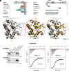
Fig. 1.
Fig. 1.
A seven-helix type MLLE domain confers specific binding. (A) Schematic representation of protein variants (molecular weight in kilo Daltons, green, RRM; orange, MLLERrm4 domains; bright blue, MLLEPab1; light blue PAM2Upa1; light red PAM2L1Upa1; dark red PAM2L2Upa1; dark gray, Ankyrin repeats 5xANK, FYVE domain, and RING domain of Upa1, cyan, His6). Sequences of PAM2 and PAM2L1,2 peptides are denoted. The following symbols are used: M3, MLLE3Rrm4; M, MLLEPab1; 5H, five helices; 7H, seven helices, G, GST tag; HS, His6-Sumo tag (figure adapted from ref. 14). (B) 3D structural models of the MLLE3Rrm4 domain, generated using TopModel, AlphaFold, and X-ray as indicated. (C) Western blot analysis of GST pull-down experiments using α-His for detection (input, respective His6-SUMO peptides). (D) Representative ITC binding curves of MLLE3Rrm4 domain (H-Rrm4-M3). KD values from two independent measurements are given (indicated data in bold).
Fig. 2.
Fig. 2.
PAM2L ligands are recognized via a distinctive interaction interface of MLLE3. (A) Crystal structures of MLLE3Rrm4–PAM2L1Upa1, MLLE3Rrm4–PAM2L2Upa1 complexes are superimposed (RMSD 0.4 Å). The PAM2L1,2Upa1 peptides are inserted into the hydrophobic pocket formed by the helices α2, 3 of MLLE3Rrm4. Models are represented as a cartoon (Left, orange, MLLE3Rrm4) and surface (Right, MLLE3Rrm4, according to electrostatic potential: blue, positively charged; red, negatively charged residues), salmon sticks, PAM2L1Upa1; ruby red sticks, PAM2L2Upa1. Key residues are labeled. (B) Interface of the MLLE3Rrm4 and PAM2L1Upa1 (Left) as well as MLLE3Rrm4 and PAM2L2Upa1 (Right). PAM2L1,2Upa1 peptides and interacting side chains of MLLE3Rrm4 are shown as sticks. Dashed lines indicate hydrogen bond interactions. PAM2L1, 2 sequences of Upa1 are indicated at the Bottom; bound residues in the crystal structure are highlighted.
Fig. 3.
Fig. 3.
A seven-helix type MLLE domain is necessary and sufficient for endosomal attachment. (A) Schematic representation of Rrm4 variants (amino acid number indicated; drawn not to scale): dark green, RRM; orange, MLLE domains; red, mKate2. (B) Hyphal growth of AB33 derivatives (6 h.p.i.; size bar, 10 μm). Growth direction is indicated by arrows. (C) Quantification of hyphal growth of AB33 derivatives shown in panel B (6 h.p.i.): Unipolarity, bipolarity, and basal septum formation were quantified (error bars, SEM; n = 3 independent experiments, >150 hyphae were counted per strain) for statistical evaluation, the percentage of uni- and bipolarity was investigated and unpaired two-tailed Student’s t test was performed (α < 0.05). (D) Micrographs (inverted fluorescence image; size bar, 10 μm) and corresponding kymographs of AB33 hyphae derivatives (6 h.p.i.) showing movement of Rrm4-Kat variants in hyphae (inverted fluorescence images; arrow length on the Left and Bottom indicates time and distance, respectively). Processive signals, aberrant microtubule staining, and accumulation of static Rrm4-Kat signals are indicated by red, yellow, and purple arrowheads, respectively.
Fig. 4.
Fig. 4.
The MLLE domain of Pab1 recognizes its PAM2 ligand in a canonical fashion. (A) Crystal structure of PAM2Upa1 bound to MLLEPab1. The PAM2Upa1 peptide wraps around the MLLEPab1. Key residues (L132 and F139) are inserted into the hydrophobic pocket formed between the helices α3,5 and between the helices α2,3. Models are represented as a cartoon; light blue sticks, PAM2Upa1 peptide; interacting side chains of MLLEPab1 are shown as sticks, and dashed yellow lines indicate hydrogen bonding. (B) Surface representation of the MLLEPab1–PAM2Upa1 complex depicting the hydrophobic binding pockets (MLLEPab1, according to electrostatic potential: blue, positively charged; red, negatively charged residues, light blue sticks, PAM2Upa1 where key residues are labeled). (C) Western blot analysis of GST pull-down experiments using α-His for detection. (D) PAM2Upa1 sequence is denoted in light blue, PAM2L2Upa1 sequence is denoted in red, and hybrid versions are denoted in light blue-red dual color). (E) Western blot analysis of GST pull-down experiments using α-His for detection.
Fig. 5.
Fig. 5.
Vps8 and Taf7 are de novo predicted interaction partners of Rrm4. (A) Schematic representation of Vps8 variants (Left, UMAG_15064) (aa number is indicated next to protein bars, drawn to scale, the bar on the top right is 200 AA; pink, PAM2L1,2; wheat, β-propeller domain; dark teal, α-solenoid domain; purple, RING domain; green, GFP). Schematic representation of Taf7 (Right, UMAG_10620) drawn to scale (Pink, PAM2L; wheat, activator interaction domain; dark teal, homolog of human TAFII250 interaction domain). (B) 3D structural model predicted using AlphaFold (Bottom) with domains depicted using the above color code; amino (N) and carboxy (C) termini are indicated. Structural model predicted using AlphaFold (Bottom) with domains depicted using the above color code, amino (N), carboxy (C) terminals are indicated. (C) Western blot analyses of GST pull-down experiments using α-His for detection. (D) De novo predicted PAM2L peptides of Vps8 and Taf7 are denoted, conserved crucial resides are shaded in black, conserved key acidic residues are shaded in gray. (E) Micrographs (inverted fluorescence image; size bar, 10 μm) and corresponding kymographs of AB33 hyphae derivatives (6 h.p.i.) depicted in SI Appendix, Fig. S6. The Kymographs show movement of Rrm4-Kat variants in hyphae (inverted fluorescence images; arrow length on the Left and Bottom indicates time and distance, respectively). Processive signals indicating the Rrm4-Kat movement, aberrant accumulation at the tip and aberrant microtubule staining are indicated by red, purple, and yellow and arrowheads, respectively.
Fig. 6.
Fig. 6.
MLLE domains of PABC1 and UBR5 differentiate between binding partners. (A) Schematic representation of protein variants drawn to scale (aa number is indicated next to protein bars; drawn to scale, the bar on the top right is 200 AA; gray, UBA, Ubr box, GST; yellow, HECT; orange, MLLEUBR5; red, PAM2L; blue, MLLEPABC1; green, RRM; cyan, MKRN1; wheat, Mkr1). (B) Comparison of PAM2 and PAM2L sequences found in Upa1 (UniProtKB ID: A0A0D1E015) with those of human proteins, such as PAIP2 (Q9BPZ3), TOB (P50616), GW182 (Q9HCJ0), MKRN1 (Q9UHC7), Mkr1 (A0A0D1E4Z6), and UBR5 (O95071). (C) Structural models of MKRN1 and Mkr1 predicted using AlphaFold with domains depicted in the color code similar to the respective labels, PAM2L motifs are shown in. (D) Western blot analysis of GST pull-down experiments using α-His for detection.
Fig. 7.
Fig. 7.
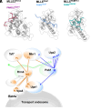
MLLE domains exhibit defined binding specificity. (A) Structures of three different MLLE domains bound to ligands. MLLE3Rrm4-PAM2L1,2Upa1, MLLEPab1-PAM2Upa1, and MLLEPABPC1-PAM2GW182 (Right, PDB ID: 3KTP). Helices 2 and 3 are labeled. (B) Model depicting the complex protein–protein interaction network based on the binding specificity of MLLE domains of Rrm4 (orange) and Pab1 (blue). The following symbols are used: M1, MLLE1; M2, MLLE2; M3, MLLE3; M, MLLE; PL, PAM2-like; P, PAM2; mRNA with poly(A) tail in green; ?, unknown proteins.

References

    1. Riquelme M., et al. , Fungal morphogenesis, from the polarized growth of hyphae to complex reproduction and infection structures. Microbiol. Mol. Biol. Rev. 82, 1–47 (2018). - PMC - PubMed
    1. Gow N. A., Hube B., Importance of the Candida albicans cell wall during commensalism and infection. Curr. Opin. Microbiol. 15, 406–412 (2012). - PubMed
    1. Lanver D., et al. , Ustilago maydis effectors and their impact on virulence. Nat. Rev. Microbiol. 15, 409–421 (2017). - PubMed
    1. Salogiannis J., Reck-Peterson S. L., Hitchhiking: A non-canonical mode of microtubule-based transport. Trends Cell Biol. 27, 141–150 (2016). - PMC - PubMed
    1. Steinberg G., The transport machinery for motility of fungal endosomes. Fungal Genet. Biol. 49, 675–676 (2012). - PubMed

LinkOut - more resources