. 2024 Nov 12;121(46):e2322759121.
doi: 10.1073/pnas.2322759121. Epub 2024 Nov 5.

VEGF inhibition increases expression of HIF-regulated angiogenic genes by the RPE limiting the response of wet AMD eyes to aflibercept

Affiliations

VEGF inhibition increases expression of HIF-regulated angiogenic genes by the RPE limiting the response of wet AMD eyes to aflibercept

Deepti Sharma et al. Proc Natl Acad Sci U S A. .

Abstract

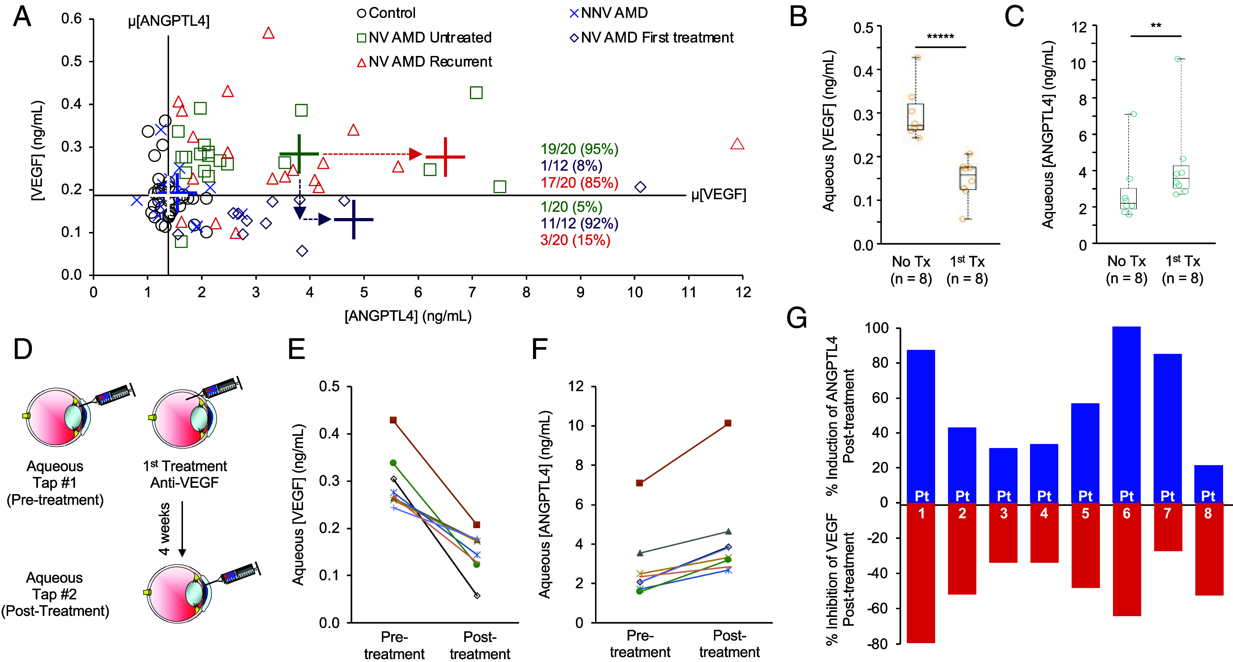
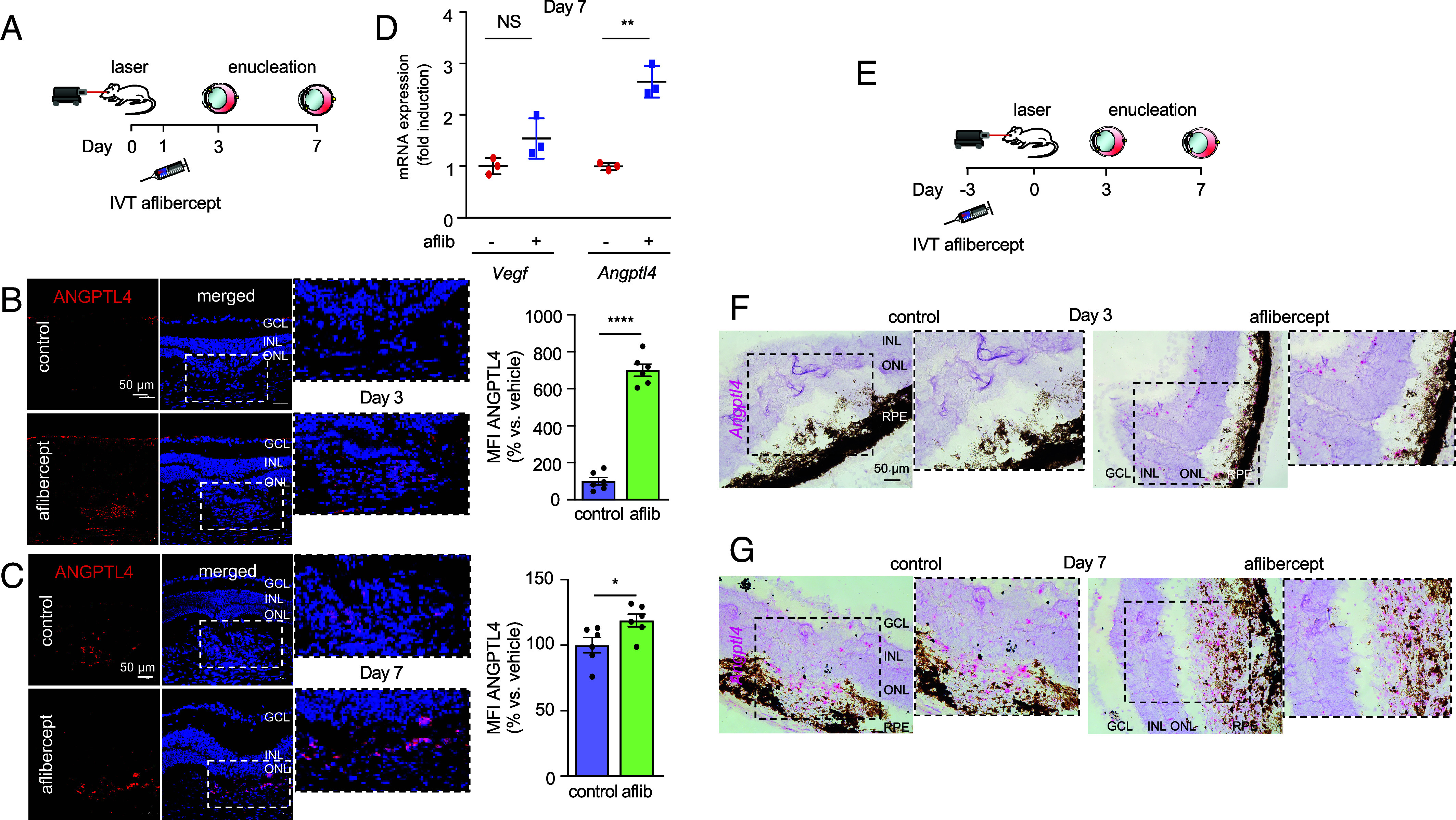
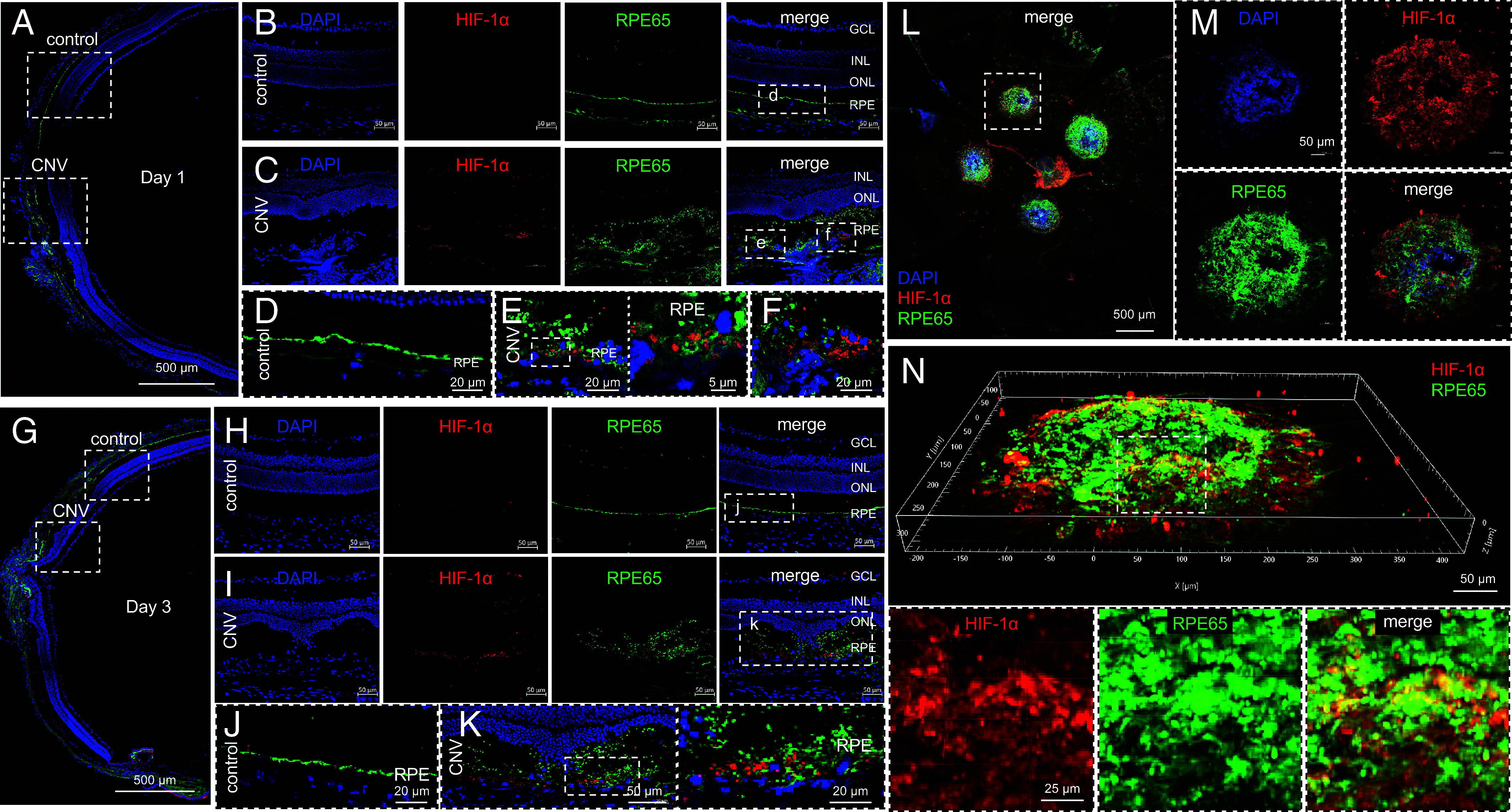
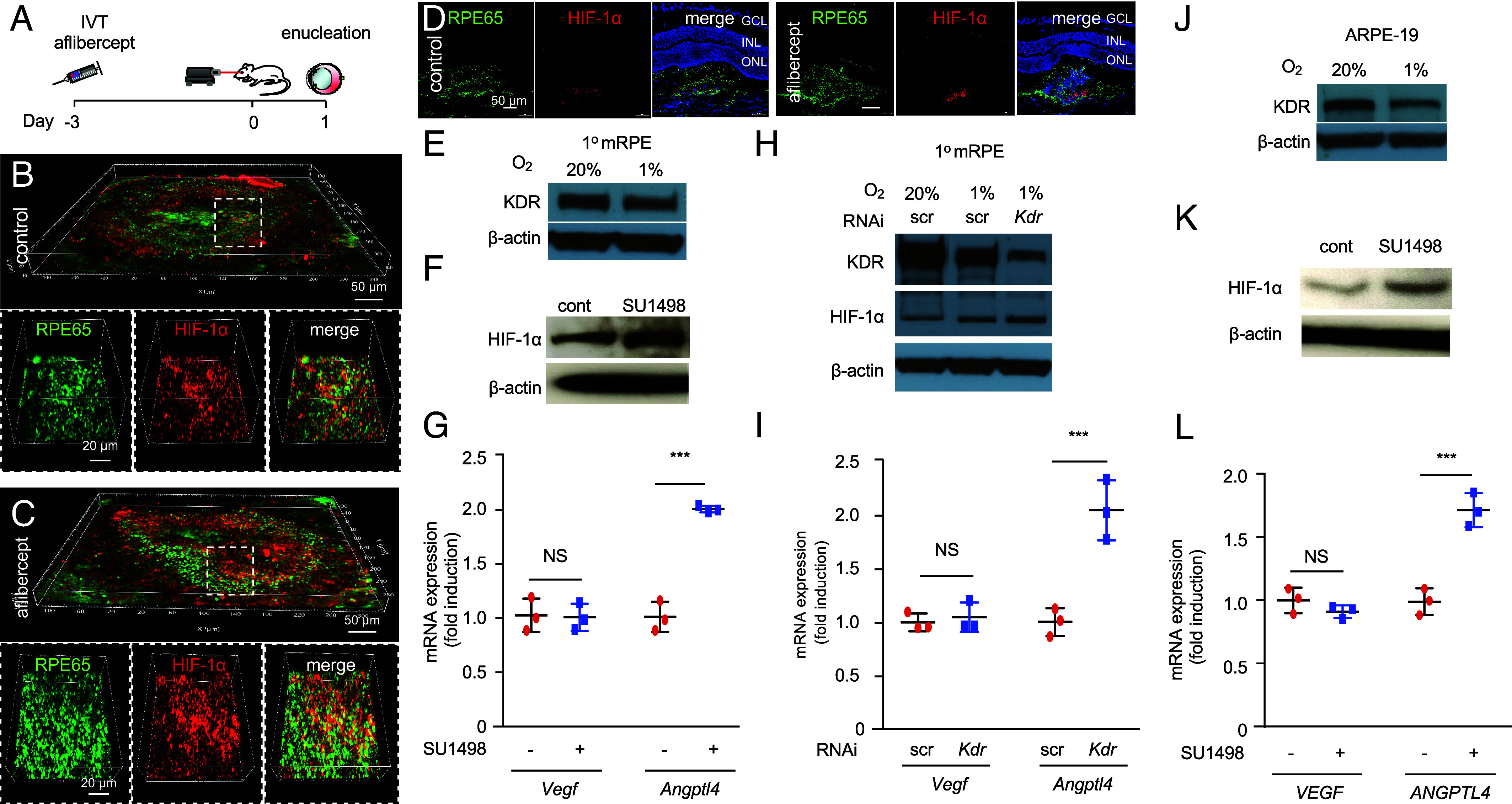
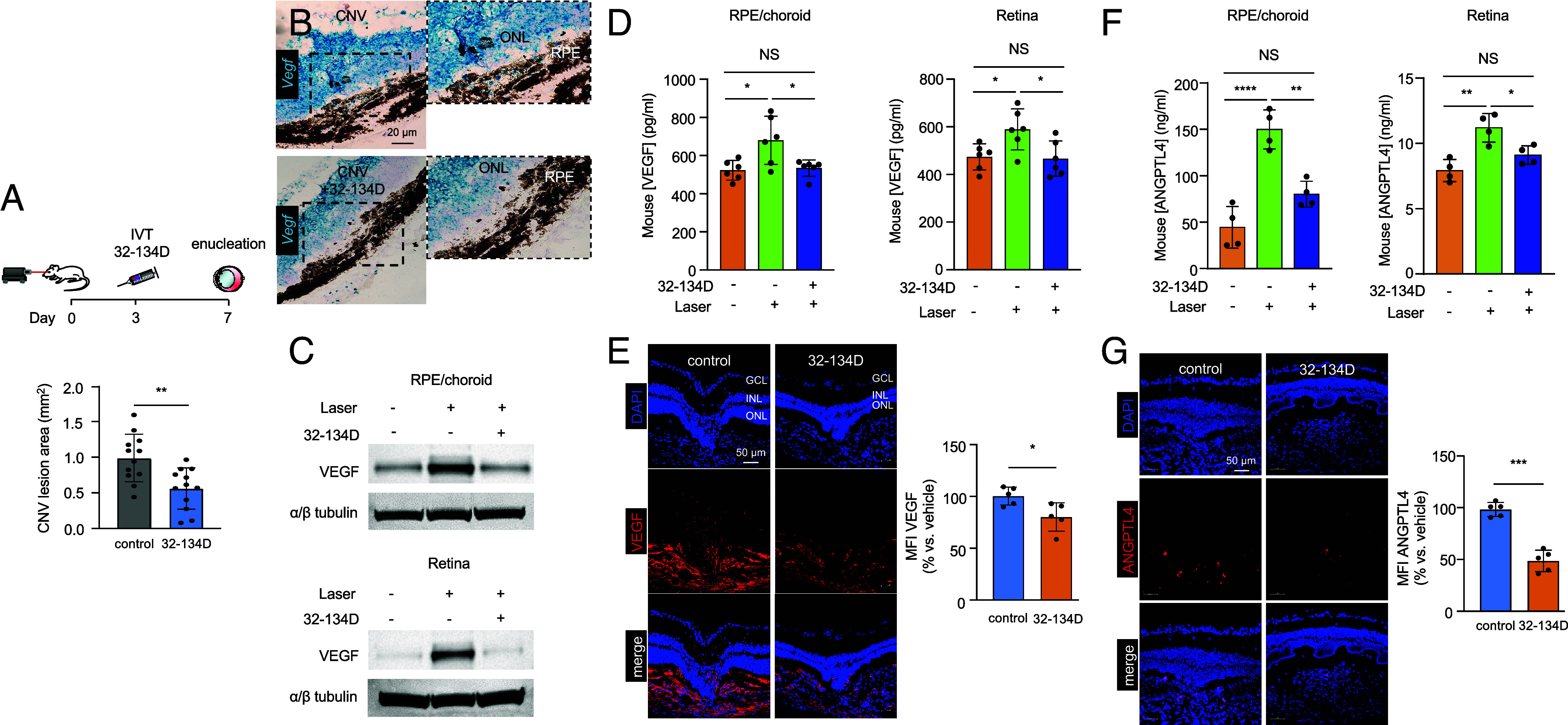
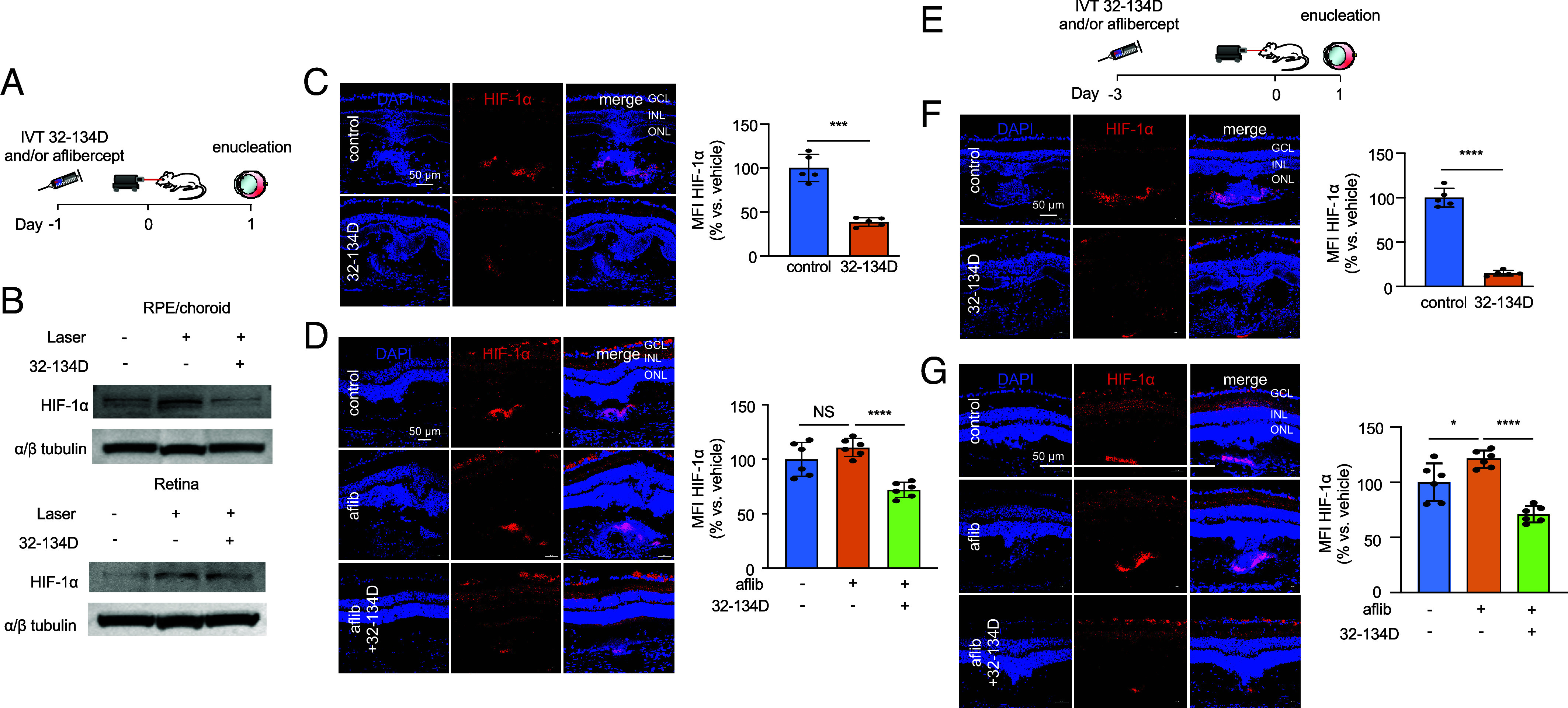
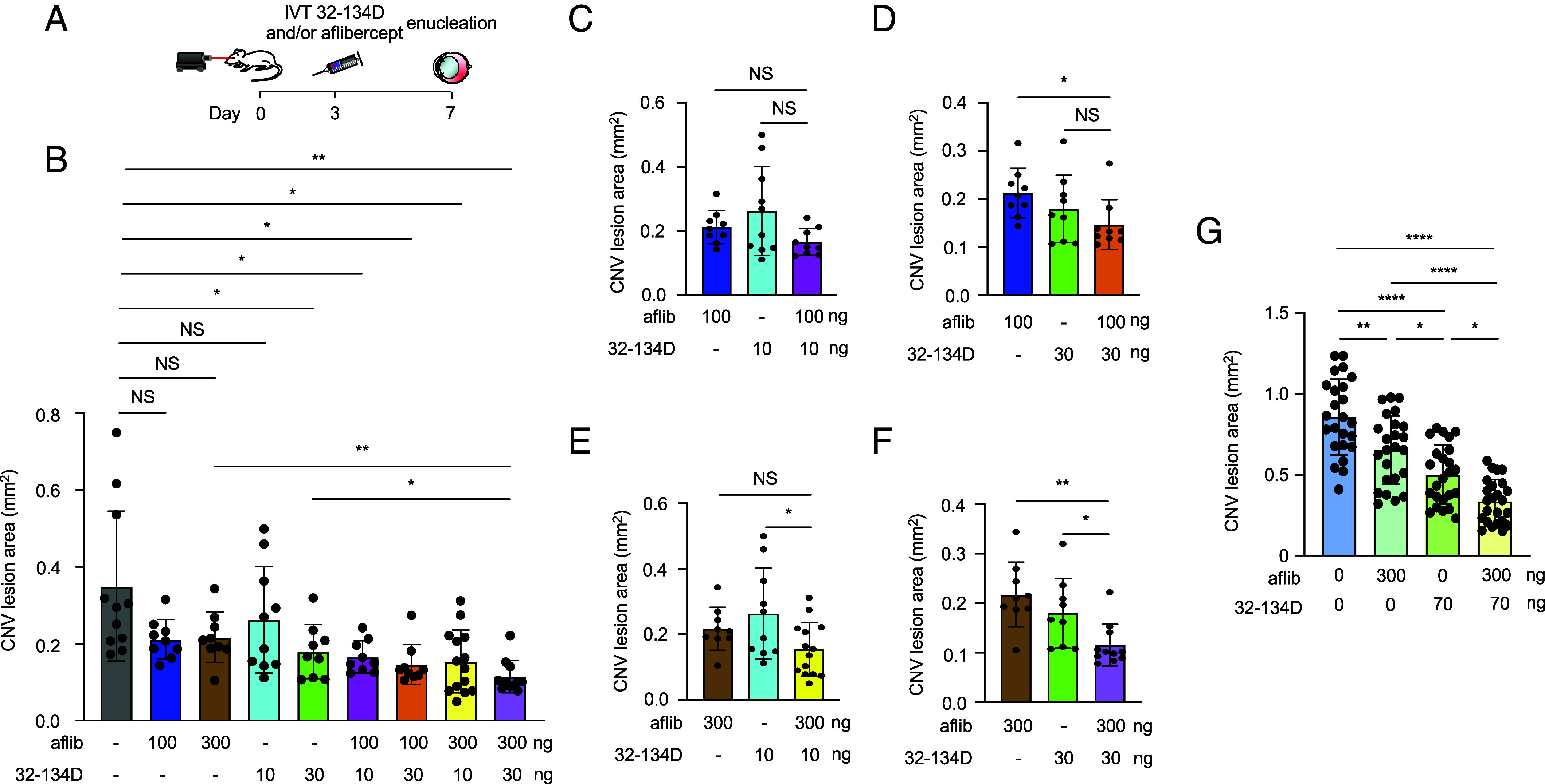
Neovascular age-related macular degeneration (nvAMD) is the leading cause of severe vision loss in the elderly in the developed world. While the introduction of therapies targeting vascular endothelial growth factor (VEGF) has provided the first opportunity to significantly improve vision in patients with nvAMD, many patients respond inadequately to current anti-VEGF therapies. It was recently demonstrated that expression of a second angiogenic mediator, angiopoietin-like 4 (ANGPTL4), synergizes with VEGF to promote choroidal neovascularization (CNV) in mice and correlates with reduced response to anti-VEGF therapy in patients with nvAMD. Here, we report that expression of ANGPTL4 in patients with nvAMD increases following treatment with anti-VEGF therapy and that this increase is dependent on accumulation of hypoxia-inducible factor (HIF)-1α in response to inhibition of VEGF/KDR signaling in the retinal pigment epithelium (RPE). We therefore explored HIF-1 inhibition with 32-134D, a recently developed pharmacologic HIF-inhibitor, for the treatment of nvAMD. 32-134D prevented the expression of both VEGF and ANGPTL4 and was at least as effective as aflibercept in treating CNV in mice. Moreover, by preventing the increase in HIF-1α accumulation in the RPE in response to anti-VEGF therapy, combining 32-134D with aflibercept was more effective than either drug alone for the treatment of CNV. Collectively, these results help explain why many patients with nvAMD respond inadequately to anti-VEGF therapy and suggest that the HIF inhibitor 32-134D will be an effective drug-alone or in combination with current anti-VEGF therapies-for the treatment of patients with this blinding disease.

Keywords: age-related macular degeneration; angiopoietin-like 4; choroidal neovascularization; hypoxia inducible factor; vascular endothelial growth factor.


Conflict of interest statement

Competing interests statement:G.L.S. and A.S. are co-founders of and hold equity in HIF Therapeutics, Inc. S.S., Y.H., G.L.S., and A.S. are inventors on provisional patent application PCT/US2022/039883. This arrangement has been reviewed and approved by the Johns Hopkins University in accordancewith its conflict of interest policies.

Figures

Fig. 1.
Fig. 1.
Aqueous levels of ANGPTL4 are increased in patients with nvAMD following treatment with anti-VEGF therapy. (A) Scatter plot comparing aqueous levels of VEGF and ANGPTL4 in NV AMD patients (untreated, following their first treatment or with recurrent CNV), compared to NNV AMD and non-AMD controls. Average (cross) VEGF (horizontal bar) and ANGPTL4 (vertical bar) levels for control (white), NN VAMD (blue), or NV AMD (untreated, green; first treatment, purple; or recurrent, red). The color matched number (percent) of patients with high VEGF/high ANGPTL4 or low VEGF/high ANGPTL4 are shown. Dashed arrows denote change in mean ANGPTL4 (horizontal) or VEGF (vertical) levels in untreated vs. first treatment or recurrent NV AMD. (B and C) Aqueous levels of VEGF (B) and ANGPTL4 (C) in NV AMD patients (treatment-naïve, No Tx; following one treatment, 1st Tx). (D) Schematic of aqueous fluid acquisition from NV AMD patients before and 4 wk after receiving a single treatment with anti-VEGF therapy. (E and F) Aqueous levels of VEGF (E) and ANGPTL4 (F) in patients with NV AMD prior to (pretreatment) and 4 wk following (posttreatment) their first anti-VEGF treatment. (G) Bar graph depicting the % change of aqueous ANGPTL4 (blue) or VEGF (red) in these patients. Statistical analyses were performed by two-tailed Student’s t test, *P < 0.05; **P < 0.01; *****P < 0.00001. NS, not significant.
Fig. 2.
Fig. 2.
Anti-VEGF therapy increases ANGPTL4 expression in mice. (A) Schematic depicting laser CNV mice treated with an injection of aflibercept (300 ng) or PBS 1 d after laser treatment. (B and C) Representative image depicting expression of ANGPTL4 protein (immunofluorescence) within CNV lesions on day 3 (B) or 7 (C) (Left) with quantitation (Right). (D) Scatter plot demonstrating expression of Vegf and Angptl4 mRNA (qPCR) in RPE/choroid lysates on day 7. (E) Schematic depicting laser CNV mice treated with an injection of aflibercept (300 ng) or PBS 3 d prior to laser treatment. (F and G) Expression of Angptl4 mRNA (in situ hybridization) within CNV lesions on day 3 (F) or 7 (G). n = 3-6 animals per group. GCL, ganglion cell layer; INL, inner nuclear layer; ONL, outer nuclear layer; RPE, retinal pigment epithelium; MFI, mean fluorescence intensity; IVT, intravitreal; aflib, aflibercept. Data are shown as means ± SD. Statistical analyses were performed by two-tailed Student’s t test, *P < 0.05; **P < 0.01; ****P < 0.0001. NS, not significant.
Fig. 3.
Fig. 3.
Expression of HIF-1α in RPE cells in laser CNV mice. (AK) Representative image depicting expression of HIF-1α (red) and RPE65 (green) in laser CNV mice on day 1 (AF) or day 3 (GK). Adjacent (nonlasered) retina is shown as controls. (LN) Representative image depicting expression of HIF-1α (red) and RPE65 (green) in flat mount (L and M) or Z-stack (N) from laser CNV mice on day 3. n = 4-6 animals per group. GCL, ganglion cell layer; INL, inner nuclear layer; ONL, outer nuclear layer; RPE, retinal pigment epithelium.
Fig. 4.
Fig. 4.
The VEGF/KDR axis regulates expression of HIF-1α expression in RPE cells. (A) Above, schematic depicting laser CNV mice treated with an injection of aflibercept (300 ng) or PBS 3 d before laser treatment. (BD) Representative images depicting expression of the RPE cell-specific marker, RPE65 (green) and HIF-1α (red) in laser CNV mice on day 1 in flat mounts (B and C) and sections (D) after treatment with PBS or aflibercept. (E) Western blot depicting expression of KDR in primary mouse RPE cells (1° mRPE) cultured in normoxia (20% O2) or hypoxia (1% O2) for 24 h. (F and G) Effect of KDR inhibition using SU1498 (1 µM; 24 h) on HIF-1α accumulation (F) and Vegf and Angptl4 mRNA expression (qPCR; H) in 1° mRPE. Vehicle (DMSO) was used as a control. (H) Western blot depicting expression of KDR and HIF-1α protein in lysates from 1° mRPE cultured in 20% O2 or 1% O2 for 24 h in the presence of siRNA targeting Kdr or scrambled (scr) control for. (I) Vegf and Angptl4 mRNA expression (qPCR) in 1° mRPE in the presence of siRNA targeting Kdr or scrambled (scr) control. (J) Western blot depicting expression of KDR in ARPE-19 cells cultured in 20% or 1% O2 for 24 h. (K and L) Effect of KDR inhibition using SU1498 (1 µM; 24 h) vs. vehicle (DMSO) control on HIF-1α accumulation (F) and Vegf and Angptl4 mRNA expression (qPCR; H) in ARPE-19 cells. Data are shown as means ± SD. Statistical analyses were performed by two-tailed Student’s t test with Welch’s Correction, *P < 0.05; **P < 0.01; ***P < 0.001; ****P < 0.0001; NS, not significant.
Fig. 5.
Fig. 5.
32-134D inhibits ANGPTL4 and VEGF expression and CNV lesion size in laser CNV mice. (A) Above, schematic depicting laser CNV in 13-to 15-wk-old mice treated with an injection of 32-134D (70 ng) or vehicle (DMSO; control) 3 d after laser treatment. Below, analysis of CNV lesion size on choroid flat mounts on day 7. (B) Expression of Vegf mRNA expression (in situ hybridization) within CNV lesions following treatment with 32-134D or vehicle. (C and D) Expression of VEGF (C, ELISA; D, WB) in RPE/choroid or neurosensory retina lysates from laser CNV mice treated with 32-134D or vehicle. (E–F) Expression of VEGF (E) or ANGPTL4 (F, ELISA; G) by immunofluorescence (Left) with quantitation (Right) on day 7 in laser CNV mice treated with 32-134D or vehicle. n = 5-6 animals. GCL, ganglion cell layer; INL, inner nuclear layer; ONL, outer nuclear layer; RPE, retinal pigment epithelium; MFI, mean fluorescence intensity; IVT, intravitreal. Data are shown as means ± SD. Statistical analyses were performed by two-tailed Student’s t test with Welch’s Correction (A, E, and G) or one-way ANOVA with Bonferroni’s multiple-comparison test (D and F). *P < 0.05; ** P < 0.01; ***P < 0.001; ****P < 0.0001; NS, not significant.
Fig. 6.
Fig. 6.
32-134D prevents aflibercept-induced increase in HIF-1α accumulation in laser CNV mice. (A) Schematic depicting laser CNV mice treated with an injection of aflibercept (300 ng) and/or 32-134D (70 ng) 1 d before laser treatment. (B) Expression of HIF-1α protein (WB) in RPE/choroid or neurosensory retina lysates from laser CNV mice treated with 32-134D or vehicle. (C and D) Expression of HIF-1α (immunofluorescence; Left) with quantitation (Right) within CNV lesions on day 1 in mice treated with 32-134D (C) or aflibercept and/or 32-134D (D). (E) Schematic depicting laser CNV mice treated with an injection of aflibercept (300 ng) and/or 32-134D (70 ng) 3 d before laser treatment. (F and G) Expression of HIF-1α (immunofluorescence; Left) with quantitation (Right) within CNV lesion on day 1 in mice treated with 32-134D (F) or aflibercept and/or 32-134D (G). n = 5-6 animals. GCL, ganglion cell layer; INL, inner nuclear layer; ONL, outer nuclear layer; IVT, intravitreal; aflib, aflibercept. Data are shown as means ± SD. Student’s t test with Welch’s Correction (C and F) and one-way ANOVA with Bonferroni’s multiple-comparison test (D and G). *P < 0.05; ***P < 0.001; ****P < 0.0001; NS, not significant.
Fig. 7.
Fig. 7.
32-134D prevents the countertherapeutic increase in ANGPTL4 observed with aflibercept alone, thereby improving the efficacy of aflibercept. (A) Schematic depicting laser CNV mice treated with an injection of aflibercept and/or 32-134D 3 d after laser treatment. (B) Combined effect of a subthreshold or threshold doses of aflibercept and/or 32-134D on the CNV lesions size on day 7 in 9- to 12-wk-old mice. (CF) Data from Panel B presented to highlight the effect of combining subthreshold and threshold doses of aflibercept and/or 32-134D. (G) Additive effect of a half maximal inhibitory concentration (IC50) of aflibercept and/or 32-134D on the CNV lesions size on day 7 in 13- to 15-wk-old mice. n = 4-5 animals. IVT, intravitreal; aflib, aflibercept. Data are shown as means ± SD. Statistical analyses were performed in a two-tailed Student’s t test with Welch’s Correction and one-way ANOVA with Bonferroni’s multiple-comparison test (BG). *P < 0.05; **P < 0.01; ***P < 0.001; ****P < 0.0001; ns, not significant.

References

    1. Nguyen D. H., Luo J., Zhang K., Zhang M., Current therapeutic approaches in neovascular age-related macular degeneration. Discov. Med. 15, 343–348 (2013). - PubMed
    1. Jager R. D., Mieler W. F., Miller J. W., Age-related macular degeneration. N. Engl. J. Med. 358, 2606–2617 (2008). - PubMed
    1. Babapoor-Farrokhran S., et al. , Pathologic vs. protective roles of hypoxia-inducible factor 1 in RPE and photoreceptors in wet vs. dry age-related macular degeneration. Proc. Natl. Acad. Sci. U.S.A. 120, e2302845120 (2023). - PMC - PubMed
    1. Kovach J. L., Schwartz S. G., Flynn H. W. Jr., Scott I. U., Anti-VEGF treatment strategies for wet AMD. J. Ophthalmol. 2012, 786870 (2012). - PMC - PubMed
    1. Solomon S. D., Lindsley K., Vedula S. S., Krzystolik M. G., Hawkins B. S., Anti-vascular endothelial growth factor for neovascular age-related macular degeneration. Cochrane Database Syst. Rev. 3, CD005139 (2019). - PMC - PubMed

MeSH terms

Substances