. 2025 Jan;65(1):94-105.
doi: 10.1002/uog.29122. Epub 2024 Nov 5.

Ultrasound assessment of the pelvic sidewall: methodological consensus opinion

Affiliations

Ultrasound assessment of the pelvic sidewall: methodological consensus opinion

D Fischerova et al. Ultrasound Obstet Gynecol. 2025 Jan.

Abstract

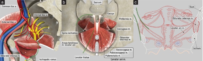
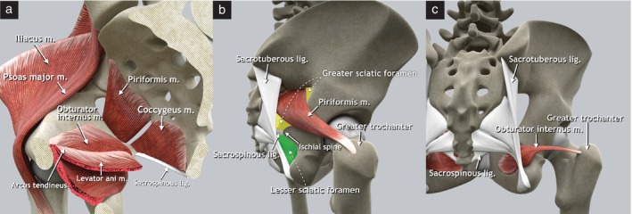
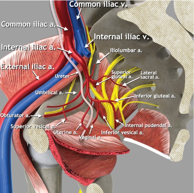
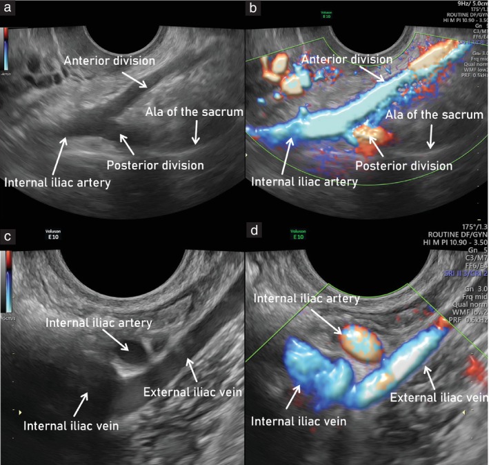
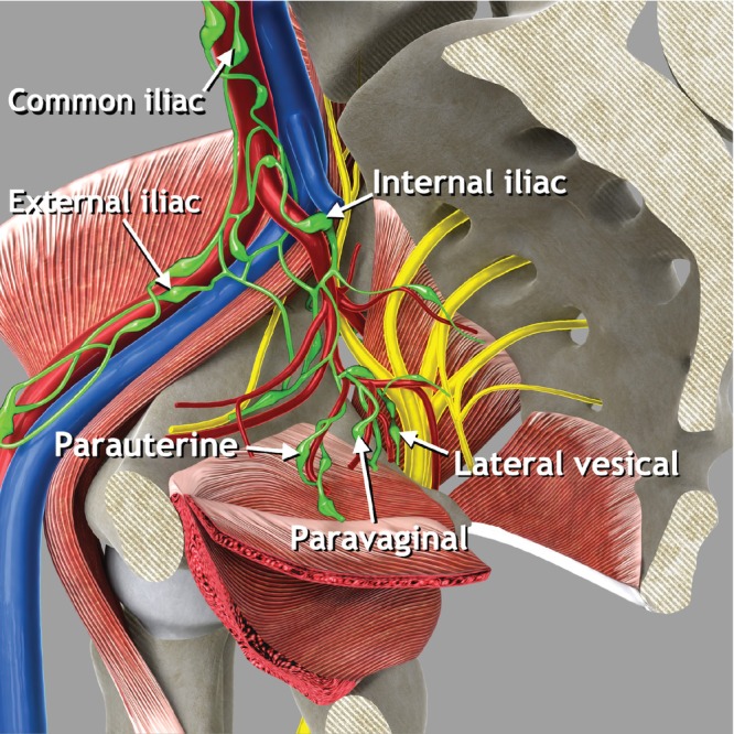
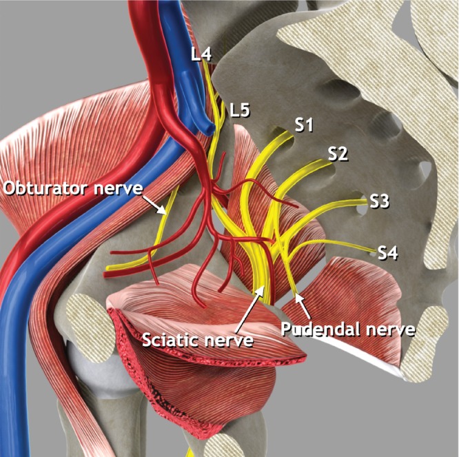
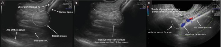
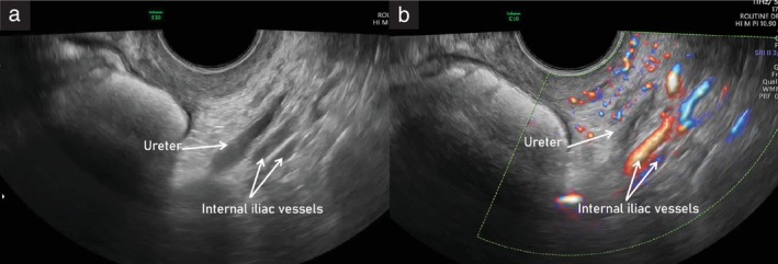
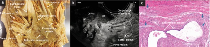
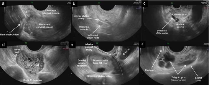
A standardized methodology for the ultrasound evaluation of the pelvic sidewall has not been proposed to date. Herein, a collaborative group of gynecologists and gynecological oncologists with extensive ultrasound experience presents a systematic methodology for the ultrasonographic evaluation of structures within the pelvic sidewall. Five categories of anatomical structures are described (muscles, vessels, lymph nodes, nerves and ureters). A step-by-step transvaginal ultrasound (or, when this is not feasible, transrectal ultrasound) approach is outlined for the evaluation of each anatomical landmark within these categories. Accurate assessment of the pelvic sidewall using a standardized approach improves the detection and diagnosis of non-gynecological pathologies that may mimic gynecological tumors, reducing the risk of unnecessary and even harmful intervention. Furthermore, it plays an important role in completing the staging of malignant gynecological conditions. Transvaginal or transrectal ultrasound therefore represents a viable alternative to magnetic resonance imaging in the preoperative evaluation of lesions affecting the pelvic sidewall, if performed by an expert sonographer. A series of videoclips showing normal and abnormal findings within each respective category illustrates how establishing a universally applicable approach for evaluating this crucial region will be helpful for assessing both benign and malignant conditions affecting the pelvic sidewall. © 2024 The Author(s). Ultrasound in Obstetrics & Gynecology published by John Wiley & Sons Ltd on behalf of International Society of Ultrasound in Obstetrics and Gynecology.

Keywords: anatomy; clinical cases; endometriosis; gynecological tumor; pelvic sidewall; transvaginal ultrasound.


Figures

Figure 1
Figure 1
Pelvic sidewall boundaries. Schematic diagrams showing boundaries on right hemipelvis: (a) medial view, with superior and inferior boundaries indicated (green dotted lines); (b) superior view, demonstrating anterior and posterior boundaries (green dotted lines); and (c) coronal view, demonstrating medial and lateral boundaries (green dotted lines). a., artery; m., muscle.
Figure 2
Figure 2
Pelvic sidewall muscles. (a) Schematic diagram of right hemipelvis, medial view. The arcus tendineus is a thickening of the fascia of the obturator internus muscle that serves as the origin of the iliococcygeus muscle. (b) Schematic diagram demonstrating insertion of piriformis muscle on apex of the greater trochanter of the femur and location of greater (yellow) and lesser (green) sciatic foramina (lateral view of right os coxae). The piriformis muscle divides the greater sciatic foramen into supra‐ and infrapiriform foramina. (c) Schematic diagram demonstrating insertion of obturator muscle onto the greater trochanter of the femur in dorsal view. lig., ligament; m., muscle.
Figure 3
Figure 3
Ultrasound assessment of pelvic sidewall muscles. Schematic diagrams showing transvaginal approach to evaluate pelvic sidewall muscles: (a) obturator internus and levator ani muscles; (b) piriformis muscle; and (c) and coccygeus muscle. m., muscle.
Figure 4
Figure 4
Pelvic sidewall vessels. Schematic diagram showing main pelvic sidewall vessels, with focus on anterior and posterior branches of the internal iliac artery. a., artery; v., vein.
Figure 5
Figure 5
Main vascular bifurcations in the pelvis (left side). Ultrasound images showing: (a,b) branching of internal iliac artery into its anterior and posterior parts (forming a V shape) on grayscale (a) and power Doppler (b) assessment; and (c,d) interiliac bifurcation, showing external and internal iliac veins, on grayscale (c) and power Doppler (d) assessment. To obtain the entire course of the external iliac vessels, the probe should be rotated 90° from transverse (a,b) to longitudinal (c,d) plane.
Figure 6
Figure 6
Pelvic parietal (iliac) and visceral lymph nodes. Schematic diagram showing pelvic parietal lymph nodes and some examples of pelvic visceral lymph nodes. Pelvic parietal (iliac) lymph nodes are divided into three groups: external iliac nodes (including obturator nodes), internal iliac nodes (including sacral nodes) and common iliac nodes. Some examples of visceral lymph nodes which can be assessed while evaluating the pelvic sidewall are shown: parauterine, paravaginal and lateral vesical lymph nodes.
Figure 7
Figure 7
Pelvic sidewall nerves. Schematic diagram showing main pelvic sidewall nerves and nerve roots. L, lumbar nerve root; S, sacral nerve root.
Figure 8
Figure 8
Ultrasound images of left sacral plexus. (a) Anatomic structures of the greater sciatic foramen with sacral plexus (dotted circle). (b) Transverse section of the nerves, exhibiting honeycomb‐like echotexture (dotted circles). (c) Longitudinal section of the nerves, exhibiting bundle‐of‐straw echotexture (dotted parallel lines); note, the sacral plexus can be traced up to the anterior sacral foramen (dotted oval) to identify the sacral nerve roots. m., muscle.
Figure 9
Figure 9
Abdominal and pelvic course of the ureter. Schematic diagrams demonstrating: (a) right hemipelvis, medial view, showing pelvic course of the ureter; (b) abdominal course of the ureter (purple), coronal view; and (c) pelvic course of the ureter, transverse view. a., artery, lig., ligament.
Figure 10
Figure 10
Ultrasound images showing course of the left ureter within the pelvic sidewall, running parallel to the internal iliac vessels, on grayscale (a) and power Doppler (b) imaging. The ureteric dilatation that can be observed in the grayscale image (a) is due to physiological peristalsis.
Figure 11
Figure 11
(a) Cadaveric specimen of left hemipelvis, showing sacral roots (S) and sacral plexus (dotted oval), leaving the pelvis through the greater sciatic foramen. (b) Ultrasound image showing left sacral plexus (dotted oval) infiltrated by a nodule of deep endometriosis (DE) with spiculated margins (dashed line) developing laterally from the cervix. Fibers of the sacral nerves are indicated (black arrows). (c) Corresponding histological image following laparoscopic excision of the lesion, showing perineural endometriosis: the endometrial glands are surrounded by connective tissue and stroma, nerve fibers are indicated (blue arrows) and striated muscle fibers (piriformis muscle) are visible in upper part of the specimen. Lat., lateral; m., muscle; Med., medial.
Figure 12
Figure 12
Ultrasound images illustrating various pathologies involving the pelvic sidewall. (a) Recurrent cervical cancer extending laterally on the right side, disrupting part of the obturator internus muscle and the iliac bone; (b) infiltrated pelvic parietal lymph node, which appears rounded with loss of normal sonographic architecture due to infiltration by cervical cancer, located along the course of the internal iliac branches; (c) right hydroureter due to locally advanced cervical cancer infiltrating the proximal third of the ureteric course; (d) Tarlov (perineural) cysts protruding from the right anterior sacral foramen of the first sacral vertebra (S1), presenting as multilocular cystic masses with thin septations; (e) pelvic‐nerve‐sheath tumor (Schwannoma) arising from the S1 sacral root and protruding into the greater sciatic foramen, presenting as a solid lesion with cystic areas; and (f) tailgut cysts (cystic hamartomas) located along the anterior surface of the sacral bone, presenting as multiloculated cystic masses with low‐level intracystic fluid (mucoid content).

Similar articles

Cited by

References

    1. Szabo G, Bokor A, Fancsovits V, et al. Clinical and ultrasound characteristics of deep endometriosis affecting sacral plexus. Ultrasound Obstet Gynecol. 2024;64:104‐111. - PubMed
    1. Guerriero S, Condous G, Rolla M, et al. Addendum to the consensus opinion from the International Deep Endometriosis Analysis (IDEA) group: sonographic evaluation of the parametrium. Ultrasound Obstet Gynecol. 2023;64:275‐280. - PubMed
    1. Timor‐Tritsch IE. Sliding organs sign in gynecological ultrasound. Ultrasound Obstet Gynecol. 2015;46:125‐126. - PubMed
    1. Fischerova D, Santos G, Wong L, et al. Imaging in gynecological disease (26): clinical and ultrasound characteristics of benign retroperitoneal pelvic peripheral‐nerve‐sheath tumors. Ultrasound Obstet Gynecol. 2023;62:727‐738. - PubMed
    1. Ramirez PT, Frumovitz M, Abu‐Rustum NR. Abdominal and pelvic anatomy. Principles of Gynecologic Oncology Surgery. Elsevier; 2021:3‐49.

Publication types

LinkOut - more resources