Review
. 2024 Dec 3;26(12):euae279.
doi: 10.1093/europace/euae279.

Ventricular arrhythmias in association with athletic cardiac remodelling

Affiliations
Review

Ventricular arrhythmias in association with athletic cardiac remodelling

Paolo D'Ambrosio et al. Europace. .

Abstract

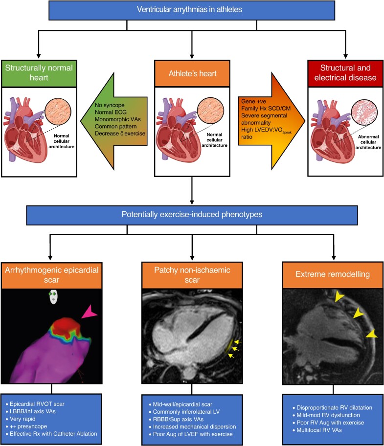
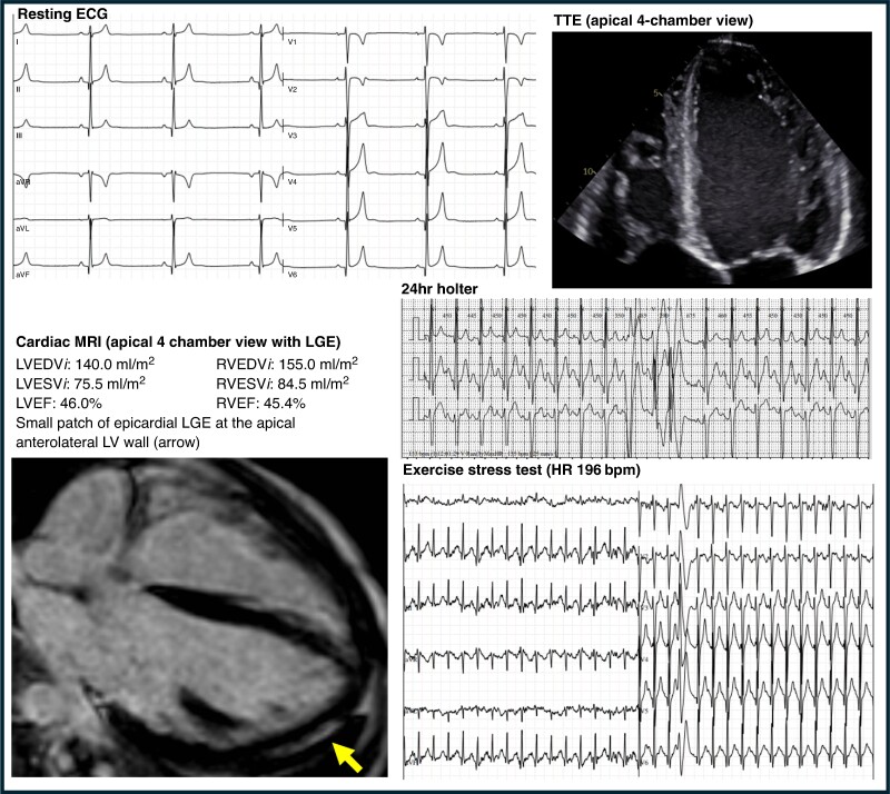
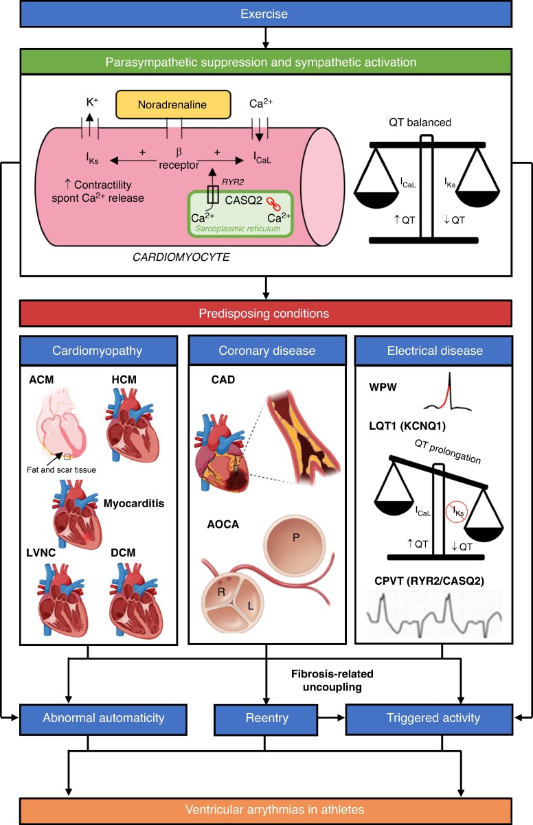
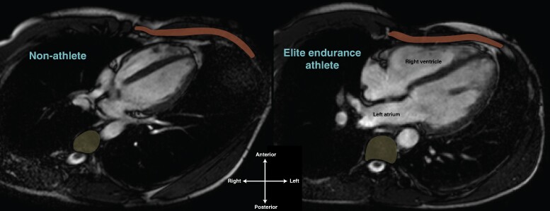
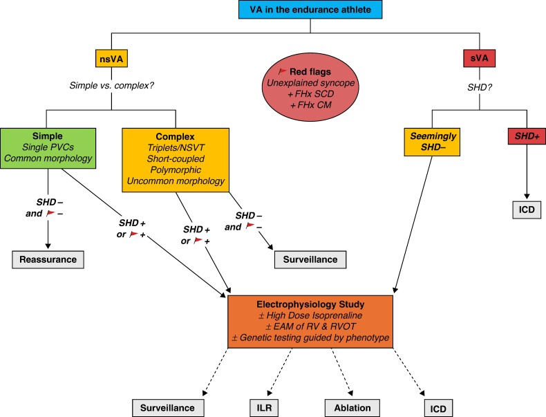
Athletes are predisposed to atrial arrhythmias but the association between intense endurance exercise training, ventricular arrhythmias (VAs), and sudden cardiac death is less well established. Thus, it is unclear whether the 'athlete's heart' promotes specific arrhythmias or whether it represents a more general pro-arrhythmogenic phenotype. Whilst direct causality has not been established, it appears possible that repeated exposure to high-intensity endurance exercise in some athletes contributes to formation of pro-arrhythmic cardiac phenotypes that underlie VAs. Theories regarding potential mechanisms for exercise-induced VAs include repeated bouts of myocardial inflammation and stretch-induced cellular remodelling. Small animal models provide some insights, but larger animal and human data are sparse. The current clinical approach to VAs in athletes is to differentiate those with and without structural or electrical heart disease. However, if the athlete's heart involves a degree of pro-arrhythmogenic remodelling, then this may not be such a simple dichotomy. Questions are posed by athletes with VAs in combination with extreme remodelling. Some markers, such as scar on magnetic resonance imaging, may point towards a less benign phenotype but are also quite common in ostensibly healthy athletes. Other clinical and invasive electrophysiology features may be helpful in identifying the at-risk athlete. This review seeks to discuss the association between athletic training and VAs. We will discuss the potential mechanisms, clinical significance, and approach to the management of athletes with VAs.

Keywords: Arrhythmogenic cardiomyopathy; Athlete; Athlete’s heart; Electroanatomic mapping; Electrophysiology study; Endurance athlete; Non-ischaemic LV scar; Premature ventricular complex; Sudden cardiac death; Ventricular arrhythmias; Ventricular tachycardia.


Conflict of interest statement

Conflict of interest: none declared.

Figures

Graphical Abstract
Graphical Abstract
Ventricular arrhythmias in athletes. VAs can be found in athletes with structurally normal hearts, those with underlying structural/electrical heart disease or athlete’s heart. Various factors can help differentiate benign athletic remodelling from structural disease. Under the umbrella term of athlete’s heart are potentially exercise-induced phenotypes including arrhythmogenic epicardial scar, patchy non-ischaemic scar (e.g. non-ischaemic LV scar), and extreme remodelling (e.g. exercise-induced arrhythmogenic cardiomyopathy). ECG, electrocardiogram; VAs, ventricular arrhythmias; SCD, sudden cardiac death; CM, cardiomyopathy; LVEDV, left ventricular end-diastolic volume; VO2Peak, absolute peak VO2; RVOT, right ventricular outflow tract; LBBB, left bundle branch block; Rx, treated; LV, left ventricle; RBBB, right bundle branch block; Aug, augmentation; LVEF, left ventricular ejection fraction; RV, right ventricle. Image created with BioRender.com.
Figure 1
Figure 1
Twenty-three-year-old male elite cyclist with sudden cardiac death during competition. Investigations are presented for an asymptomatic elite cyclist after an initial screening 12-lead electrocardiogram (ECG), top left. T-wave inversion in V2 (a common but potentially abnormal athletic trait) prompted investigation with a transthoracic echocardiogram (TTE) on which moderate to severe chamber dilation and low-normal systolic function was identified (top right). A 24 h Holter monitor showed 370 premature ventricular complexes (PVCs) per 24 h, two couplets and one triplet [shown is a fusion beat followed by two left bundle branch block (LBBB) morphology PVCs]. Exercise stress test showed three PVCs at peak exercise [shown is one of these PVCs with right bundle branch block (RBBB) morphology]. Cardiac magnetic resonance (MRI) demonstrated athletic remodelling with dilatation of all chambers, mildly reduced LV and RV function and a small area of epicardial late gadolinium enhancement (LGE, scar) in the apical anterolateral LV wall (arrow). The athlete remained asymptomatic until suffering a cardiac arrest during competition several months after these investigations were completed. LVEDVi, left ventricular end-diastolic volume indexed to body surface area; LVESVi, left ventricular end-systolic volume indexed to body surface area; RVEDVi, right ventricular end-diastolic volume indexed to body surface area; RVESVi, right ventricular end-systolic volume indexed to body surface area; LVEF, left ventricular ejection fraction; RVEF, right ventricular ejection fraction; HR, heart rate.
Figure 2
Figure 2
A schematic of some of the proposed mechanisms for VAs in athletes. Exercise activates the sympathetic nervous system (SNS) and suppresses the parasympathetic nervous system. Binding of noradrenaline to cardiomyocytes and physiological adaptation of Ca2+ homeostasis promotes increased triggered activity. SNS activation also leads to abnormal automaticity. Concomitant activation of IKs ensures the QT is balanced. Increase in cardiac impulse formation via the above mechanisms can lead to idiopathic VAs in structurally normal hearts. Reentry is promoted by underlying myocardial fibrosis associated with cardiomyopathies and coronary anomalies. Some idiopathic VAs (e.g. LV fascicular VT) are also caused by reentry. Reentry itself can lead to triggered activity via fibrosis-related uncoupling. The combination of abnormal cardiac impulse formulation and reentry can lead to malignant VAs and SCD in athletes with predisposing conditions. Electrical diseases also cause VAs via other mechanisms. Long QT Syndrome type 1 (LQT1) syndrome is caused by pathological variants in the KCNQ1 gene whilst catecholaminergic polymorphic ventricular tachycardia (CPVT) is caused by pathogenic variants in Ryanodine Receptor 2 (RYR2, autosomal dominant, 50%) and Calsequestrin 2 (CASQ2, autosomal recessive, 2%) genes. Please note that this schematic does not include other proposed mechanisms for VAs in athletes such as stretch-activated ion channel expression and stretch-mediated gene expression. K+, potassium; Ca2+, calcium; β Receptor, β adrenergic receptor; IKs, slow delayed rectifier potassium channel; ICaL, L-type Ca2+ channel; ACM, arrhythmogenic cardiomyopathy; HCM, hypertrophic cardiomyopathy; LVNC, left ventricular non-compaction cardiomyopathy; DCM, dilated cardiomyopathy; CAD, coronary artery disease; AOCA, anomalous origin of the coronary arteries; WPW, Wolff–Parkinson–White syndrome. Image created with BioRender.com.
Figure 3
Figure 3
The athlete's heart within the thorax. Horizontal long axis cardiac MRI of a non-athlete (left) and elite endurance athlete’s heart (right), both 24-year-old males. Compared to the non-athlete, note the significantly greater proportion of the thorax inhabited by the athlete’s heart and the intimate relationship between the right ventricle and the chest wall (anterior, rust) and the left atrium and thoracic vertebrae (posterior, yellow). The right ventricle and left atrium of the athlete’s heart appear to be deformed in shape compared to the non-athlete.
Figure 4
Figure 4
Management of VAs in athletes. nsVA, non-sustained ventricular arrhythmia—any VA lasting <30 s; sVA, sustained ventricular arrhythmia—ventricular tachycardia lasting ≥30 s and/or requiring intervention and/or ventricular fibrillation. Common premature ventricular complex (PVC) morphology: outflow tract/infundibular (LBBB morphology and inferior axis) and/or fascicular (typical RBBB morphology, superior/inferior axis, and QRS < 130 ms). Uncommon PVC morphology: all other types of PVCs. Short-coupled PVCs: PVC with interval from onset of preceding QRS to onset of PVC < 350 ms. SHD, structural heart disease; ICD, implantable cardioverter defibrillator; NSVT, non-sustained ventricular tachycardia; FHx SCD, family history of premature sudden cardiac death (male < 40 years, female < 50 years); FHx CM, family history of cardiomyopathy; EAM, electroanatomic mapping; RV, right ventricle; RVOT, right ventricular outflow tract; ILR, implantable loop recorder.

References

    1. Kokkinos P, Myers J, Faselis C, Panagiotakos DB, Doumas M, Pittaras A et al. Exercise capacity and mortality in older men: a 20-year follow-up study. Circulation 2010;122:790–7. - PubMed
    1. Baldesberger S, Bauersfeld U, Candinas R, Seifert B, Zuber M, Ritter M et al. Sinus node disease and arrhythmias in the long-term follow-up of former professional cyclists. Eur Heart J 2008;29:71–8. - PubMed
    1. Claessen G, Colyn E, La Gerche A, Koopman P, Alzand B, Garweg C et al. Long-term endurance sport is a risk factor for development of lone atrial flutter. Heart 2011;97:918–22. - PubMed
    1. Grimsmo J, Grundvold I, Maehlum S, Arnesen H. High prevalence of atrial fibrillation in long-term endurance cross-country skiers: echocardiographic findings and possible predictors—a 28–30 years follow-up study. Eur J Prev Cardiol 2010;17:100–5. - PubMed
    1. Aizer A, Gaziano JM, Cook NR, Manson JE, Buring JE, Albert CM. Relation of vigorous exercise to risk of atrial fibrillation. Am J Cardiol 2009;103:1572–7. - PMC - PubMed

MeSH terms

LinkOut - more resources