

Analysis of clinicopathological factors associate with the visibility of early gastric cancer in endoscopic examination and usefulness of linked color imaging: A multicenter prospective study
- PMID: 39499715
- PMCID: PMC11537390
- DOI: 10.1371/journal.pone.0312385
Analysis of clinicopathological factors associate with the visibility of early gastric cancer in endoscopic examination and usefulness of linked color imaging: A multicenter prospective study
Erratum in
-
Correction: Analysis of clinicopathological factors associate with the visibility of early gastric cancer in endoscopic examination and usefulness of linked color imaging: A multicenter prospective study.PLoS One. 2025 Apr 8;20(4):e0322354. doi: 10.1371/journal.pone.0322354. eCollection 2025. PLoS One. 2025. PMID: 40198597 Free PMC article.
Abstract
Background: This study investigated clinicopathological factors associated with the visibility of early gastric cancer and the efficacy of linked color imaging.
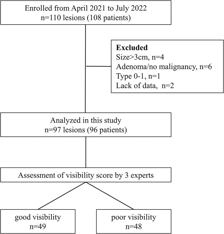
Methods: Patients with early gastric cancer who underwent endoscopic treatment between April 2021 and July 2022 were enrolled. All cases underwent white light imaging and linked color imaging. Three experts evaluated lesion visibility using a visual analog scale. A mean score ≥3 on white light imaging was defined as "good visibility", and <3 as "poor visibility". We extracted patient information and endoscopic and pathological data for the lesion and background mucosa, analyzed factors associated with the visibility of early gastric cancer, and compared visibility between white light imaging and linked color imaging.
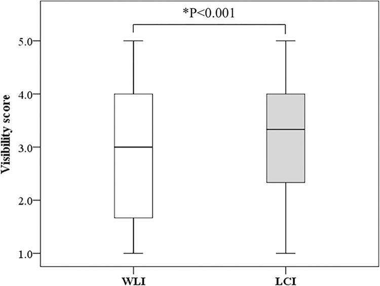
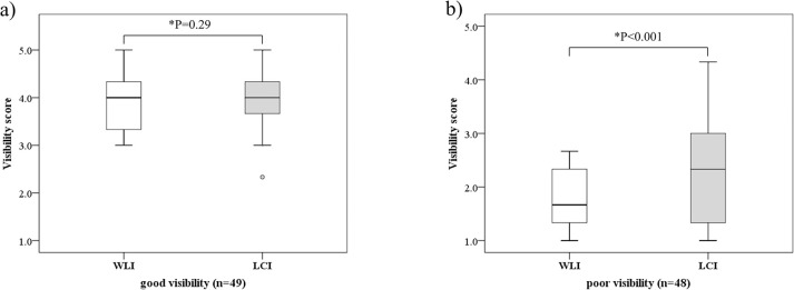
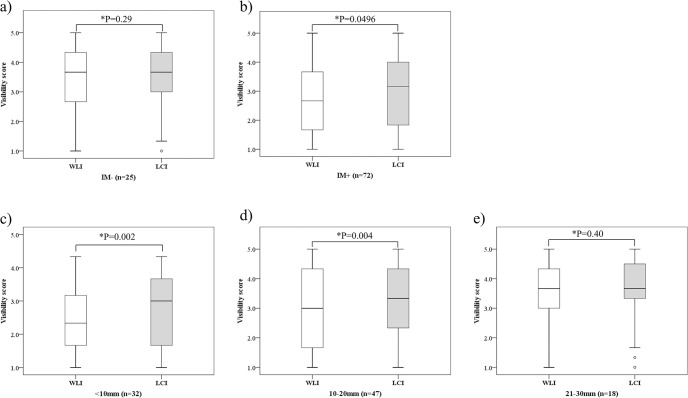
Results: Ninety-seven lesions were analyzed, with good visibility in 49 and poor visibility in 48. Multivariate analysis revealed small lesion size (odds ratio 1.89) and presence of endoscopic intestinal metaplasia (odds ratio 0.49) as significantly associated with the poor visibility of early gastric cancer. Mean visibility score was significantly higher for linked color imaging (P<0.001). Mean score for linked color imaging was significantly higher in the poor visibility group (P<0.001), but not significantly different in the good visibility group (P = 0.292). Mean score was significantly higher with linked color imaging in cases with endoscopic intestinal metaplasia (P = 0.0496) and lesions <20 mm in diameter (<10 mm, P = 0.002; 10-20 mm, P = 0.004).
Conclusions: Lesion size and endoscopic intestinal metaplasia are associated with the visibility of early gastric cancer in white light imaging. Linked color imaging improves visibility of gastric cancer with these factors.
Copyright: © 2024 Fukuda et al. This is an open access article distributed under the terms of the Creative Commons Attribution License, which permits unrestricted use, distribution, and reproduction in any medium, provided the original author and source are credited.
Conflict of interest statement
The authors have declared that no competing interests exist.
Figures
References
-
- World Health Organization Fact Sheets on Cancer. https://www.who.int/news-room/fact-sheets/detail/cancer. Accessed July 10, 2023.
-
- Shibagaki K, Mishiro T, Fukuyama C, Takahashi Y, Itawaki A, Nomura S, et al.. Sporadic foveolar-type gastric adenoma with a raspberry-like appearance in Helicobacter pylori-naïve patients. Virchows Arch. 2021;479(4):687–95. - PubMed
Publication types
MeSH terms
LinkOut - more resources
Full Text Sources
Medical
Miscellaneous
