Observational Study
. 2024 Dec 10;103(11):e210008.
doi: 10.1212/WNL.0000000000210008. Epub 2024 Nov 5.

Association of Cerebrovascular Reactivity With 1-Year Imaging and Clinical Outcomes in Small Vessel Disease: An Observational Cohort Study

Affiliations
Observational Study

Association of Cerebrovascular Reactivity With 1-Year Imaging and Clinical Outcomes in Small Vessel Disease: An Observational Cohort Study

Emilie Sleight et al. Neurology. .

Abstract

Background and objectives: In patients with cerebral small vessel disease (SVD), impaired cerebrovascular reactivity (CVR) is related to worse concurrent SVD burden, but less is known about cerebrovascular reactivity and long-term SVD lesion progression and clinical outcomes. We investigated associations between cerebrovascular reactivity and 1-year progression of SVD features and clinical outcomes.

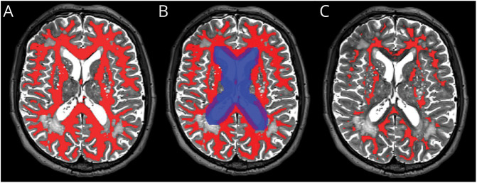
Methods: Between 2018 and 2021, we recruited patients from the Edinburgh/Lothian stroke services presenting with minor ischemic stroke and SVD features as part of the Mild Stroke Study 3, a prospective observational cohort study (ISRCTN 12113543). We acquired 3T brain MRI at baseline and 1 year. At baseline, we measured cerebrovascular reactivity to 6% inhaled CO2 in subcortical gray matter, normal-appearing white matter, and white matter hyperintensities (WMH). At baseline and 1 year, we quantified SVD MRI features, incident infarcts, assessed stroke severity (NIH Stroke Scale), recurrent stroke, functional outcome (modified Rankin Scale), and cognition (Montreal Cognitive Assessment). We performed linear and logistic regressions adjusted for age, sex, and vascular risk factors, reporting the regression coefficients and odds ratios with 95% CIs.

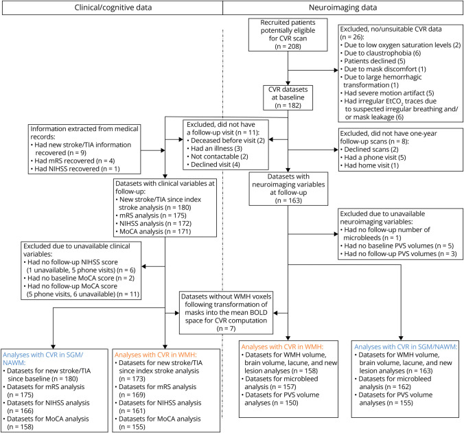
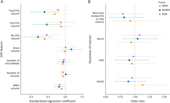
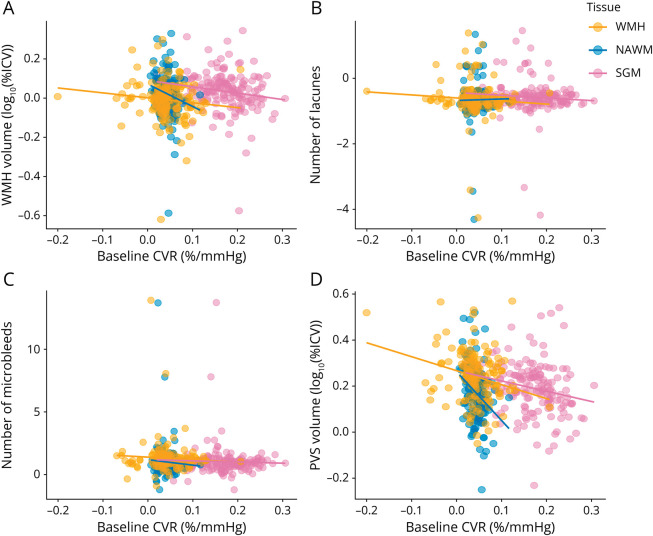
Results: We recruited 208 patients of whom 163 (mean age and SD: 65.8 ± 11.2 years, 32% female) had adequate baseline CVR and completed the follow-up structural MRI. The median increase in WMH volume was 0.32 mL with (Q1, Q3) = (-0.48, 1.78) mL; 29% had a recurrent stroke or incident infarct on MRI. At 1 year, patients with lower baseline cerebrovascular reactivity in normal-appearing tissues had increased WMH (regression coefficient: B = -1.14 [-2.13, -0.14] log10 (%ICV) per %/mm Hg) and perivascular space volumes (B = -1.90 [-3.21, -0.60] log10 (%ROIV) per %/mm Hg), with a similar trend in WMH. CVR was not associated with clinical outcomes at 1 year.

Discussion: Lower baseline cerebrovascular reactivity predicted an increase in WMH and perivascular space volumes after 1 year. CVR should be considered in SVD future research and intervention studies.


Conflict of interest statement

M.S. Stringer was part-funded by Siemens Healthineers, administered by the University of Edinburgh. The other authors report no conflicts. Go to Neurology.org/N for full disclosures.

Figures

Figure 1
Figure 1. Example of Tissue Processing for the CVR Analysis: NAWM Mask in a Participant's T2W Space
(A) NAWM mask (red mask) before processing. (B) The original NAWM mask (red mask) was eroded by 1 mm in all directions, and voxels within the dilated mask of the ventricles (blue mask) were removed. (C) The final NAWM mask (red mask) before registration to the participant's mean BOLD space. BOLD = blood oxygen level-dependent; NAWM = normal-appearing white matter.
Figure 2
Figure 2. Flow Diagram of Study Recruitment, Data Collection, and Analysis
The blue numbers correspond to number of data sets related to CVR analyses in SGM and NAWM, whereas the yellow ones correspond to number of data sets related to CVR analyses in WMH. BOLD = blood oxygen level-dependent; CVR = cerebrovascular reactivity; EtCO2 = end-tidal CO2; MoCA; Montreal Cognitive Assessment; mRS = modified Rankin score; NAWM = normal-appearing white matter; NIHSS = NIH Stroke Scale; PVS = perivascular space; SGM = subcortical gray matter; WMH = white matter hyperintensity.
Figure 3
Figure 3. Standardized Regression Coefficients and Odds Ratios Between Parameter of Interest at 1-Year Follow-Up and Baseline CVR
(A) Standardized regression coefficients between CVR and quantitative SVD features. (B) Odds ratios between CVR and stroke severity, cognition, recurrent strokes, and new infarcts. CVR was computed in SGM (pink), NAWM (blue), and WMH (yellow). All models were adjusted for the corresponding parameter of interest at baseline (except for new strokes/lesions), age, sex, and vascular risk factors. Models related to NIHSS, modified Rankin scale, MoCA, and new strokes/lesions were also adjusted for baseline WMH volume. The dots represent the mean standardized coefficients and the horizontal lines the associated 95% CIs. The vertical dashed line emphasizes a zero-valued coefficient for linear models and an odds ratio of 1 for ordinal logistic regressions. BG = basal ganglia; CSO = centrum semiovale; CVR = cerebrovascular reactivity; MoCA = Montreal Cognitive Assessment; mRS = modified Rankin Score; NAWM = normal-appearing white matter; NIHSS = NIH Stroke Scale; PVS = perivascular space; SGM = subcortical gray matter; SVD = small vessel disease; WMH = white matter hyperintensity.
Figure 4
Figure 4. Relationships Between SVD Features After 1 Year and Baseline CVR
SVD features shown are (A) WMH volume, (B) number of lacunes, (C) number of microbleeds, and (D) BG PVS volumes. SVD features of interest at 1-year follow-up were adjusted for the corresponding SVD features of interest at baseline, age, sex, and vascular risk factors. The results are shown for adjusted CVR in SGM (pink), NAWM (green), and WMH (blue). The regression lines are shown in the plots. BG = basal ganglia; CVR = cerebrovascular reactivity; NAWM = normal-appearing white matter; PVS = perivascular space; SGM = subcortical gray matter; SVD = small vessel disease; WMH = white matter hyperintensity.

References

    1. Georgakis MK, Duering M, Wardlaw JM, Dichgans M. WMH and long-term outcomes in ischemic stroke: a systematic review and meta-analysis. Neurology. 2019;92(12):e1298-e1308. doi:10.1212/WNL.0000000000007142 - DOI - PubMed
    1. Sweeney MD, Montagne A, Sagare AP, et al. . Vascular dysfunction—the disregarded partner of Alzheimer's disease. Alzheimers Dement. 2019;15(1):158-167. doi:10.1016/j.jalz.2018.07.222 - DOI - PMC - PubMed
    1. Gorelick PB, Scuteri A, Black SE, et al. . Vascular contributions to cognitive impairment and dementia: a statement for healthcare professionals from the American Heart Association/American Stroke Association. Stroke. 2011;42(9):2672-2713. doi:10.1161/STR.0b013e3182299496 - DOI - PMC - PubMed
    1. Wardlaw JM, Smith C, Dichgans M. Small vessel disease: mechanisms and clinical implications. Lancet Neurol. 2019;18(7):684-696. doi:10.1016/S1474-4422(19)30079-1 - DOI - PubMed
    1. Bath PM, Wardlaw JM. Pharmacological treatment and prevention of cerebral small vessel disease: a review of potential interventions. Int J Stroke. 2015;10(4):469-478. doi:10.1111/ijs.12466 - DOI - PMC - PubMed

Publication types